1806
The Feasibility of 3D MR Fingerprinting using Cartesian sampling for isotropic 1mm3 resolution at 0.55T1Department of Radiology, University of Michigan, Ann Arbor, MI, United States, 2Department of Electrical and Computer Engineering, University of Michigan, Ann Arbor, MI, United States, 3Department of Biomedical Engineering, University of Texas at Austin, Austin, TX, United States, 4Oden Institute for Computational Engineering and Sciences, University of Texas at Austin, Austin, TX, United States
Synopsis
The purpose of this work is to assess the feasibility of performing MR Fingperprinting with 3D pseudorandom Cartesian sampling for an isotropic spatial resolution of 1mm3 at 0.55T in the brain.
Introduction
The purpose of this work is to assess the feasibility of performing MRF with 3D pseudorandom Cartesian sampling for an isotropic spatial resolution of 1mm3 at 0.55T in the brain. MR Fingerprinting (MRF)1 is a quantitative imaging technique which can be used to estimate multiple tissue properties efficiently and accurately2,3. Most MRF studies up to date have been performed at the field strength of 1.5T or 3T. A recently introduced 0.55T scanner with modernized gradient and RF hardware provides a potential platform for implementing advanced imaging acquisition and reconstruction with lower cost compared to higher field scanners4. However, the inherently lower signal-to-noise ratio at 0.55 T makes it challenging to achieve the high spatial resolutions required for some clinical applications. Previous work has shown that it is possible to quantify T1 and T2 with a spatial resolution of 1.2×1.2×5 mm3 using 3D MRF acquisition with a stack-of-spirals trajectory at 0.55T5. This work seeks to generate maps with an isotropic spatial resolution of 1 mm3 by moving to a pseudorandom Cartesian sampling approach in conjunction with MRF.Methods
The proposed 3D MRF-FISP acquisition with a pseudorandom Cartesian sampling was implemented on a 0.55T scanner (MAGNETOM Free.Max, Siemens Healthcare, Erlangen, Germany); note the reduced gradient performance of this system compared with higher field scanners (maximal gradient amplitude of 15 mT/m and a maximal slew rate of 40 mT/m/ms). All experiments were performed with a 12-channel head receiver array; MRF maps in the ISMRM/NIST system phantom and the brains of five subjects were collected in this IRB-approved study.A total of 500 time points were acquired with flip angles described previously2, a TR of 5.9 ms, and an echo time of 2.9 ms. Each time point was highly undersampled with a pseudorandom Poisson-disc sampling pattern; for each 3D volume, 283 readout lines with a bandwidth of 605 Hz/pixel were acquired. For a matrix size of 192×192×192, the approximate undersampling rate was ~100 for each time point. The total scan time was 14 minutes.
The reconstruction consisted of two steps. First, an iterative low-rank reconstruction similar to that applied in other MRF applications6 was performed to compress the highly undersampled images to 6 coefficient images. Then, tissue properties were retrieved by finding the dictionary entry with the highest cross-correlation to the coefficient images. The dictionary of potential signal evolutions was calculated using a Bloch equation simulation and consisted of 31,605 entries with T1 of 10 – 5000 ms, T2 of 2 – 800 ms.
Results
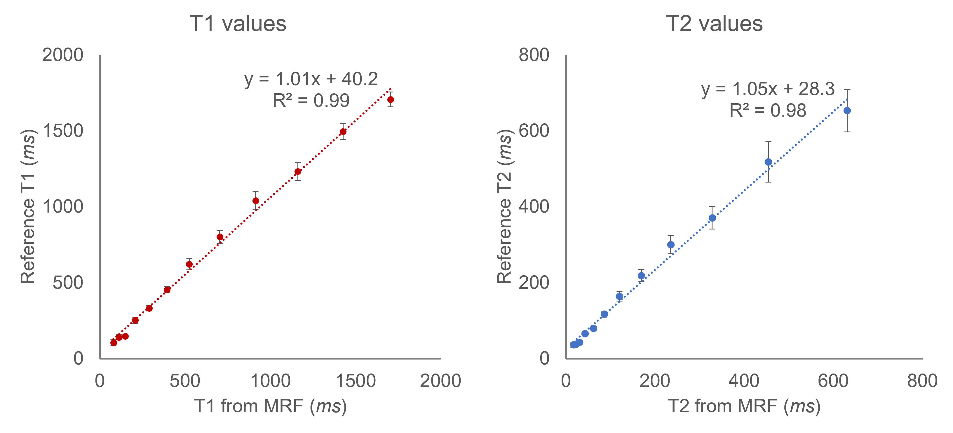
Figure 1 shows correlation curves comparing the T1 and T2 values derived from the proposed method to the standard values measured by the spin-echo methods in the ISMRM/NIST MR system phantom. The MRF measurements from the proposed approach are in good agreement with reference values.Figure 2 shows T1 and T2 maps from one slice of the 3D volume collected in the brain acquired with the proposed method. T1 and T2 values of various regions of interest are shown in the following Table, which are in the range of previously reported values from 3D spiral MRF-FISP at 0.55T5.
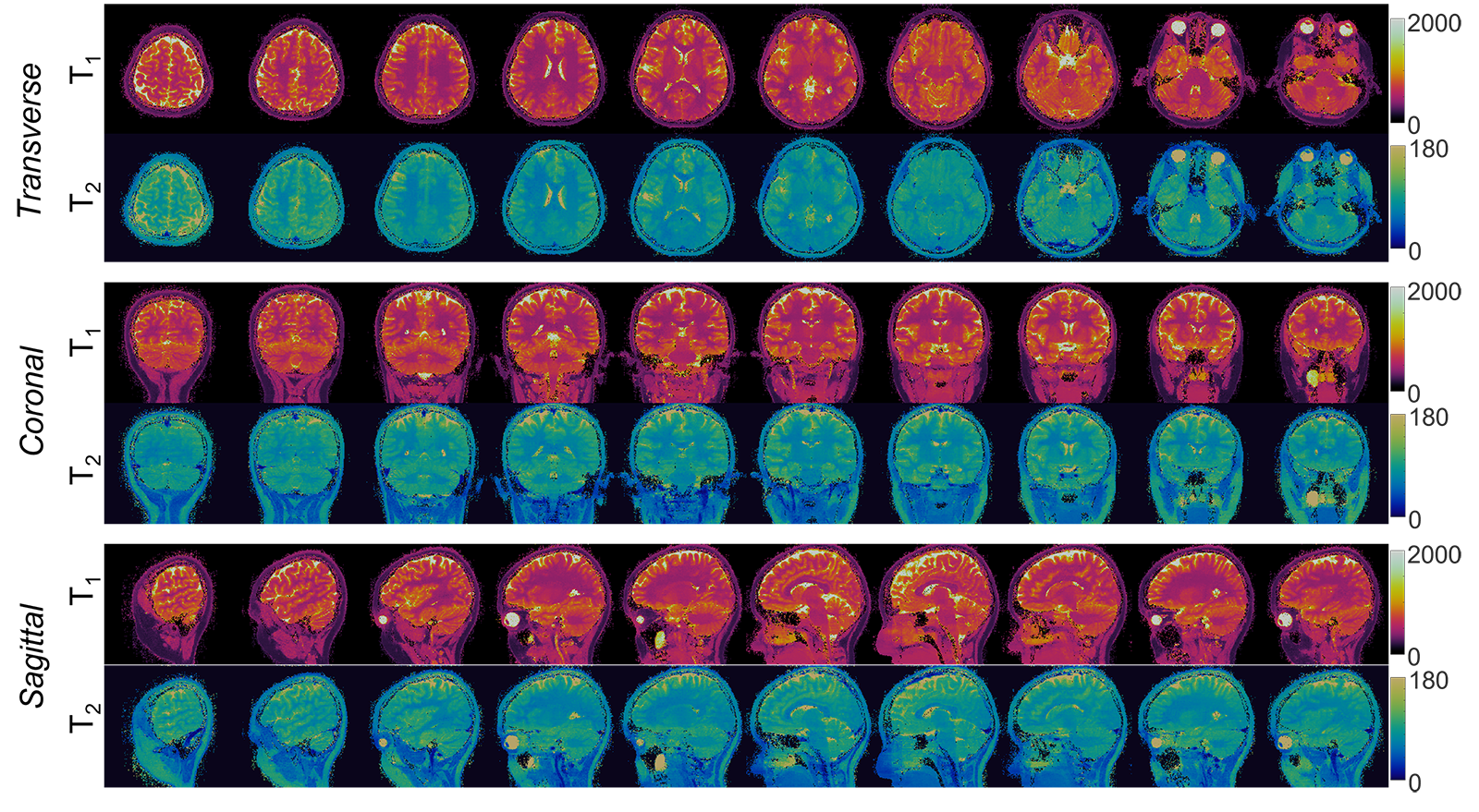
Figure 3 shows reformatted T1 and T2 maps in transverse, sagittal, and coronal planes, which demonstrate high quality T1 and T2 maps for the whole brain.
Discussion and Conclusions
In summary, this study shows that it is feasible to measure high-resolution (1mm3 isotropic) T1 and T2 maps simultaneously in the brain using MRF at 0.55T. Compared to the non-Cartesian read-outs often used in MRF, the proposed method provides a flexible acquisition scheme based on a Cartesian acquisition. Additionally, this Cartesian sampling does not require high-performance gradients, enabling these measurements to be made on the 0.55T scanner with its weaker gradient performance. Further analysis on the comparison between the Cartesian and non-Cartesian acquisitions will be performed to find the most efficient acquisition strategy for this field strength and gradient specifications.Acknowledgements
This work was supported by Siemens Healthcare and NIH grants R37CA263583 and R01CA208236.References
1. Ma D, Gulani V, Seiberlich N, Liu K, Sunshine JL, Duerk JL, Griswold MA. Magnetic resonance fingerprinting. Nature. 2013 Mar 14;495(7440):187–192. PMCID: PMC3602925
2. Jiang Y, Ma D, Keenan KE, Stupic KF, Gulani V, Griswold MA. Repeatability of magnetic resonance fingerprinting T1 and T2 estimates assessed using the ISMRM/NIST MRI system phantom. Magn Reson Med. 2017;78(4):1452–1457. PMCID: PMC5408299
3. Körzdörfer G, Kirsch R, Liu K, Pfeuffer J, Hensel B, Jiang Y, Ma D, Gratz M, Bär P, Bogner W, Springer E, Lima Cardoso P, Umutlu L, Trattnig S, Griswold M, Gulani V, Nittka M. Reproducibility and Repeatability of MR Fingerprinting Relaxometry in the Human Brain. Radiology. 2019;292(2):429–437. PMID: 31210615
4. Campbell-Washburn AE, Ramasawmy R, Restivo MC, Bhattacharya I, Basar B, Herzka DA, Hansen MS, Rogers T, Bandettini WP, McGuirt DR, Mancini C, Grodzki D, Schneider R, Majeed W, Bhat H, Xue H, Moss J, Malayeri AA, Jones EC, Koretsky AP, Kellman P, Chen MY, Lederman RJ, Balaban RS. Opportunities in Interventional and Diagnostic Imaging by Using High-Performance Low-Field-Strength MRI. Radiology. 2019 Nov;293(2):384–393. PMCID: PMC6823617
5. Feasibility of MR fingerprinting using a high-performance 0.55 T MRI system - ScienceDirect [Internet]. [cited 2021 Nov 9]. Available from: https://www.sciencedirect.com/science/article/pii/S0730725X21000904
6. Zhao B, Setsompop K, Adalsteinsson E, Gagoski B, Ye H, Ma D, Jiang Y, Ellen Grant P, Griswold MA, Wald LL. Improved magnetic resonance fingerprinting reconstruction with low-rank and subspace modeling. Magn Reson Med. 2018;79(2):933–942. PMCID: PMC5641478
Figures