1788
Predicting artifacts in maximum intensity projections of high b-value DWI of the breast using neural networks.1Institute of Radiology, University Hospital Erlangen, Friedrich-Alexander-Universität Erlangen-Nürnberg (FAU), Erlangen, Germany, 2Pattern Recognition Lab, Department of Computer Science, Friedrich-Alexander University Erlangen Nuremberg, Erlangen, Germany
Synopsis
DWI acquisitions in MRI are prone to artifacts caused e.g. by insufficient fat saturation and can significantly impede the diagnostic assessment. We investigated the ability to predict the occurrence of these artifacts in maximum intensity projections (MIPs) of high b-value DWI using a neuronal network that analyses T2w images, which were acquired prior to the DWI sequence. An AuRoC of 0.83 with sensitivity of 0.82 and specificity of 0.61 was achieved. Investigation of the regions of the T2w images most important for the decision of the neural network showed a good correlation with artifact-affected areas in the DWI MIP images.
Introduction
Maximum intensity projections (MIPs) of high b-value DWI acquisitions were recently shown to decrease the initial reading time for radiologists in breast MRI1-3. However, such MIP DWI images are sensitive to artifacts, which can potentially cover lesions making them undetectable. As improper adjustments – such as a bad shim – may be a major cause of these artifacts, a prediction of artifacts might be highly valuable for optimizing or adapting the adjustment process. This should be possible as imperfect adjustments also leave a fingerprint in morphological images. The effect size is much reduced in comparison to that observed in EPI DWI images and the image alterations might be hardly noticeable to the human eye. But it is reasonable to assume that well-trained neural networks should be able to discern them.In this work we propose a method to predict the occurrence of the high intensity artifacts in DWI MIP’s using a neural network which analyses a previously acquired T2-weigthed fat-saturated volume.
Materials and Methods
This IRB approved retrospective study included patients undergoing standard breast MRI at 1.5T Aera, 3T Skyra Fit or 3T Vida MRI systems (Siemens Healthineers, Erlangen Germany), with a multichannel breast coil. The full diagnostic protocol included T1w, T2w, DCE and a multi-b-value DWI sequence (b-values of 50, 750 and 1500 s/mm2). MIPs were derived from DWI acquisitions acquired with the highest b-value (1500 s/mm2). The MIPs were labelled for the presence of high intensity artifacts presumably caused by bad shimming or insufficient fat saturation by an experienced radiologist (10 years experience). Overall n=1099 patient datasets were used for this work.A 3D-DensetNet2014 network was trained using as input data the volumetric 3D T2 weighted fat saturated acquisitions. These T2 weighted acquisitions were acquired directly before the DWI acquisition and used the same shimming and adjustments volumes. The network was trained using 989 datasets from which 791 were used as the training dataset and 198 were used as the validation dataset using a stratified split. The networks were trained using the binary cross-entropy loss function. The ADAM5 optimizer was used together with an joined cosine annealing scheduling of the learning rate from 5 sup> images were derived from the networks and overlaid on the labelled MIP images. For the evaluation of the performance, the sensitivity, specificty and AuRoC were used.
Results and Discussion
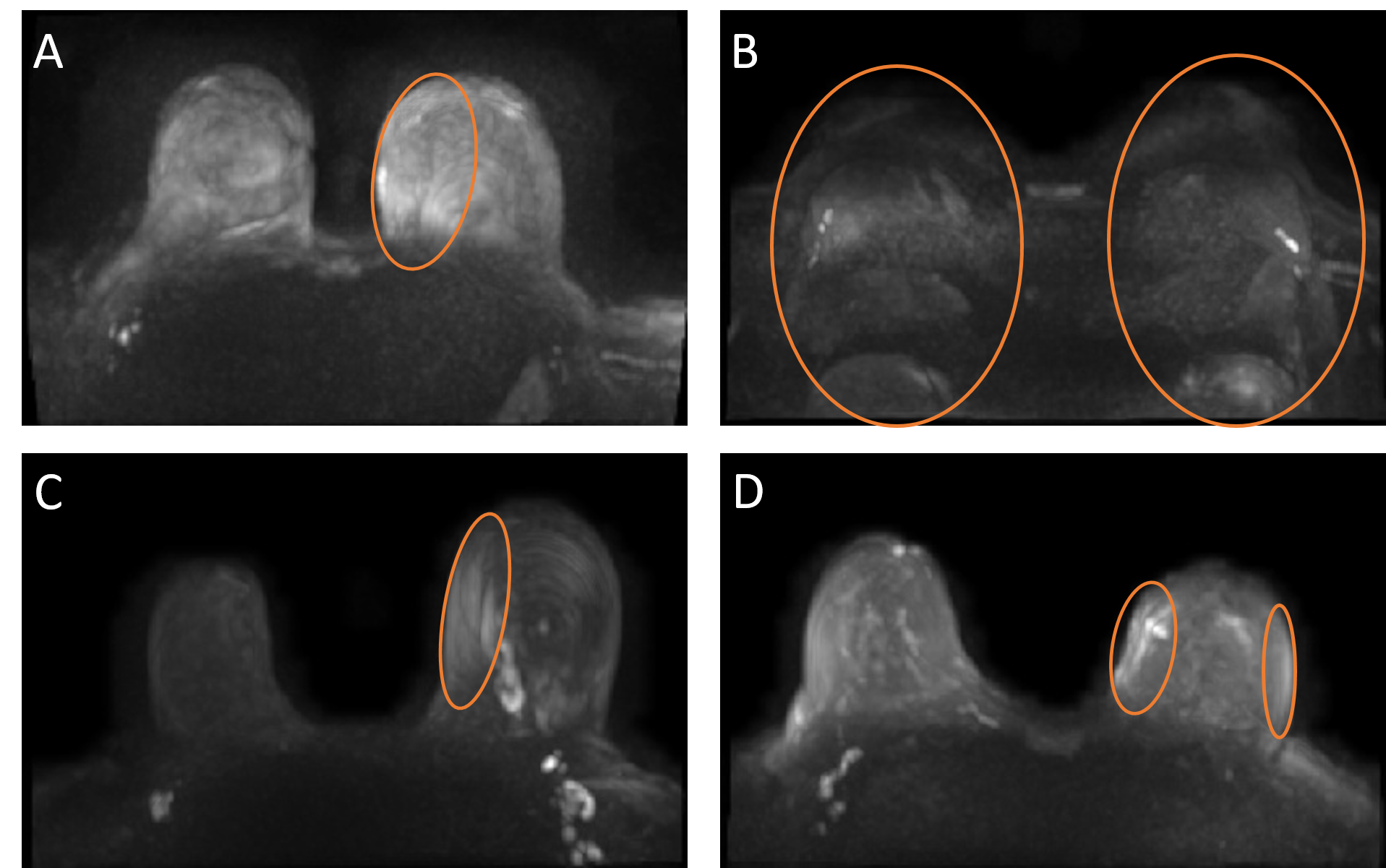
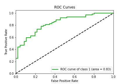
Representative cases of the acquisitions with artifacts, which were labeled as the positive class (class 1) are presented in Figure 1. Localized increased intensities and phase encoding artifacts can be observed in these cases.Figure 2 shows the RoC of the positive class of the 3D-DenseNet201 model. A sensitivity of 0.82 and specificity of 0.61 could be achieved with an Accuracy of 0.74 and AuRoC of 0.83 . Figure 2 shows an overlay of the GradCam of the predicted class over the labelled DWI MIP for representative true positive (Fig 2A-D), true positive (Fig 2E-H), false negative (Fig 2I-L) and true negative (Fig 2M-P) cases. The true positive and false positive cases show that the most important regions for the prediction of an artifact appear to be regions of high intensity in the DWI MIP image. A major hematoma (2E) or cases with an extremely dense fibro-glandular tissues (2G-H) can generate false prediction of an artifact occurring in the DWI MIP.
At the same time, the false negative and true negative cases indicate that for the prediction of an artifact free DWI MIP, the most important features of the T2 weighted acquisition correspond to a similar tissue intensity in both breasts.
The biggest limitation of the study is the potentially limited generalizability, as the study was performed on data acquired from a single vendor and with a single type of T2 weighted sequence. Additionally, the labelling was performed by a single radiologists. Both of these aspects should be improved in future works.
Conclusion
The applied DenseNet201 neural network demonstrated the capability to predict artifacts occurring in the DWI sequence by analyzing the T2w images, which are commonly acquired before the DWI sequence. Further research is necessary to assess whether such a prediction approach might help to trigger additional adjustments before the scan in order to reduce the occurrence of image artifacts in DWI.Acknowledgements
The financial support of BMBF GoBioInitial project "SMART SELECT MR" is gratefully acknowledged.References
1. Amornsiripanitch N, Bickelhaupt S, Shin HJ, et al. Diffusion-weighted MRI for Unenhanced Breast Cancer Screening. Radiology. 2019;293(3):504-520.
2. Bickelhaupt S, Laun FB, Tesdorff J, et al. Fast and Noninvasive Characterization of Suspicious Lesions Detected at Breast Cancer X-Ray Screening: Capability of Diffusion-weighted MR Imaging with MIPs. Radiology. 2016;278(3):689-697.
3. Partridge SC, Rahbar H, Murthy R, et al. Improved diagnostic accuracy of breast MRI through combined apparent diffusion coefficients and dynamic contrast-enhanced kinetics. Magn Reson Med. 2011;65(6):1759-1767.
4. Huang G, Liu Z, Van Der Maaten L, Weinberger KQ. Densely connected convolutional networks. Paper presented at: Proceedings of the IEEE conference on computer vision and pattern recognition2017.
5. Kingma DP, Ba J. Adam: A method for stochastic optimization. arXiv preprint arXiv:14126980. 2014.
6. Chattopadhay A, Sarkar A, Howlader P, Balasubramanian VN. Grad-cam++: Generalized gradient-based visual explanations for deep convolutional networks. Paper presented at: 2018 IEEE winter conference on applications of computer vision (WACV)2018.
Figures