1774
Hybrid diffusion imaging in chronic traumatic brain injury: Longitudinal investigation and correlation with clinical outcome1Neurosurgery, Thomas Jefferson University Hospital, Philadelphia, PA, United States, 2Mechanical Engineering, Villanova University, Villanova, PA, United States, 3Radiology, Thomas Jefferson University Hospital, Philadelphia, PA, United States, 4Marcus Institute of Integrative Health, Villanova, PA, United States
Synopsis
Traumatic brain injury (TBI) is a major cause of death and disability worldwide. Currently, non-invasive and longitudinal assessment in TBI is still limited. In the current study, we applied an advanced hybrid diffusion imaging sequence (HYDI) to capture both diffusion tensor imaging and neurite orientation dispersion and density imaging to develop more sensitive biomarkers associated with longitudinal outcomes after TBI. We both contrasting and complementary correlates between diffusion metrics and clinical outcomes, suggesting HYDI to have potential clinical impact for treatment monitoring after TBI.
INTRODUCTION
Chronic consequences of traumatic brain injury (TBI) may contribute to increased emotional and cognitive deficits, primarily due to diffusion axonal injury. Diffusion tensor imaging (DTI) is a more recent method of assessing axonal integrity, and has been used to determine cognitive deficits in TBI.[1] Hybrid diffusion imaging (HYDI) is a comprehensive diffusion sequence sensitive to different diffusivities and multiple diffusion-weighting directions.[4] The HDYI sequence is advantageous as multiple models can be fit using a single acquisition.[2] While neurite orientation/dispersion density imaging (NODDI) has been shown to be a more sensitive and specific biomarker than DTI for white matter changes [3] no study to date has implemented both NODDI and DTI in a prospective longitudinal study for diagnosis, prognosis, and treatment monitoring of patients with chronic TBI (cTBI). In this prospective longitudinal study, we aim to investigate changes in white matter using hybrid diffusion imaging (HYDI) and their correlations with improvements in neuropsychological assessments post-injury in patients experiencing chronic symptoms after TBI. The primary objective of this study was to explore the long-term prognostic significance of white matter microstructural changes using both NODDI and DTI.METHODS
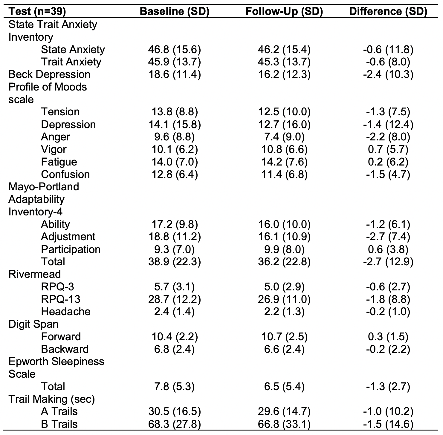
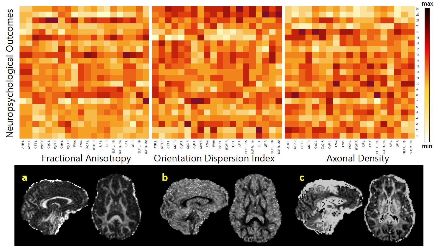
Participants. 39 patients aged 49.85 ±13.39 (9 male, 29 female) with chronic traumatic brain injury were studied with extensive imaging and clinical battery as part of a longitudinal prospective imaging study. The planned time period between baseline and follow-up examinations was 3 months in which patients received one of three treatment arms: waitlist control, N-ACETYL CYSTEINE (NAC), or nutritional counseling. After three months patients returned and received the same imaging and neuropsychological examination. Improvements in neuropsychological scores were calculated by subtracting the baseline values from the follow-up exams.Hybrid diffusion imaging. At both the baseline and follow-up visit, all patients were scanned using HYDI using a 3T Siemens Biograph MR PET-MR scanner with a 32-channel head coil. Eddy current and motion correction were applied to all diffusion scans. DTI parameter maps were calculated using the FSL Diffusion Toolbox (DTIFIT). NODDI components of the analysis were analyzed including the orientation dispersion index (ODI), axonal density (VIC). Diffusion maps were aligned to the MNI152 template in standard space using a nonlinear registration. A mean skeletonized image was created for all subjects in standard space and thinned to generate a mean white matter skeleton representative of all tracts common to the entire group of scans. The JHU atlas (https://neurovault.org/collections/264/) was overlayed on each of the skeletonized maps, and singular values for each of the 20 segmented tracts were obtained by averaging the diffusion values within each ROI. The aligned volumes were projected onto the skeleton by filling the skeleton with values from the nearest relevant tract center. Output images were visually inspected for accuracy. Changes in diffusion metrics were quantified by subtracting the baseline skeletonized map from those of the follow-up scan for each individual patient.
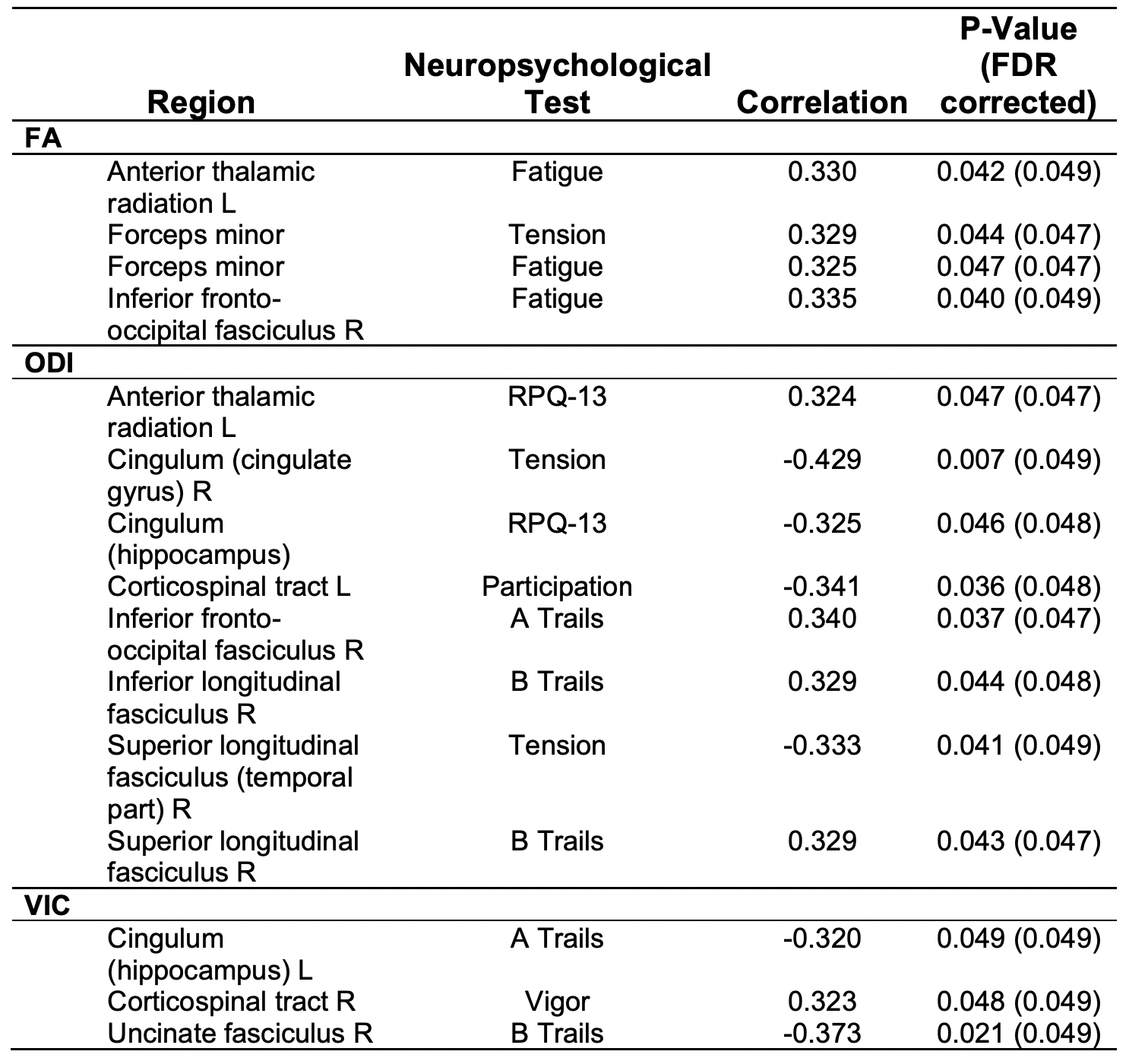
Statistical Analysis. Spearman rank partial correlation coefficients between FA, VIC, and ODI were calculated for all subjects controlling for age. FDR correction was applied to remove spurious correlations. A significance value was determined using a Student’s t distributions, with the linear correlation being considered significant if p < 0.05.
RESULTS
Neuropsychological outcomes and results. Patients showed varying improvement in tests of neuropsychological and cognitive function. Most notably, patients showed on average a 1 second improvement in the time it took to complete trail making, a neuropsychological test of visual attention.Correlation with diffusion metrics. Significant DTI and NODDI correlations with neuropsychological improvement are further presented in Table 2. Overall, NODDI showed more sensitivity in detecting improved outcomes in patients, particularly in cognitive associated such as RPQ-13 and trail making. Increased FA values in several major tracts showed longitudinal evidence of recovery and were positively associated with better performance in fatigue and tension, whereas ODI and VIC showed significant correlation between tension, participation, vigor, RPQ-13, and trail making. A histogram representing the correlation results between DTI and NODDI metrics with improvements in neuropsychological testing is displayed in Figure 1.
DISCUSSION AND CONCLUSIONS
This study may be the first to implement a prospective, longitudinal evaluation of cTBI patients using HYDI to compare DTI and NODDI associations with outcomes after TBI. Our results support our baselines findings [2] which showed that DTI and NODDI provide both different and complementary information for detecting cognitive deficits in patients with cTBI. Our longitudinal findings suggest NODDI to be a more sensitive biomarker than DTI, with ODI and VIC being more sensitive in detecting improvements after TBI. Our findings further validate prior studies which suggest an elevation of free water fraction after injury, followed by a decline in axonal density months after trauma.[3] These new hypotheses warrant testing in larger cohorts with better statistical power to further determine imaging-cognition relationships. Overall, we suggest HYDI to be a beneficial sequence to better characterize, diagnose, and monitor longitudinal symptoms in cTBI.Acknowledgements
No acknowledgement found.References
[1] Kraus, M. F., Susmaras, T., Caughlin, B. P., Walker, C. J., Sweeney, J. A., & Little, D. M. (2007). White matter integrity and cognition in chronic traumatic brain injury: a diffusion tensor imaging study. Brain, 130(10), 2508-2519s
[2] Muller, J., Middleton, D., Alizadeh, M., Zabrecky, G., Wintering, N., Bazzan, A. J., ... & Mohamed, F. B. (2021). Hybrid diffusion imaging reveals altered white matter tract integrity and associations with symptoms and cognitive dysfunction in chronic traumatic brain injury. NeuroImage: Clinical, 30, 102681.
[3] Palacios, E. M., Owen, J. P., Yuh, E. L., Wang, M. B., Vassar, M. J., Ferguson, A. R., ... & TRACK-TBI Investigators. (2020). The evolution of white matter microstructural changes after mild traumatic brain injury: a longitudinal DTI and NODDI study. Science advances, 6(32), eaaz6892.
[4] Wu YC, Mustafi SM, Harezlak J, Kodiweera C, Flashman LA, McAllister TW. Hybrid diffusion imaging in mild traumatic brain injury. Journal of neurotrauma. 2018 Oct 15;35(20):2377-90.
Figures