1747
MULTI-CONTRAST ATHEROSCLEROSIS CHARACTERIZATION (MATCH) AND MULTI-SEQUENCE MRI IN QUANTIFYING CAROTID PLAQUE COMPOSITION1CARIM School for Cardiovascular Diseases, Maastricht University, Maastricht, Netherlands, Maastricht, Netherlands, 2Department of Radiology and Nuclear Medicine, Maastricht University medical center (MUMC+), Maastricht, Netherlands, 3Department of Nuclear Medicine, Georg-August University Göttingen, Gottingen, Germany, 4Department of Clinical Neurophysiology, Maastricht University medical center (MUMC+), maastricht, Netherlands, 5Department of Neurology, Maastricht University Medical Center+ (MUMC+), maastricht, Netherlands, 6Department of Radiology, University of Southern California, Los Angeles, CA, United States
Synopsis
Multi-contrast Atherosclerosis Characterization (MATCH) was developed to quantify of carotid atherosclerotic plaque composition within 5 minutes’ scan time. Twenty symptomatic patients with ≥2 mm carotid plaque underwent 3.0 Tesla conventional multi-sequence and MATCH MRI. Excellent agreement was obtained for scoring a Lipid-rich necrotic core (LRNC) intraplaque hemorrhage (IPH) on the MATCH images while fair for calcifications. No significant differences between MATCH and multi-sequence MRI were found in volume of LRNC, IPH and calcifications. We demonstrated excellent agreement between MATCH and multi-sequence MRI for the identification and quantification of LRNC and IPH within significant shorter time.
Background
Multi-sequence magnetic resonance imaging (MRI) is commonly used for the quantification of carotid atherosclerotic plaque composition (1). Limitations are long scan time and image misregistration errors. Multi-contrast Atherosclerosis Characterization (MATCH) was developed to overcome these limitations (2).Aim
To compare MATCH with multi-sequence MRI for the quantification of carotid plaque components.Methods
Twenty symptomatic patients with ≥2 mm carotid plaque underwent 3.0 Tesla multi-sequence and MATCH MRI. Image quality was scored on a 5-point scale based on vessel wall signal-to-noise ratio (SNR) and visibility of vessel wall and substructures (1, poor and 5, excellent) (3). The effective vessel wall SNR and contrast-to-noise ratio (CNR) between IPH and the muscle were calculated by dividing SNR and CNR by the acquisition time. Image analysis of the MATCH images was performed independently of that of the multi-sequence images (Figure 1). A Cohen’s kappa test was used to determine agreement in the detection of plaque components using multi-sequence versus MATCH MRI. The sensitivity and specificity of MATCH in identifying plaque components were calculated using multi-sequence MRI as the reference standard.Results
One total or nearly occluded artery was excluded. The mean quality scores of the MATCH images were lower than multi-sequence images (2.4±0.5 versus 3.7±0.4; p<0.05), respectively. The overall effective SNR of MATCH was higher than the multi-sequence protocol except for the hyper T1w images (Figure 3 1). The mean effective CNR of intraplaque hemorrhage (IPH) on MATCH was significantly higher than that of magnetization prepared rapid acquisition gradient echo (MPRAGE) (7.2 ±4.3 versus 4.7±2.8; p=0.007). The scan time for MATCH and multi-sequence MRI was 7 and 39 minutes, respectively. Excellent intraobserver agreement was obtained for scoring a Lipid-rich necrotic core (LRNC) (k=0.89) and IPH (k=0.91) on the MATCH images while fair intraobserver agreement was observed for calcifications (k=0.32). The sensitivity and specificity of scoring LRNC and IPH were >83% and >96% while for calcifications the sensitivity and specificity were 76% and 50%, respectively (Figure 4). No significant differences between MATCH and multi-sequence MRI were found in the volume of LRNC, IPH, and calcifications. There was a small but significant difference in the total volume of the vessel wall and total volume of fibrous tissue (Figure 2).Conclusion
We demonstrated excellent agreement between MATCH and multi-sequence MRI for the identification and quantification of LRNC and IPH. There was only fair agreement for scoring the presence of calcifications. Although MATCH images showed a lower mean image quality score, short scan time and perfect co-registration are major advantages of MATCH.Acknowledgements
No acknowledgement found.References
1. Saba L, Yuan C, Hatsukami TS, et al. Carotid Artery Wall Imaging: Perspective and Guidelines from the ASNR Vessel Wall Imaging Study Group and Expert Consensus Recommendations of the American Society of Neuroradiology. AJNR Am J Neuroradiol 2018;39:E9-E31. 2. Fan Z, Yu W, Xie Y, et al. Multi-contrast atherosclerosis characterization (MATCH) of carotid plaque with a single 5-min scan: technical development and clinical feasibility. J Cardiovasc Magn Reson 2014;16:53. 3. Yuan C, Mitsumori LM, Ferguson MS, et al. In vivo accuracy of multispectral magnetic resonance imaging for identifying lipid-rich necrotic cores and intraplaque hemorrhage in advanced human carotid plaques. Circulation 2001;104:2051-6.Figures
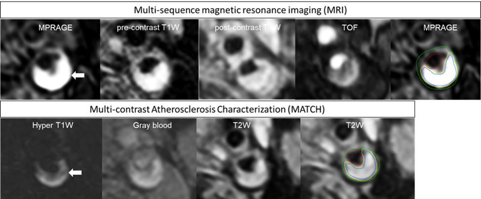
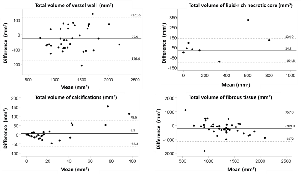


Figure 4. Concordance between identification of intraplaque hemorrhage (IPH), lipid-rich necrotic core (LRNC), and calcifications (CA) on MATCH versus multi-sequence images.
*4 carotids were excluded (contraindication of contrast injection)