1735
Brain-to-brain interaction in parent-child eye contact1Columbia University, New York, NY, United States, 2NASA, Houston, TX, United States
Synopsis
Parent-child interaction is a significant part of human life, and a large portion of such interaction is carried out by eye contact. However, how the dyadic brain networks enable eye contact is not well understood. Here, we take advantage of fMRI hyperscan and concurrent eye tracking to measure the BOLD responses and pupil sizes during eye contact between parent and child. Our initial analysis begins to reveal some of the brain networks supporting the parent-child bond. A better understanding of such mechanisms may have a significant impact on human social life, and improve parent-child interaction therapy for many psychiatric diseases.
INTRODUCTION
Parent-child interaction is a significant part of human life, and a large portion of such interaction is carried out by eye contact. However, how the dyadic brain networks enable eye contact is not well understood. Here, we take advantage of fMRI hyperscan1 and concurrent eye tracking2 to measure the BOLD responses and pupil sizes during eye contact between parent and child who are lying in two MRI scanners. The goal of this study is to better understand the brain dynamics of the parent-child bond.MEHTODS
Parent-child fMRI hyperscan was performed in two Siemens (Erlangen, Germany) Prisma 3T scanners. The functional scans on both scanners were synchronized by a PC on the scanners’ subnet. The real-time visualization between parents and children in different scanners was carried out by two in-bore high-resolution video cameras (MRC Inc., Germany). A total of 26 parent-child pairs participated in the experiment, 21 datasets were successfully acquired.During each functional session, the dyadic face videos, eye-tracking data, and physiological data were also synchronized and recorded. The fMRI protocol consists of one structural T1 MPRAGE (TR 2.3s TE 2.26ms TI 900ms voxel size 1x1x1mm3 BW 200Hz/pixel), three multiband gradient-echo EPI (TE 30ms, TR 1s, voxel size 3x3x3mm3 MB factor 3 BW 1660Hz/pixel measurement 400), and one dual-echo field mapping.
Parent-child pairs performed three tasks in the three functional scans: 1) in each 40s block, dyads open and close their eyes simultaneously for 20s respectively; 2) in each 40s block, dyads open and close their eyes alternately for 20s respectively, where children open their eyes first; 3) the same as the second task, but parents open their eye first.
The functional data were processed by software FSL (Cambridge, UK). The group-level GLM and t-test comparisons between eye contact (task1) and non-eye-contact (task2, task3) were performed by FEAT in FSL. Given that children’s more severe motion, the stage 4 (with sinc interpolation) motion correction was used, and any volume with framewise displacement > 0.2mm was treated as outliers. In addition, the dyadic facial muscle movements in the face video were analyzed by the software FaceReader (Noldus, the Netherlands). The eye contact times and pupil size were analyzed by DataViewer (SR Research, Canada).
RESULTS
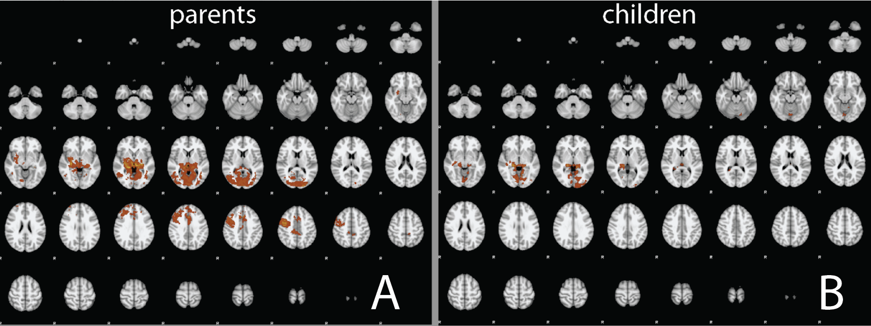
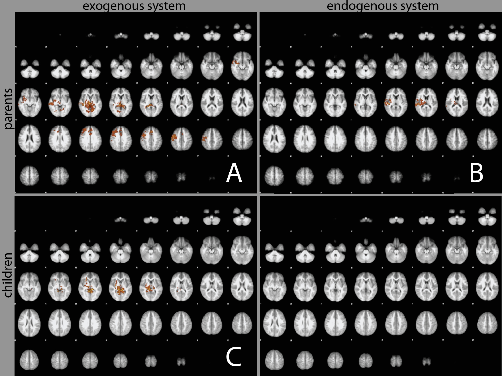
The group averages of BOLD activations of parents and children from task1 are shown in Fig. 1, A, and B, where p<0.05, z-score>2.3. The second-order group analysis, the t-test comparison for both children (task1 – task2, task2 – task1) and parents (task1 – task3, task3 – task1) are in Fig. 2, where p<0.05, z-score>1.6. Here, task1-task2 and task1-task3 are attributed as an exogenous system; task2-task1 and task3-task1 are endogenous systems3. Based on Harvard-Oxford Atlas, for the exogenous system, both parents and children have similar activations (bilateral thalamus, right putamen, and right hippocampus) in subcortices, but dramatic differences in cortical areas, as shown in Fig.2 A, C. For the endogenous system, only parents have activations, mostly in the right insular, posterior superior temporal, Heschl’s gyrus and planum temporal, as well as right thalamus and putamen, see Fig. 2B, D. In concurrent with fMRI task1, the averaged time course of pupil sizes of parents and children is shown in Fig. 3, in which parents and children demonstrated distinguished patterns.DISCUSSION
In our initial analysis, we modeled the brain responses to eye contact with a dual system. In which exogenous system represents reflexive activation and endogenous system represents more reflective activation. The results in Fig. 2 suggest that in parent-child eye contact, both exogenous systems are active, although the parents have richer content. Meanwhile, only parents’ endogenous systems are activated, whereas children’s are not. These results are consistent with the psychological theory that during eye contact, parents are mostly in attunements whereas children are most in a reward/punish state.Pupil sizes in parent-child eye contact reveal the asymmetric relations in parent and children communication. Fig. 3 is clear evidence of the dramatic differences when parent and child eye meet. Combining the pupil size analysis and fMRI, the frontal eye field in the superior frontal gyrus is correlated to pupil size, the relations between the eye field and other cortical regions, such as insular, posterior cingulate, etc. may reveal the brain networks that support eye contact.
CONCLUSION
In searching for a better understanding of parent-child interaction, we have set up a platform to observe the brain network activations and pupil size variations during their eye contact. Our initial analysis begins to reveal some of the brain networks supporting the parent-child bond. A better understanding of such mechanisms may have a significant impact on human social life, as well as improving parent-child interaction therapy for many psychiatric diseases.Acknowledgements
This work is supported by the grant NSF 1926789References
1 Koike, T., Sumiya, M., Nakagawa, E., Okazaki, S. & Sadato, N. What Makes Eye Contact Special? Neural Substrates of On-Line Mutual Eye-Gaze: A Hyperscanning fMRI Study. eNeuro 6, doi:10.1523/ENEURO.0284-18.2019 (2019).
2 DiNuzzo, M. et al. Brain Networks Underlying Eye's Pupil Dynamics. Front Neurosci 13, 965, doi:10.3389/fnins.2019.00965 (2019).
3 Lee, R. F. Dual logic and cerebral coordinates for reciprocal interaction in eye contact. PLoS One 10, e0121791, doi:10.1371/journal.pone.0121791 (2015).
Figures