1732
A separation between motor and sensory somatotopic maps in the human cerebellum1Spinoza Centre for Neuroimaging, Amsterdam, Netherlands
Synopsis
We investigated the organisation of the cerebellar digit map with three distinct tasks (flexing/extending/stroking) using B1-shimmed, high-resolution fMRI at 7T. For all tasks, the positions of the COG of the digit representations formed an orderly progression in at least one of the lobules. The distance between the motor task activations for flexing and extending of the digits were smaller than from either motor task to stroking clusters, indicating a separation between somatosensory and motor clusters in cerebellar lobule V.
Introduction
The human cerebellum is an important part of the sensory and motor networks, though largely unexplored compared to the neocortex, due to its highly-foliated cortex and challenging B1 profile at higher fields. There is a sensory somatotopic digit map in the anterior and posterior lobules of the cerebellum1 which has been suggested to overlap with cerebellar motor digit representations2. By comparison, in the primary motor cortex, flexing and extending are known to result in distinct digit maps3. The precise sensory-motor digit organisation in the cerebellar cortex has not yet been explored in adequate spatial resolution. Here, we investigated the organisation of the cerebellar digit map with three distinct tasks (flexing/extending/stroking) using RF-shimmed, high-resolution fMRI at 7T.Methods
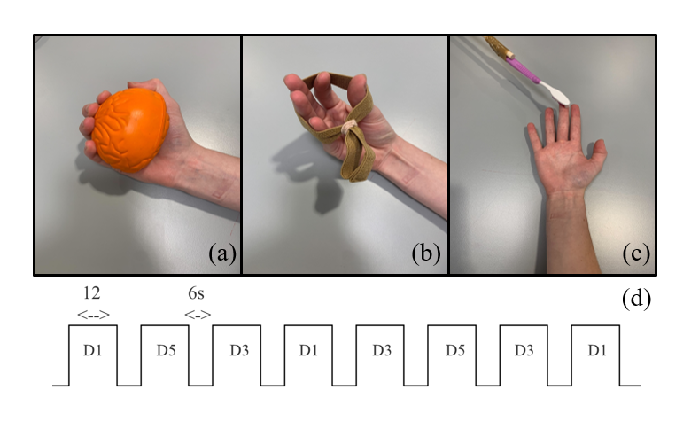
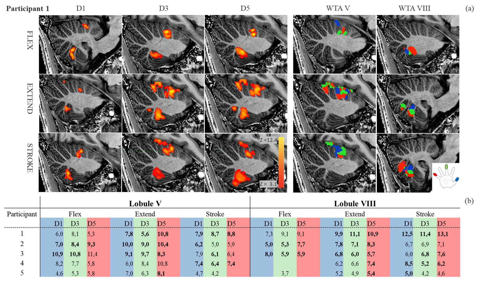
Five right-handed participants (22-42years) were scanned in a 7T Achieva (Phillips) with an 8Tx/32Rx (Nova Medical) whole-head coil. An RF-shim was calculated following B1 mapping. A 3D-EPI slab covering the cerebellum (1mm isotropic, TR/TE=3300ms/21ms, SENSEy/z= 2.6/3.27, FOV=182x60x186mm, partial-fouriery/z =0.8, flip-angle=20°) was used for functional acquisitions. Each session consisted of three 15-minute functional runs in which right-hand digit 1, 3 or 5 was flexed into a ball (motor), extended against an elastic band (motor) or stroked with a toothbrush (sensory); 12s ON and 6s OFF (Fig-1). fMRI data were motion/distortion-corrected, the cerebellum was segmented and a GLM (finger-stimulus>rest) was fitted (FSL, SPM12 and ANTs). Dominant clusters (p<0.05) in lobule V and VIII for each of the three tasks and digits were identified by visual inspection. When the cluster extended into the left hemisphere of the cerebellum, the zstat threshold was raised to 4 (n=2). For each cluster, the peak z-stat value and the centre of gravity (COG) and direction of the somatotopic gradient in the individual space were extracted. A winner-takes-all (WTA) map was generated for each task and participant. For each participant, the median distance between COGs of each digit were calculated for each task, as well as the distance between groups of COGs for the different tasks.Results
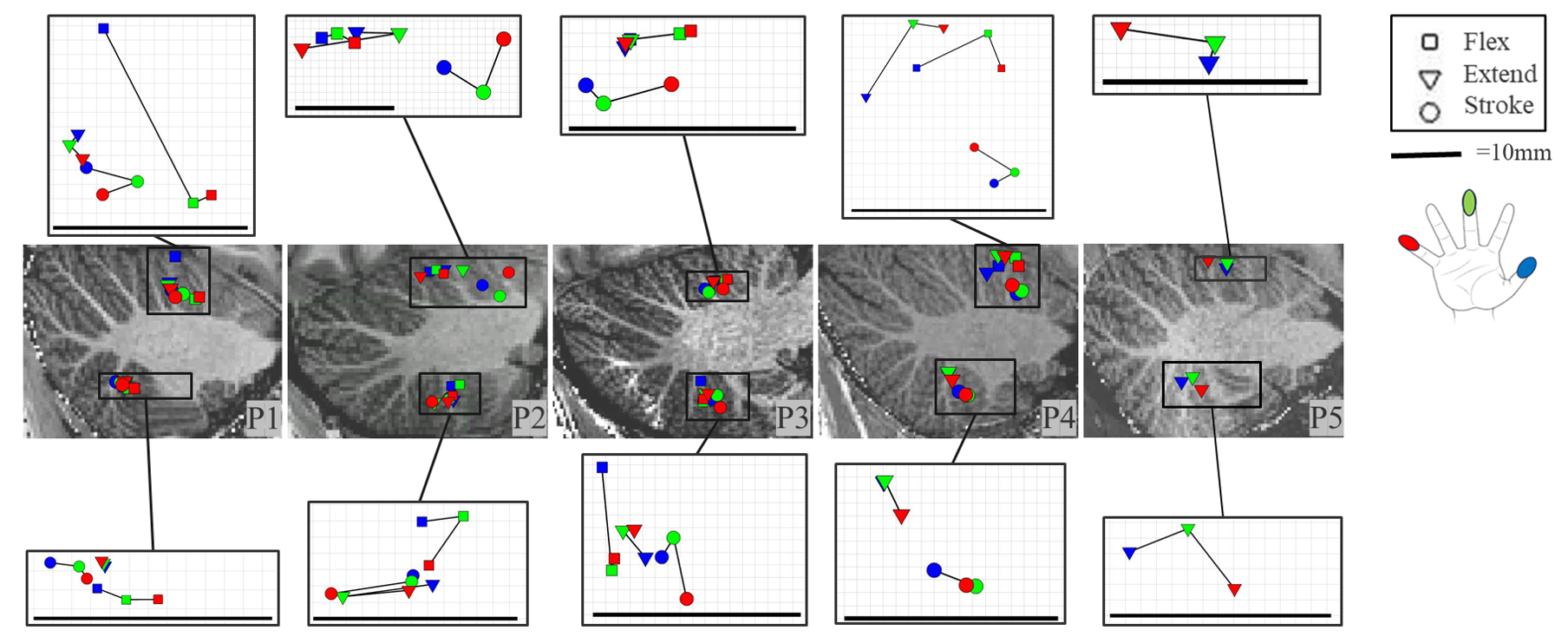
Stroking, flexing and extending typically resulted in individual-digit significant clusters in both lobule V (anterior lobe) and lobule VIII (posterior lobe) (Fig-2a). The winner takes all (WTA) maps presented all three digits in a semi-orderly progression, but not in consistent direction, for most subjects and tasks. While a posterior-anterior (P-A) progression can be seen in Figure 2a in lobule V for all tasks, a P-A direction is found for flexing in lobule VIII and a superior-inferior (S-I) direction for extending and stroking. Interestingly, we found consistent bilateral activation in lobule VIII during the extending task (Fig-2b), but not for flexing or stroking.In lobule V, during the flexing task, a consistent organisation in the P-A direction was found in the COG positions for all participants (Fig-3), though not for extending and stroking. In lobule VIII, P-A gradients were found for two participants in all tasks and a left-right gradient for 2/3/3 participants for flexing, extending and stroking respectively. The S-I gradient was inconsistent for all participants and tasks.
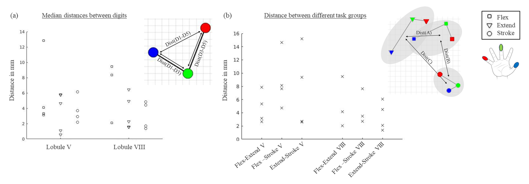
The distinct tasks resulted in separated series of COGs within the same lobules. Median distances between digit COGs were largest for the flexing task (Fig-4a). Between task groups, the largest distances were found between the two motor and stroking task in lobule V (Fig-4b). In lobule VIII, groups of COGs were closer together.
Discussion
All three tasks reliably engaged the cerebellar motor regions in lobule V and VIII at single-digit level. More consistently organised activation in lobule V and larger distances between tasks compared to lobule VIII indicate that the digit regions cover more cortical surface in the anterior lobe, though both regions demonstrated digit maps.The flexing and stroking tasks showed mostly unilateral results. In comparison, the extending task resulted in bilateral clusters. This could be due to the higher complexity of the task, as more difficult movements have been shown to yield more bilateral activation in M14.
Larger distances between the stroking task and the two motor tasks could potentially indicate a separation between motor and sensory organisation in the cerebellum similar the neocortex, where distinct sensory (S1) and motor digit maps (M1) exist3. A separation between flexing and extending, as has been reported in M13, was not apparent in this cerebellar data, though flexing demonstrated a more distinct digit progression compared to extending.
With the current 1mm spatial resolution, individual digit representations could be distinguished, but areas responding to different tasks were close and overlapping. To further investigate the intralobular organisation of lobule VIII, a higher spatial resolution and/or surface-based approaches might be required.
Conclusion
To conclude, we employed an RF-shimmed high-resolution 7T fMRI acquisition to demonstrate that digit-maps exist both in the anterior (Lobule V) and posterior (Lobule VIII) part of the cerebellum, across motor and sensory tasks. We found a separation between motor and sensory digit maps, indicating a similar organisation to the neocortex.Acknowledgements
This study was supported by an NWO TTW VIDI grant (VI.Vidi.198.016)References
1.Van der Zwaag W, Kusters R, Magill A, et al. Digit somatotopy in the human cerebellum: A 7T fMRI study. Neuroimage. 2013;67:354-362. doi:10.1016/j.neuroimage.2012.11.041
2.Wiestler T, McGonigle DJ, Diedrichsen J. Integration of sensory and motor representations of single fingers in the human cerebellum. J Neurophysiol. 2011;105(6):3042-3053. doi:10.1152/JN.00106.2011
3.Huber L, Finn ES, Handwerker DA, et al. Sub-millimeter fMRI reveals multiple topographical digit representations that form action maps in human motor cortex. Neuroimage. 2020;208:116463. doi:10.1016/J.NEUROIMAGE.2019.116463
4.Chettouf S, Rueda-Delgado LM, de Vries R, Ritter P, Daffertshofer A. Are unimanual movements bilateral? Neurosci Biobehav Rev. 2020;113:39-50. doi:10.1016/J.NEUBIOREV.2020.03.002
Figures