1725
Model-based reconstruction of IVIM-DTI in skeletal muscle: Improving IVIM-DTI parameter map quality in low-perfused tissues1Department of Biomedical Engineering and Physics, Amsterdam UMC, Amsterdam, Netherlands, 2Institute of Medical Engineering, Graz University of Technology, Graz, Austria, 3Department of Radiology and Nuclear Medicine, Amsterdam UMC, Amsterdam, Netherlands
Synopsis
IVIM and DTI techniques are both sensitive to a variety of muscle disorders. Their combination can give a more complete description of ordered diffusion processes and perfusion. Model-based reconstruction can overcome many short-comings of image-based fitting such as Rician noise bias or low SNR. In this study we investigate the feasibility of model-based IVIM-DTI reconstruction in skeletal muscle, a low-perfused tissue. Results were compared to image-based least-square fitting. The parameter maps show more anatomical detail with model-based reconstruction. The mean parameter values are comparable to image-based fitting. Model-based reconstruction is sensitive to subtle changes after exercise in the underlying parameters.
Introduction
Diffusion tensor imaging (DTI) and intravoxel-incoherent motion (IVIM) can provide microstructural information of the underlying tissue. In skeletal muscle, those techniques are sensitive to exercise1, injury2,3 and disease4 and their combination is desirable to obtain directional diffusion and perfusion information simultaneously. However, this faces some challenges. First, the scan time increases significantly compared to pure DTI or IVIM due to the need for additional b-values and diffusion directions. Second, due to the low perfusion fraction of skeletal muscle (approx. 3-10%)5 fitting of the IVIM component is challenging and especially the D* maps often show a high variance, making interpretation difficult. Third, the low T2-value of skeletal muscle (approx. 32ms at 3T)6 yields low SNR in diffusion images and the Rician noise introduces noise-dependent bias for image-based fitting.Model-based reconstruction has the potential to overcome those limitations. It was shown that model-based reconstruction for IVIM and combined IVIM-DTI fitting is feasible in well-perfused abdominal organs7. The aim of this work is to test the feasibility and performance of model-based reconstruction in low-perfused muscle tissue and improve the quality of IVIM-DTI parameter maps.
Methods
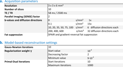
Five healthy participants (3 female, mean age 29.4 years) underwent MRI examination of the calves with a 3T MRI scanner (Philips Ingenia). Detailed scan parameters for the IVIM-DTI scan are listed in Table 1. One subject performed plantar flexion exercise until exhaustion after the first scan in the scanner to stimulate the gastrocnemius muscle. The IVIM-DTI scan was repeated directly after the exercise.The model-based reconstruction was performed on the raw k-space data using PyQMRI8. The workflow of image-based versus model-based reconstruction is illustrated in Figure 1. PyQMRI uses a total generalized variation (TGV) regularization and an iteratively regularized Gauss-Newton approach. The Gauss-Newton steps are solved with a Primal-Dual algorithm. The settings used in this work are listed in Table 1. The IVIM-DTI model was used for fitting with S0 the signal at b=0s/mm², f perfusion fraction, D* pseudo-diffusion coefficient, $$$\underline{\underline{D}}$$$ diffusion tensor and $$$\underline{g}$$$ the gradient directions:
$$S(b)=S_{0}\cdot(f\cdot e^{-bD^*}+(1-f)\cdot e^{-b\underline{g}^T \underline{\underline{D}}\underline{g}})\quad\quad[1]$$
For comparison, a voxel-wise fit in image space was performed. The k-space data was reconstructed with ReconFrame (Gyrotools, Switzerland) and fitted with in-house developed software in Matlab R2021a (The MathWorks, USA) using a nonlinear least-square algorithm. We considered a free fit to equation [1], fitting the unknowns f, D* and $$$\underline{\underline{D}}$$$ simultaneously, the fairest comparison to PyQMRI. The current gold-standard for IVIM-DTI fitting is a two-step fit, where first the diffusion tensor is estimated from data at b≥200s/mm² and then the tensor is fixed to estimate f and D*, and was included for comparison.
Segmentations were drawn in the right and left tibialis anterior (TA) and gastrocnemius medialis (GM) muscle. The mean values of the IVIM-DTI outcome measure (mean diffusivity (MD), fractional anisotropy (FA), perfusion fraction (f) and pseudo-diffusion coefficient (D*)) were calculated for all fitting methods.
Results
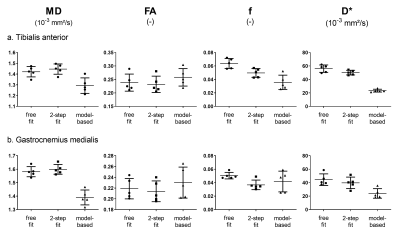
Parameter maps of the model-based reconstruction and conventional image-based fits are shown in Figure 2. The model-based reconstruction derived parameter maps show more anatomical detail, which is especially visible in the MD and f maps. The FA maps obtained from model-based and image-based fitting are similar, however, in regions with little signal, such as the tibia bone, the conventional fit returns high FA values while model-based reconstruction favors lower values. The D* maps obtained by conventional fitting exhibit several outliers, visible as noisy pixels in the maps. This is not observed with model-based reconstruction. However, the D* map is prone to over-regularization and details are smoothed out. Conversely, the quality of the D* maps from the reference fit approaches is poor, making it hard to see any details on those either.The mean values of MD, FA, f and D* in the TA and GM muscle are displayed in Figure 3. MD values are lower with model-based reconstruction while FA values tend to be higher. Perfusion fraction is lower in the TA and similar for GM. Values for D* were lower with model-based reconstruction.
After plantar-flexion exercise until exhaustion, an increase in all parameters can be observed in the GM muscle, both with image-based fitting and model-based reconstruction (Figure 4).
Discussion
We are the first to perform model-based reconstruction of IVIM-DTI in low-perfused muscle tissue and showed that our approach provides more anatomical detail in the parameter maps compared to conventional fitting. This is especially beneficial for the perfusion-derived parameters f and D* which suffer from poor image quality with conventional fitting. The bias in the parameter values between model-based and conventional fitting will be investigated in more detail with simulations. However, both the values found with model-based reconstruction and conventional fitting are within the range of values reported in literature5. The lower values of D* might be a result of over-regularization or of limited information available to accurately fit D* due to the rapid loss of signal. The results after exercise show that model-based IVIM-DTI reconstruction can depict subtle changes in the underlying microstructure.Conclusion
Model-based reconstruction for IVIM-DTI fitting can be used to substantially improve parameter map quality of low-perfused tissue. Currently, mean values differ from conventional fitting but are in line with literature.Acknowledgements
No acknowledgement found.References
1. Mastropietro A, Porcelli S, Cadioli M, et al. Triggered intravoxel incoherent motion MRI for the assessment of calf muscle perfusion during isometric intermittent exercise. NMR Biomed. 2018;31(6):1-13. doi:10.1002/nbm.3922
2. Zaraiskaya T, Kumbhare D, Noseworthy MD. Diffusion tensor imaging in evaluation of human skeletal muscle injury. J Magn Reson Imaging. 2006;24(2):402-408. doi:10.1002/jmri.20651
3. Hooijmans MT, Monte JRC, Froeling M, et al. Quantitative MRI Reveals Microstructural Changes in the Upper Leg Muscles After Running a Marathon. J Magn Reson Imaging. 2020. doi:10.1002/jmri.27106
4. Federau C, Kroismayr D, Dyer L, Farshad M, Pfirrmann C. Demonstration of asymmetric muscle perfusion of the back after exercise in patients with adolescent idiopathic scoliosis demonstrated using intravoxel incoherent motion (IVIM) MRI. NMR Biomed. 2019;(March):6-13. doi:10.1002/nbm.4194
5. Englund EK, Reiter DA, Shahidi B, Sigmund EE. Intravoxel Incoherent Motion Magnetic Resonance Imaging in Skeletal Muscle: Review and Future Directions. J Magn Reson Imaging. 2021. doi:10.1002/jmri.27875
6. Gold GE, Han E, Stainsby J, Wright G, Brittain J, Beaulieu C. Musculoskeletal MRI at 3.0 T: Relaxation Times and Image Contrast.; 2004.
7. Rauh S, Maier O, Gurney-Champion OJ, et al. Model-based reconstruction for IVIM and combined IVIM-DTI fitting: Initial experience. Proc Intl Soc Mag Reson Med . 2021.
8. Maier O, Spann SM, Bödenler M, Stollberger R. PyQMRI : An accelerated Python based Quantitative MRI toolbox. J Open Source Softw. 2020;5(56):2727. doi:10.21105/joss.02727
Figures


