1713
Fast 3D Isotropic High-Resolution MRI of Mouse Brain Using a Variable Flip Angle RARE Sequence With T2 Compensation @9.4T1Medical Physics Group, Institute of Diagnostic Radiology, Jena University Hospital, Jena, Germany
Synopsis
Some applications require T2-weighted mouse brain MRI with isotropic high resolution (100µm3), but at 9.4T the short T2 of mouse brain limits the echo train length of RARE sequences and exacerbates T2-decay artifacts. Adapting a RARE sequence by using variable flip-angles allows a reduction in scan time from 19min to 7-9min with similar image quality. However, long RARE echo trains and strong imaging gradients can unintentionally cause substantial diffusion weighting. To minimize the diffusion effects crusher gradients were optimized and the variable flip angles were kept above 40°.
Introduction
We aim to achieve artifact-free, isotropic high-resolution images of the mouse brain for neurological applications1. While 9.4T renders T1-based grey-white-matter contrast mostly useless, T2-weighted images achieve excellent anatomical contrast and can be efficiently acquired by 2D-RARE sequences. However, 2D acquisitions limit the achievable isotropic resolution due to minimal slice thickness and SAR. Using 3D acquisitions increases the acquisition time (TA), particularly since at 9.4T the short T2 of brain tissue limits echo train length with standard 180° refocusing pulses. The use of refocusing pulses with constant reduced flip angles was shown2 to prolong the echo train to facilitate anatomic mouse brain scans within 20-40min.Our aim here was to reduce the acquisition time for T2w-3D-RARE by applying variable flip angles (VFAs) to shape the echoes' signal envelope3-4while preserving the image quality.
Material & Methods
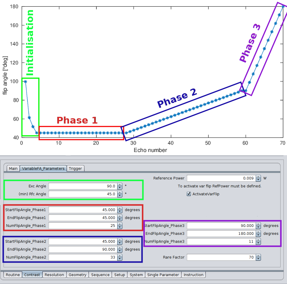
All data were acquired on a Bruker BioSpec 94/20USR with 660mT/m gradients using a helium-cooled 2-channel transceiver quadrature surface coil (CryoProbe, Bruker Biopsin, Ettlingen). All mice were scanned in vivo under Isoflurane anesthesia.The proposed varFlipRARE sequence uses three initialization RF pulses4 to drive the magnetization into a pseudo-steady-state matching the target FA<< 90°. Subsequently, three acquisition phases can be adjusted separately, each allowing for a linear transition from one FA to another within a given number of echoes (Figure 3). Typically, the first phase comprised the first 1/3 of echoes, repeating the target FA or slightly increasing it. In the second phase, the FA increased to ~90° over 1/2 of the total echoes, followed by the last phase, which ramped the FA up to 160-180°. Usually, k0 was acquired at the center of the echo train.
The general scan parameters for all varFlipRARE acquisitions were TA=7min 15s, TR=2200ms, isotropic resolution 100µm3, ΔTE=2.45ms, TF=70, BW=150kHz. The excitation and refocusing pulses were non-selective hard pulses with duration 128µs (exc) and 80µs (rfc) corresponding to 10kHz bandwidth to minimize echo spacing. The phase encoding gradients were 430µs long and the read and slice crusher gradients were played out during the same time frame at 10% (approx. 66mT/m).
To explore further possibilities of applying VFAs, a faster sequence with RARE factor TF=140 with TA=4min was used, setting the target FA to 30-35°, following similar VFA increase patterns as stated above. The second variation aimed to increase the resolution to 76µm3 with TF=86 and TA=24min, ΔTE=3ms, TR=2700ms, and 2 averages.
For reference, a RARE acquisition2 with slab-selective 90° excitation and 125° refocusing RF pulses was used with TA=19min, TR=1600ms, isotropic resolution 100µm3, ΔTE=5ms, TF=20, BW=150kHz.
All sequences used full Nyquist sampling of the 18x14x10mm3 FoV and were acquired in coronal orientation, i.e. read out head-feet, phase left-right, and slice ventral-dorsal direction. All shown images were reconstructed with T2-compensation and Gibbs-ringing-filter for basic artifact reduction2. SNR and CNR values were calculated from the mean signal in ROIs and the standard deviation in artifact-free background areas.
Results
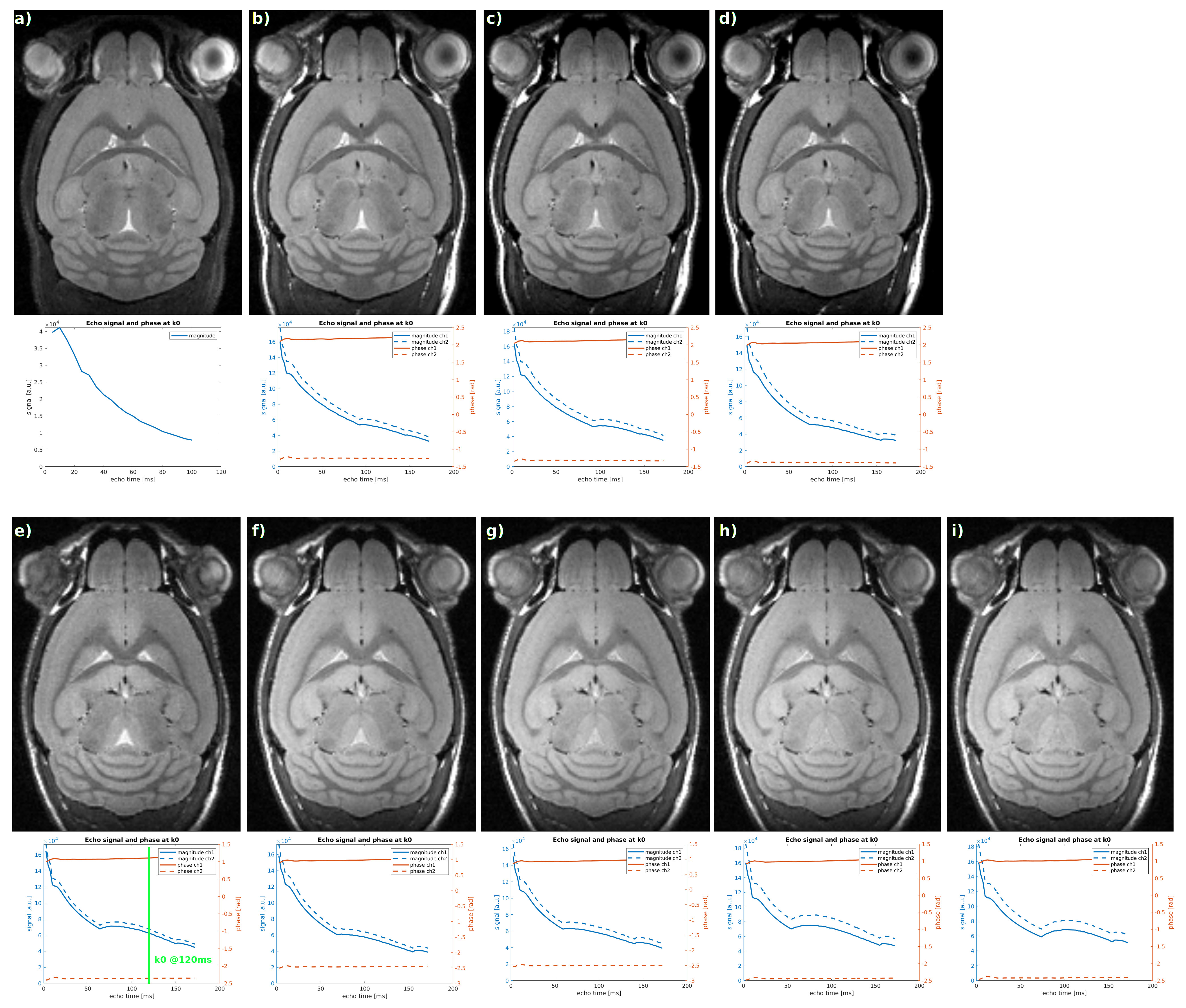
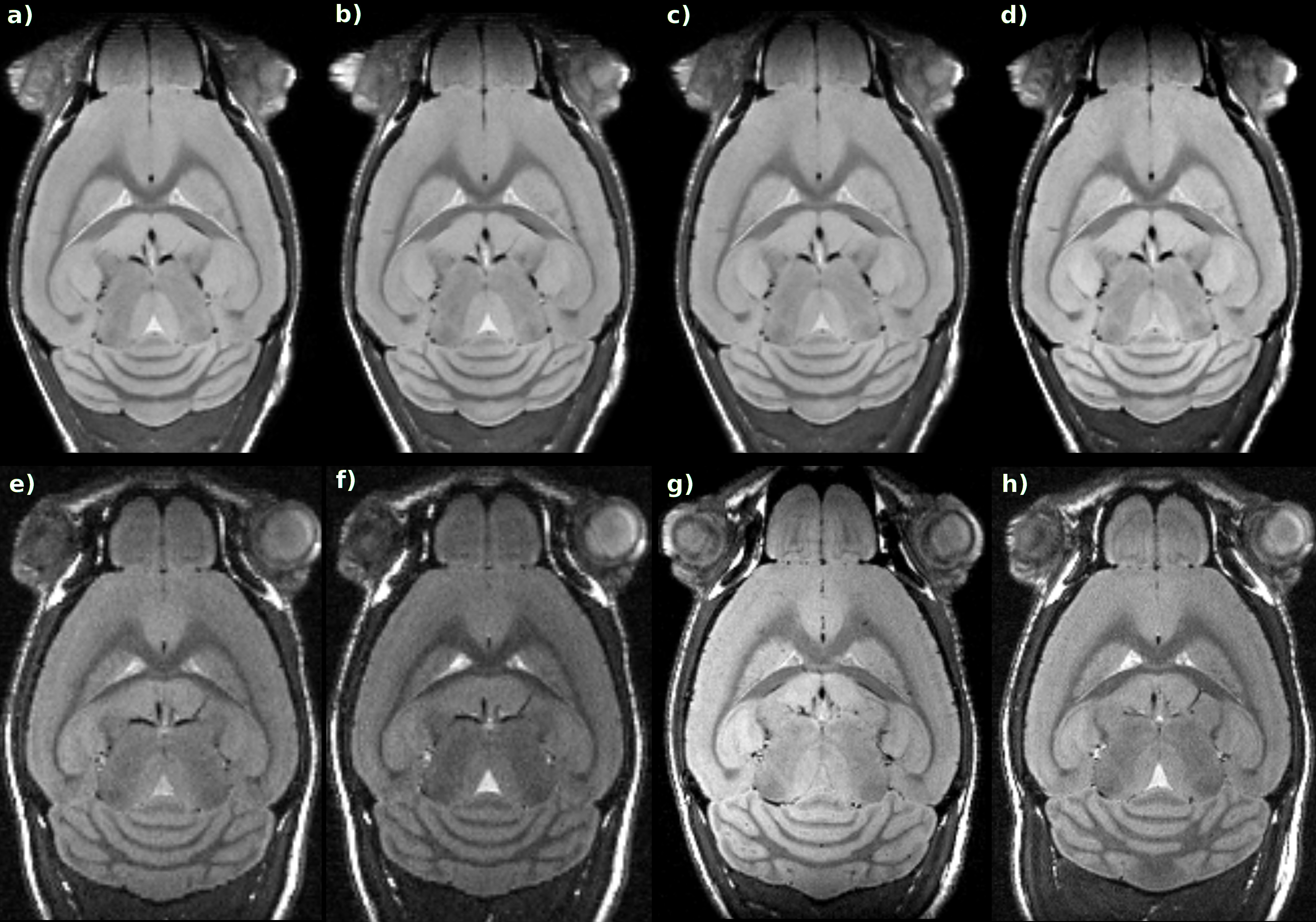
Figure 1 presents the reference image (a) and images acquired with the faster varFlipRARE sequence (b-i) for different FA schemes. With a low target FA=30° (h, i) a distinctly lower signal for the ventricles(Vs) was observed. For a higher target, FA≈40° (f) or when k0 was shifted to 120ms (FA≈35°, e) the T2- and ventricle-contrast was improved. The contrast could be further enhanced by immediately ramping up to ~50° from the target FA≈40° (c,d) in the first phase. To further improve ventricle signal TR was increased to 2700ms (TA=9min, b). The SNR grey matter (GM)=35, white matter(WM)=27, V=51 and CNR CGM-WM=9, CV-GW=15, CV-WM=24 in Fig 1b and c both match the reference image in GM and WM, the ventricle contrast is 15-20% lower. The signal envelope along the echo train is given below each MR image for the 2 channels of the CryoProbe, providing more insights into the actual signal behavior.To assess the effect of the crusher gradients in read and slice direction Figure 2 shows an image comparison. In 2b) no crusher was used at all, leading to some ripple artifacts in the frontal olfactory bulb. Increasing the slice-crusher to 10% amplitude doesn't create a visual difference. Increasing the read-crusher to 10% removes the ripple artifacts, but at 20% the frontal bulbous and ventricles lose signal.
Figure 2e-f shows the resulting images using TF=140. The low ventricle contrast in e) was improved by a faster increase of the target FA (f). The higher resolution scans with 76µm are shown in Figure 2g-h.
Discussion
The use of VFAs together with faster non-selective acquisitions reduced the scan time from 19min to 7-9min achieving similar image contrast and quality as the reference. Reducing the scan time further to TA=4min leads to SNR below 10 due to unintentional diffusion weighting as has been described before5. Simulations5 showed that the use of FAs lower than 40° strongly enhances the effective b-value and 99% of the b-contribution arises from the readout gradients, leading to water-signal suppression. Increasing the read crushers to remove artifacts will further increase the diffusion weighting and has to be carefully chosen.Acknowledgements
We would like to thank Matthias Weigel for some enlightening discussions on RARE sequences and VFAs. The small animal scanner was provided by a large system grant from the DFG (INST 1757/18-1). This work was supported by the German Research Foundation (DFG) within the Research Unit Programme FOR5151 "QuaLiPerF (Quantifying Liver Perfusion–Function Relationship in Complex Resection - A Systems Medicine Approach)" grant number 436883643.References
1 Gaser C, Schmidt S, Metzler M et al. Deformation- based brain morphometry in rats. Neuroimage. 2012, Oct 15;63(1):47-53.
2 Krämer M, Herrmann K-H, Schmidt S et al. Reduction of ringing artifacts for high-resolution 3D RARE imaging at high magnetic field strength. ISMRM 2017, #5082.
3 Hennig J, Weigel M, and Scheffler K. Multiecho sequences with variable refocusing flip angles: Optimization of signal behavior using smooth transitions between pseudo steady states (TRAPS). Magn. Reson. Med. 2003, 49: 527-535.
4 Hennig J, Weigel M, and Scheffler K. Calculation of flip angles for echo trains with predefined amplitudes with the extended phase graph (EPG)‐algorithm: Principles and applications to hyperechoic and TRAPS sequences. Magn. Reson. Med. 2004, 51: 68-80. 5 Weigel M and Hennig J. Diffusion sensitivity of turbo spin-echo sequences. Magn. Reson. Med. 2012, 67: 1528-1537.
Figures