1710
More Efficient SPGR 3D Abdominal Imaging Using an Interleaved Randomized Spoiler1Radiology, Stanford University, Stanford, CA, United States, 2Electrical Engineering, Stanford University, Stanford, CA, United States
Synopsis
3D SPGR is commonly used clinically for contrast-enhanced body imaging, but requires time for a gradient spoiler every TR. This consumes up to 40% of scan time, even for a non-cartesian trajectories with relatively long readouts, such as cones. Removing the spoiler can save time but causes severe artifacts. In this study, we propose a novel approach of applying interleaved, randomized spoilers to a motion-robust 3D-cones sequence. The feasibility of the approach to shorten acquisition time without introducing image artifacts was validated on both healthy subjects and patients.
Introduction
SPGR in contrast-enhanced is standard in abdominal imaging, but gradient spoiling itself is time-consuming. This inefficiency is mitigated with long-readout non-cartesian trajectories, but remains a significant issue. For example, the three-dimensional (3D) cones trajectory has a relatively long readout and exhibits good motion properties, which can be quite useful in abdominal imaging that suffers from extensive motion1–3. Despite the long readout, the spoiling gradient in 3D-cones sequence can still consume up to 40% time of the TR. One possible approach to improve the scan efficiency is by removing these gradients. However, it will introduce severe image artifacts arising from unspoiled transverse magnetization. Here, we propose a novel approach exploiting interleaved-randomized spoilers (IRS) in the 3D-cones sequence, to reduce these artifacts while maintaining scan time reduction. We will first demonstrate the feasibility of the approach to shorten acquisition time without introducing image artifacts on both phantom and healthy subjects, which will then be further validated in free-breathing exams in patients.Methods
IRS Turbo-cones:In 3D-cones sequence, a rewinder and spoiler gradient will be played at the end of each TR to avoid image artifact arising from the unspoiled transverse magnetization signal (Fig. 1A). Removing these two gradients can substantially increase the scan speed (Fig. 1B), but at the cost of severe artifacts. To effectively destroy the residual transverse magnetization, an interleaved-randomized spoiler (IRS) was implemented (Fig. 1C). Specifically, the spoiler gradient is played every M acquisition blocks, and the magnitude of the gradient is randomized between half and full of the largest gradient available. In doing so, only N/M spoilers are required, assuming N acquisition blocks are needed to achieve full sampling, which can substantially shorten the scan times. The randomized spoiler can prevent the residual transverse magnetization from reaching a steady-state, which cannot form a conceivable signal in the image. Therefore, the image artifact can be effectively reduced while shortening the acquisition time.
Phyllotaxis K-space Trajectory Ordering:
In addition to the IRS mechanism, we have employed a phyllotaxis k-space trajectory ordering in analogy to the arrangement of leaves on a stem (Fig. 2). Our current 3D-cones sequence uses a golden angle k-space trajectory ordering, where the cones interleave rotates ~222.5º each time to achieve a uniformly sampled k-space. In contrast, the phyllotaxis trajectory ordering rotates the cones interleave sequentially for a few lines and then rotates a large golden angle (~137.5º) for the next set of readout lines. In doing so, the final k-space trajectory will also uniformly sample the k-space as in the golden angle ordering. Additionally, because the readout gradient itself can partially serve as the spoiling gradient, the readout gradients in the set of sequentially rotated cones interleaves together can achieve better spoiling effect than in a set of large golden angle rotation.
Data Acquisition and Analysis:
An IRS turbo-cones sequence (Fig. 1) was implemented on a 3T GE MR750/Premier scanner (GE Healthcare, Waukesha, Wisconsin). The feasibility of the sequence was first validated on a water phantom. Then, with IRB approval, free-breathing abdominal images were acquired using IRS turbo-cones from six healthy human subjects and four patients (4y to 34y, mean age: 19.9±12.7 years old). Images were also acquired using default 3D-cones and turbo-cones as a comparison. In patients, post-contrast images were acquired after administration of ferumoxytol as intravenous contrast, which remains in the blood pool for many hours, and thus enables comparison between successively run sequences.
The key sequence parameters for both phantom imaging and in-vivo imaging were: slice thickness=3mm, FOV=36cm×36cm×18cm, matrix=320×320×120, flip angel=15º, TE=0.6ms, TR was 5ms, 2.7ms and 2.7ms, and acquisition time was 2:20, 1:34 and 1:52 for default 3D-cones, turbo-cones and IRS turbo-cones, respectively. After acquiring the k-space data, images were reconstructed offline using a custom Python program based on the gridding algorithm (non-uniform FFT) provided in BART4.
Results
Phantom images acquired using turbo-cones sequence without any spoiler gradient exhibit severe artifact, whereas the images acquired using IRS turbo-cones were free from artifact (Fig. 3). A similar result was observed in T1-weighted liver images of healthy subjects, where IRS turbo-cones showed similar image quality as default 3D-cones sequence despite a shorter acquisition time of about 20% (Fig. 4). The feasibility of IRS turbo-cones was further validated on patients (Fig. 5). Again, the contrast-enhanced liver and bowel images acquired using IRS turbo-cones were very close to the images acquired using default cones but with 17.3% reduced acquisition time.Discussion and Conclusion
Application of an interleaved-randomized spoiler to 3D-cones results in a 20% scan time reduction with minimal residual artifact in free-breathing abdominal imaging. The results from this study demonstrate that the interleaved randomized spoiler together with the phyllotaxis trajectory ordering is a viable approach that can effectively destroy transverse magnetization while shortening the TR. The saved acquisition time could be translated to higher spatial resolution or faster scans. The same approach can also be extended to other trajectories such as cartesian or radial SPGR based sequences and is not limited to 3D-cones. Our next step is to determine the optimal series of spoilers that would yield the highest scan efficiency by performing Bloch simulations. In conclusion, IRS turbo-cones can provide accelerated abdominal imaging without compromising image quality.Acknowledgements
This work was supported in part by NIH R01EB009690 and NIH U01EB029427, and GE Healthcare. We thank Drs. Adam Bush and Zhitao Li for helpful discussions.References
1. Gurney, P. T., Hargreaves, B. A. & Nishimura, D. G. Design and analysis of a practical 3D cones trajectory. Magn. Reson. Med. 55, 575–582 (2006).
2. Chen, B. et al. Three-dimensional ultrashort echo time cones (3D UTE-Cones) magnetic resonance imaging of entheses and tendons. Magn. Reson. Imaging 49, 4–9 (2018).
3. Kee, Y. et al. Free‐breathing mapping of hepatic iron overload in children using 3D multi‐echo UTE cones MRI. Magn. Reson. Med. 85, 2608–2621 (2021).
4. Uecker, Martin et al. mrirecon/bart: version 0.7.00. (Zenodo, 2021). doi:10.5281/ZENODO.592960.
Figures
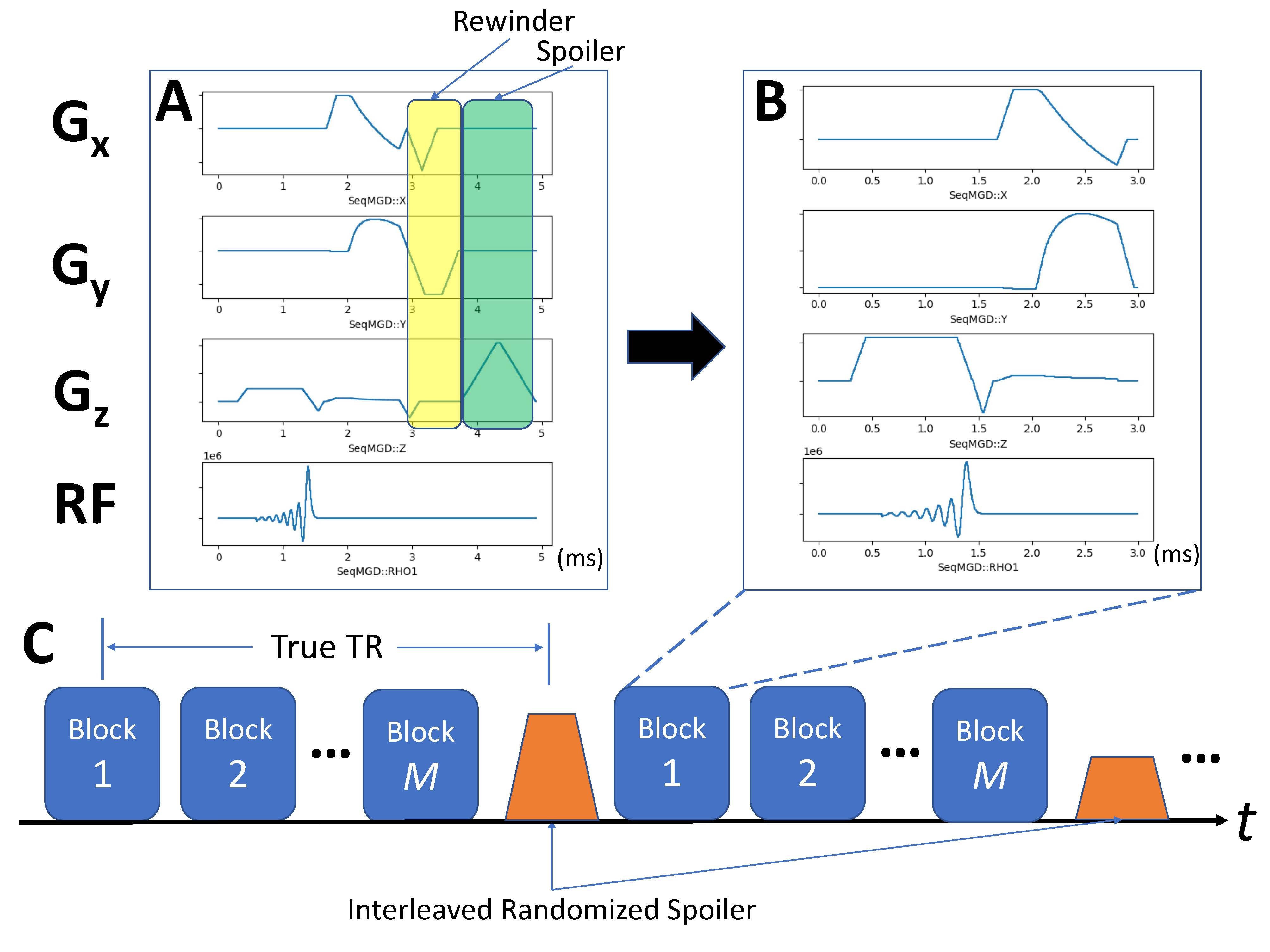
Figure 1: Illustration of the proposed Interleaved-Randomized Spoiler (IRS) mechanism. Removing the rewinder and spoiler gradients in the conventional 3D-cones sequences (A) can substantially shorten the acquisition time, which leads to “turbo-cones” (B) but with severe artifacts. In IRS turbo-cones sequence (C), the spoiler gradient amplitude is randomized and played every M acquisition blocks. In doing so, only N/M spoilers are required assuming N acquisition blocks to achieve full sampling, which can effectively avoid image artifact while shortening the scan time.
Figure 2: Phyllotaxis ordering of the k-space trajectory used in this study (animated GIF). A set of cones interleaves first rotates sequentially, then rotates a large golden angle (~137.5 degree) for the next set of readout lines, which is very similar to the arrangement of leaves on a stem. The final k-space trajectory is uniformly sampled, as in the golden angle ordering, but can more effectively destroy the residual transverse magnetization.

Figure 3: Phantom images acquired using the default cones (A), turbo-cones (B) and interleaved randomized spoiler (IRS) turbo-cones (C), respectively. The residual transversal magnetization can be effectively destroyed using IRS as demonstrated on the phantom images.
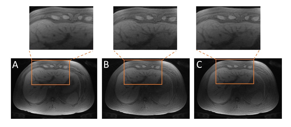
Figure 4: Liver images acquired from a healthy subject using the default cones (A), turbo-cones (B) and interleaved randomized spoiler (IRS) turbo-cones (C), respectively. The IRS turbo-cones provides similar image quality compared with the fully spoiled 3D cones sequence, despite a 20% reduction of the acquisition time. The image from unspoiled turbo-cones is corrupted by artifact.
