1699
BPE XD-GRASP: Using GROG-BPE for improved respiratory motion compensation in dynamic contrast enhanced golden-angle radial MRI1Medical Image Processing Research Group (MIPRG), Department of Electrical & Computer Engineering, COMSATS University Islamabad, Islamabad, Pakistan, 2Department of Electrical Engineering, University of Gujrat, GUJRAT, Pakistan, 3Service of Radiology, Geneva University Hospitals and Faculty of Medicine, University of Geneva, Geneva, Switzerland
Synopsis
This work aims at incorporating the GROG facilitated Bunch Phase Encoding (BPE) in XD-GRASP reconstruction framework for improved motion compensation in free-breathing Dynamic Contrast Enhanced golden-angle radial MRI data. In the proposed method, the inherent randomness and added redundancy in the BPE data, is exploited by the Compressed Sensing (CS) algorithm resulting in images with higher quality and improved mitigation of respiratory motion artifacts as compared to the conventionally used XD-GRASP method.
Introduction
Golden-Angle Radial MRI with Reconstruction of Extra Motion-State Dimensions, termed as XD-GRASP has been demonstrated as an effective technique for motion compensation in free breathing dynamic contrast enhanced (DCE) MRI1. XD-GRASP employs the respiratory motion signal, directly extracted from the acquired data, to resolve the acquired radial projections along an additional motion-state dimension1. Effectuality of XD-GRASP has further been explored by devising alternate ways of constructing motion states or bins, comprising uniform binning, adaptive binning and optimal binning2. Reconstruction of the binned data is performed using Compressed Sensing (CS) algorithm3, exploiting sparsity in the extra motion-state dimension1.GRAPPA Operator Gridding facilitated Bunch Phase Encoding (GROG-BPE) has been presented in the past to reduce the scan time by creating a bunch of additional k-space points around each of the acquired source data point4. To enhance the efficacy of GROG-BPE4 with a sophisticated reconstruction algorithm like compressed sensing3, CS-GROG-BPE has recently been proposed for the reconstruction of highly undersampled static radial MRI data5. The current work, termed as BPE XD-GRASP, aims to exploit GROG-BPE for reconstructing the DCE-MRI data using XD-GRASP to achieve improved overall image quality and effective suppression of motion artifacts.
Methods
Figure 1 shows a schematic illustration for the proposed BPE XD-GRASP method in comparison with the conventional XD-GRASP1 technique. Initially, the continuously acquired multicoil golden angle radial k-space data is divided into multiple frames. Then, the radial spokes in each frame are sorted with respect to the self-navigated respiratory signal and distributed into various motion states or bins using either of the uniform, adaptive or optimal binning techniques2. After binning, the dynamic k-space data is first extrapolated to the randomly blipped BPE trajectory followed by CS algorithm (i.e. Non-linear Conjugate Gradient (NLCG))3 to generate a series of solution images along the temporal as well as motion-state dimension.Undersampled, binned dynamic golden angle radial k-space data and its trajectory are used to generate the randomly blipped BPE signal via SC-GROG6. In this proposed work, the number of bunch points $$$(NBP)$$$ and maximum distance of a calculated bunch point from its source point $$$k\_max$$$ are set to $$$NBP = 9$$$ and $$$k\_max = 0.5$$$ after performing a range of experiments. In our initial experiments, we have tested the proposed method on uniform binning.
The reconstruction results using BPE XD-GRASP (proposed method) and conventional XD-GRASP1 are compared and quantified using Signal-to-Noise Ratio (SNR)7 and Contrast-to-Noise Ratio (CNR)2.
Results and Discussion
The proposed BPE XD-GRASP method has been tested on a free breathing 3D axial liver perfusion DCE-MRI data8, acquired at golden-angle (GA) radial trajectory with 3D stack-of-stars pulse sequence on a Siemens 3T scanner with a matrix size of 512 readouts, 1100 GA radial spokes and 20 receiver coils8. The continuously acquired DCE-MRI data was divided into 11 frames and each frame with 100 radial spokes was binned into four motion states using uniform binning. Each motion state comprises of 25 radial spokes in our experiments.Figure 2a shows reconstruction results of the proposed BPE XD-GRASP method while the results of the conventional XD-GRASP1 method are shown in Figure 2b. It can be seen that images in Figure 2a have better contrast and higher clarity of hepatic vessel and other structures. It is also observed that the proposed method produces less motion artifacts in comparison with the images reconstructed by the conventional method (indicated by yellow arrows in Figure 2).
CNR and SNR values of the images reconstructed by the proposed method in Figure 2a show improvement over the images reconstructed by the conventional XD-GRASP1 method in Figure 2b e.g. for the second motion state, the image reconstructed by the proposed BPE XD-GRASP method has CNR and SNR of 15.5287 and 29.2659, respectively; whereas with the conventional XD-GRASP1 method, the reconstructed image in the second motion state has CNR and SNR values of 11.5183 and 26.8048, respectively. Similar improvement can be observed in other motion states as well. There is an average percentage improvement of 15.5 % and 4.2 % in the CNR and SNR values for the image in all the four motion states by the proposed method over the conventional XD-GRASP method1.
Conclusion
This work incorporates GROG-BPE in reconstruction pipeline of the conventional XD-GRASP method with uniform binning to achieve improved image clarity with lesser motion artifacts. Reconstruction results confirm that the proposed BPE XD-GRASP method performs better than the conventional XD-GRASP1 method, both in terms of better image quality and superior quantifying parameters.Acknowledgements
No acknowledgement found.References
1 Feng, L. et al. XD‐GRASP: golden‐angle radial MRI with reconstruction of extra motion‐state dimensions using compressed sensing. Magnetic resonance in medicine 75, 775-788 (2016).
2 Shahzadi, I., Siddiqui, M. F., Aslam, I. & Omer, H. Respiratory motion compensation using data binning in dynamic contrast enhanced golden-angle radial MRI. Magnetic resonance imaging 70, 115-125 (2020).
3 Lustig, M., Donoho, D. L., Santos, J. M. & Pauly, J. M. Compressed sensing MRI. IEEE signal processing magazine 25, 72-82 (2008).
4 Seiberlich, N. et al. Using the GRAPPA operator and the generalized sampling theorem to reconstruct undersampled non‐Cartesian data. Magnetic Resonance in Medicine: An Official Journal of the International Society for Magnetic Resonance in Medicine 61, 705-715 (2009).
5 Yumna Bilal, I. A., Muhammad Faisal Siddiqui, Hammad Omer. in ISMRM 29th Annual Meeting & Exhibition (Vancouver, 2021).
6 Seiberlich, N., Breuer, F., Blaimer, M., Jakob, P. & Griswold, M. Self‐calibrating GRAPPA operator gridding for radial and spiral trajectories. Magnetic Resonance in Medicine: An Official Journal of the International Society for Magnetic Resonance in Medicine 59, 930-935 (2008).
7 Ji, J. X., Son, J. B. & Rane, S. D. PULSAR: A Matlab toolbox for parallel magnetic resonance imaging using array coils and multiple channel receivers. Concepts in Magnetic Resonance Part B: Magnetic Resonance Engineering: An Educational Journal 31, 24-36 (2007).
8 Li Feng, R. O. Open Resources for Advanced Imaging Research, [Online]. Available: https://cai2r.net/resources/ (2021).
Figures
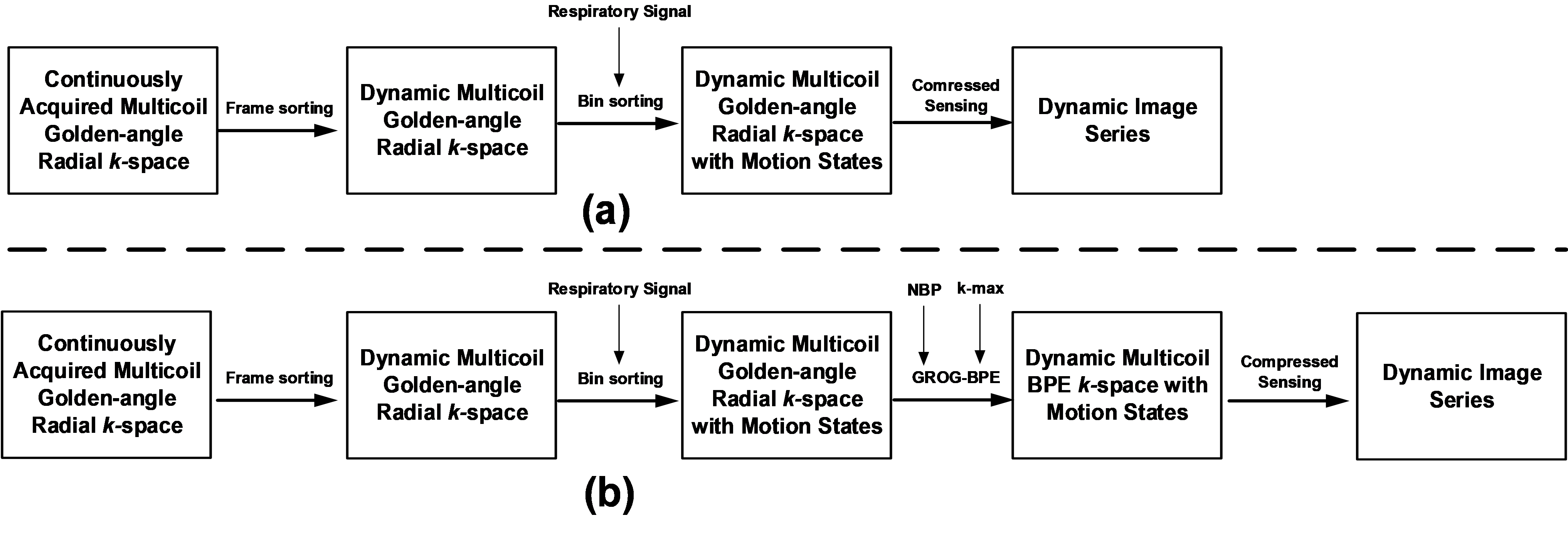
Figure 1: (a) Reconstruction pipeline of conventional XD-GRASP1. (b) Reconstruction pipeline of the proposed BPE XD-GRASP technique. After sorting the Multicoil Golden-angle radial data into frames, self-extracted respiratory signal is used to further sort each frame into multiple motion states or bins. The data is extrapolated to BPE trajectory using GROG-BPE and then compressed sensing reconstruction is performed.
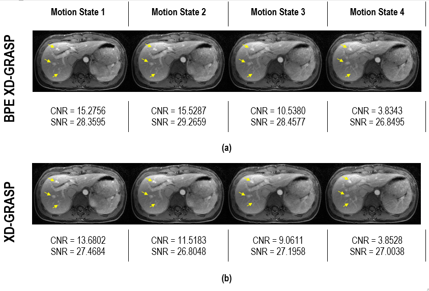
Figure 2: Reconstruction Results of the DCE liver data in venous phase. The multicoil GA radial DCE-MRI data has been divided into four motion states with uniform binning; (a) shows the images reconstructed by the proposed BPE XD-GRASP method; (b) shows the images reconstructed by the conventional XD-GRASP1 method. SNR and CNR values have been given beneath every image in (a) and (b). Yellow arrows highlight some of the regions, where images of the proposed BPE XD-GRASP method offer better clarity of structures in comparison with the images of the conventional XD-GRASP1 method.