1696
Correcting for gradient non-linearity in concurrent field monitoring1Medical Biophysics, Western University, London, ON, Canada, 2Centre for Functional and Metabolic Mapping, Western University, London, ON, Canada
Synopsis
Concurrent field monitoring (FM) via integration of field probes in a radiofrequency coil provides advantages over sequential field monitoring that include patient-specific corrections, as well as improvements to user workflow. However, specific design considerations can require placing field probes far from isocentre where gradient fields are no longer linear, reducing data integrity when fitting high order spatially varying terms. We propose a three-part correction algorithm that seeks to correct these errors and compare FM data, as well as reconstructed images, before and after correction. Correction improved integrity of FM data and enhanced quality of anatomical and diffusion weighted images.
Introduction
Field monitoring (FM) has shown great utility for correcting field perturbations up to third order in space from effects including eddy currents, heating, and mechanical vibrations1. This is performed by monitoring the spatiotemporal field evolution, which can be described as2:$$\boldsymbol{k}(t) = \boldsymbol{P}^{+}\boldsymbol{\phi}_{P}(t))\tag{1}$$
where φP(t) is the extracted probe phase, P+ is a pseudoinverse probing matrix of spherical harmonic basis functions that characterize the phase, and k(t) which are the spherical harmonic basis coefficients (k-coefficients) that describe the spatiotemporal field evolution. FM can be performed sequentially, where field cameras characterize field dynamics in an empty scanner, and the measurements are used to correct images from a subsequent identical acquisition3. Concurrent FM is performed simultaneous to patient scanning, and unlike sequential FM, patient-induced perturbations are tracked, while also improving user workflow4. This is made possible by integrating field probes into a radiofrequency (RF) coil. Depending on coil design and spatial constraints, the field probes may require placement in the non-linear region of the gradient coil. Occupying this region can have adverse effects on probe magnitudes and decay lifetimes. As a result, higher order fits become erroneous, and images reconstructed with unreliable k-coefficients degrade image quality. We propose an algorithm that aims to improve the integrity of FM data acquired with concurrent FM. Correction A accounts for gradient non-linearity (GNL) by correcting probe positions and first-order terms that are inputted into P+. This was made possible by mapping the gradient field distribution inside the scanner via a spherical-harmonic expansion, coupled with vendor-provided coefficients5. Secondly, k(t) is solved one order at a time instead of simultaneously to avoid inaccurate fitting from simultaneous fitting of all terms (Correction B). Lastly, to further penalize probes that are farther in the non-linear region, weighted least squares is performed using the following weighting scheme (Correction C):
$$W_j = 1/\left\|r_j\right\|_2^4\tag{2}$$
where W is a matrix of probe weights and inversely depends on the Euclidean distance of probes from isocentre. In this work, the performance of the correction algorithm is tested and compared to third-order sequential FM correction.
Methods
A healthy patient was scanned on a 7T head-only MRI (Siemens). Diffusion-weighted single-shot spiral acquisitions were performed with a parallel imaging acceleration factor of 4. The imaging parameters were: (FOV: 192 x 192 mm2, in-plane resolution: 1.5 x 1.5 mm2, slice thickness: 3 mm, number of slices: 10, TE/TR: 33/2,500 ms, BW: 2,170 Hz/pixel, flip angle: 70°, b = 0 s/mm2 acquisitions: 1, diffusion directions: 6, b-value: 1000 s/mm2). B0 field maps (in-plane resolution: 1.5 mm, slice thickness: 3 mm) were acquired for inclusion in a model-based image reconstruction6. FM was performed simultaneously using 16 transmit/receive 19F commercial field probes (Skope) that are integrated into a 32-channel RF head coil7. Image reconstruction was performed in MATLAB using in-house developed software that uses an iterative expanded encoding model-based reconstruction1. Images were reconstructed using up to third-order k-coefficients that were generated and corrected using the algorithm described in Figure 1. Reconstructions using subsets of the three steps of the correction were also performed to determine each component’s efficacy. For comparison to sequential FM correction, images were corrected using FM data from an identical acquisition acquired when field probes were situated on the manufacturer’s scaffold.Results
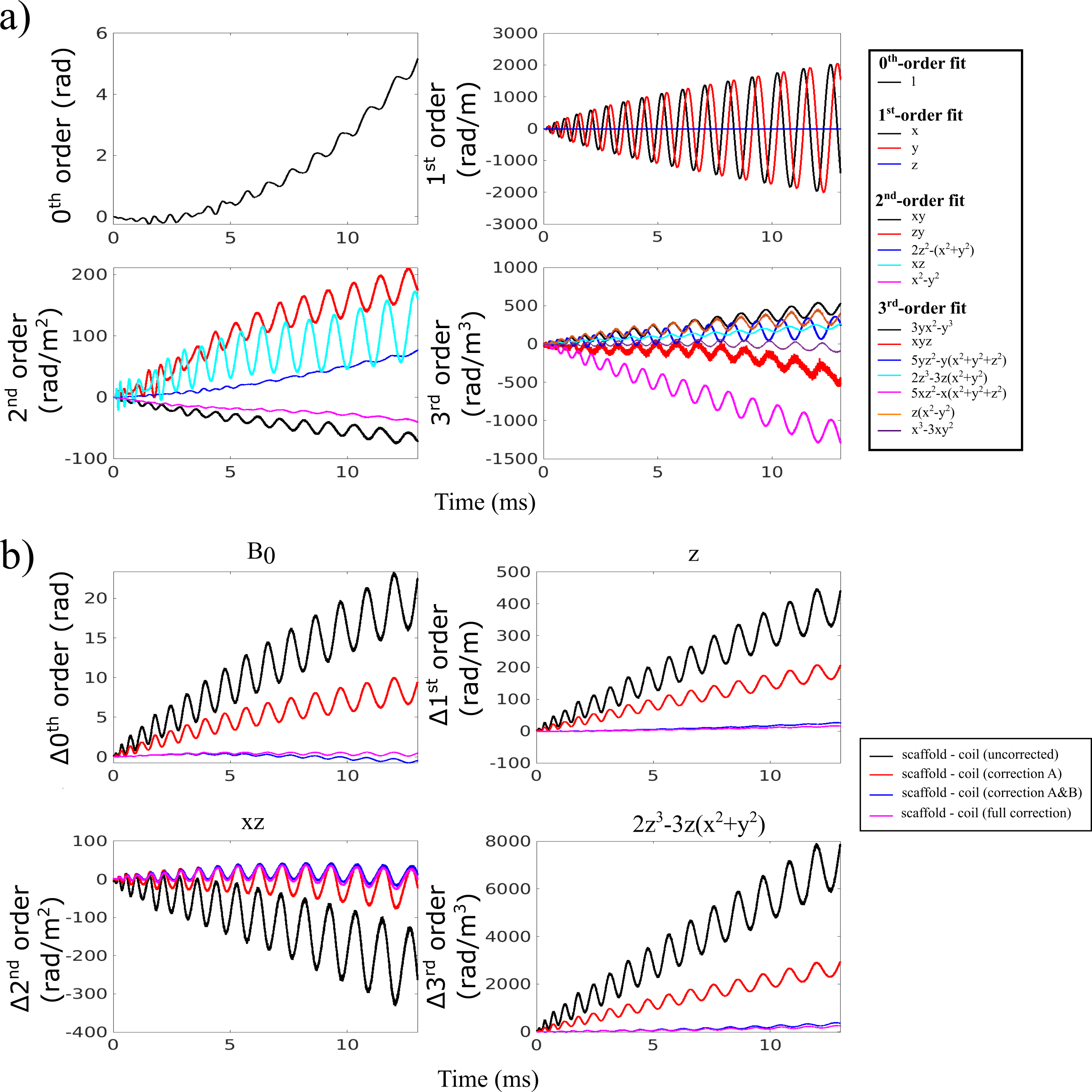
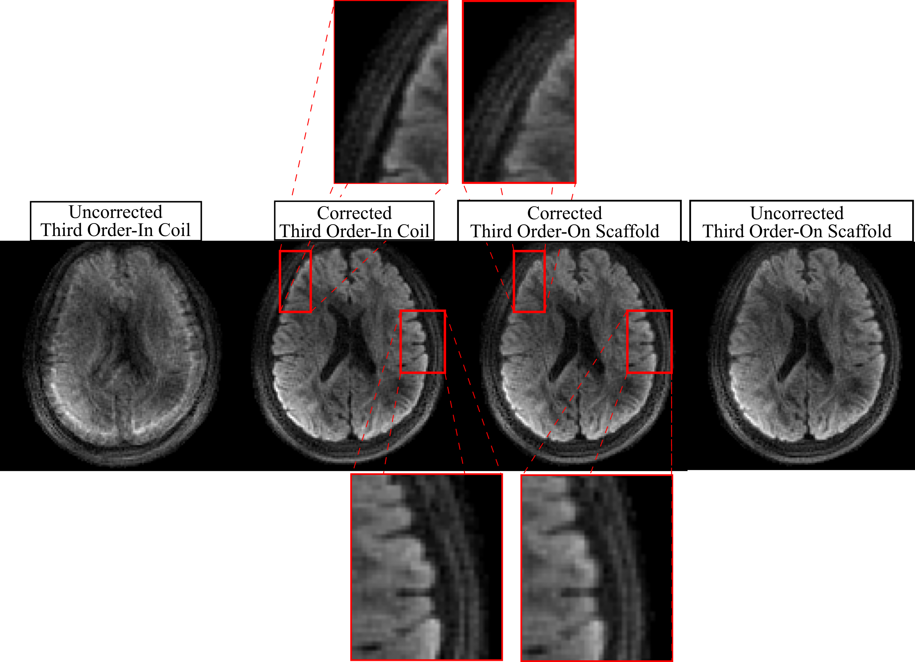
The difference in k-coefficients relative to the third-order scaffold fit (Fig. 2a) decreased significantly from the uncorrected case to correction A and also to correction A&B (Fig. 2b). Small differences were also observed when adding weighted least squares to obtain the full correction. In Figure 3, full correction showed significant improvement for a diffusion weighted image (DWI), while the correction improvement for the scaffold case was incremental. Overall reduction in blurring was observed for the corrected in-coil image relative to the corrected on-scaffold image. Figure 4 illustrates for a b = 0 image and DWI the effectiveness of each correction technique, with the addition of the first two correction steps noticeably reducing the error relative to the “gold standard” fully corrected image.Discussion
Overall, images showed less blurring for concurrent monitoring with correction compared to sequential monitoring (with or without correction) (Fig. 3). This observation shows the added benefits of concurrent FM, which corrects for time-varying effects like frequency drift due to heating. The incremental improvement to image quality of scaffold data was expected due to the scaffold’s accurate measurements. The utility of GNL correction is evidenced by the reduction in the k-coefficient difference relative to the uncorrected case. This is further demonstrated by the higher quality of GNL-corrected images (Fig. 4). The step-wise estimation of k(t) shows substantial improvement in image quality, suggesting that it provides the largest benefit of the three strategies employed. Accordingly, solving k(t) one order at a time allows for a more accurate estimation of each order, while reducing the chances of over/under-fitting lower order terms. While not having a substantial effect on correction, the weighted least squares fit nevertheless helped decrease blurring (Fig. 4).Conclusion
A correction algorithm that successfully improves the reliability of higher order concurrent FM data is presented and validated. This motivates the production of reproducible images for anatomical and functional imaging, as shown by spiral DWI presented in this work.Acknowledgements
This work has been supported by the Natural Sciences and Engineering Research Council of Canada (NSERC), Canada Research Chairs, Canada First Research Excellence Fund to BrainsCAN, and the NSERC PGS D program.References
1. Wilm BJ, Barmet C, Pavan M, Pruessmann KP. Higher order reconstruction for MRI in the presence of spatiotemporal field perturbations. Magnetic Resonance in Medicine 2011;65:1690–1701 doi: 10.1002/mrm.22767.
2. Barmet C, de Zanche N, Pruessmann KP. Spatiotemporal magnetic field monitoring for MR. Magnetic Resonance in Medicine 2008;60:187–197 doi: 10.1002/mrm.21603.
3. Dietrich BE, Brunner DO, Wilm BJ, et al. A field camera for MR sequence monitoring and system analysis. Magnetic Resonance in Medicine 2016;75:1831–1840 doi: 10.1002/mrm.25770.
4. Kennedy M, Lee Y, Nagy Z. An industrial design solution for integrating NMR magnetic field sensors into an MRI scanner. Magnetic Resonance in Medicine 2018;80:833–839 doi: 10.1002/mrm.27055.
5. Janke A, Zhao H, Cowin GJ, Galloway GJ, Doddrell DM. Use of spherical harmonic deconvolution methods to compensate for nonlinear gradient effects on MRI images. Magnetic Resonance in Medicine 2004;52:115–122 doi: 10.1002/mrm.20122.
6. Wilm BJ, Barmet C, Gross S, et al. Single-shot spiral imaging enabled by an expanded encoding model: Demonstration in diffusion MRI. Magnetic Resonance in Medicine 2017;77:83–91 doi: 10.1002/mrm.26493.
7. Gilbert KM, Dubovan P, Gati JS, Menon RS, Baron CA. Integration of a radiofrequency coil and commercial field camera for ultra-high-field MRI. bioRxiv 2021:doi: 10.1101/2021.09.27.462001.
Figures