1694
6-Minute Turbo Spin Echo MRI of the Elbow Using Combined Simultaneous Multi-Slice and Parallel Imaging Acceleration1NYU Grossman School of Medicine, New York, NY, United States
Synopsis
Elbow MRI contributes essential information in acute and chronic elbow injuries. Elbow MRI is ideally obtained in the superman position (prone position with elbow extended, arm elevated above the head, and hand pronated); however, this position can be challenging for patients due to injury-related pain and positioning-related discomfort. To shorten the exam time and reduce patient discomfort, we developed a 4-fold accelerated 5-sequence 6-minute clinical MRI protocol using combined simultaneous multi-slice and parallel imaging acceleration of turbo spin echo sequences. In this study, we compared the image quality of our conventional and novel accelerated elbow MRI protocols.
Introduction
Elbow Magnetic Resonance Imaging (MRI) contributes essential information in acute and chronic elbow injuries, providing high-resolution detail of intra-articular and periarticular anatomical structures, as well as injuries of bone, articular cartilage, synovium, ligaments, tendons, and nerves 1.Elbow MRI is ideally obtained in the superman position (prone position with elbow extended, arm elevated above the head, and hand pronated). However, this position can be challenging for patients due to injury-related pain and positioning-related discomfort. Shorter acquisition times can increase the value of MRI by improving patient tolerability and subsequently reducing motion artifacts 2.
MRI protocols typically include combinations of T1-, proton density-, and T2-weighed turbo spin echo (TSE) pulse sequences without and with fat suppression 3. The required spatial resolution for elbow MRI may result in lengthy protocols. Elbow MRI is ideally obtained in the superman position (prone position with elbow extended, arm elevated above the head, and hand pronated); however, this position can be challenging for patients due to injury-related pain and positioning-related discomfort and may lead to motion artifacts4.
Therefore, implementing novel techniques to reduce time is imperative for improving patient comfort and image quality in these cases. TSE elbow protocols are typically accelerated with parallel imaging undersampling (PAT) 6, which is clinically typically limited to a factor of two. In contrast, simultaneous multi-slice (SMS) acceleration permits simultaneously exciting several slices without reducing the number of k-space samples, allowing for a decreased penalty from under-sampling while reducing scan times 7. 2-fold PAT and 2-fold SMS acceleration can be combined to achieve 4-fold accelerated TSE MRI in the knee 8, but has not been demonstrated in the elbow.
The objective of this study was to compare our conventional 2-fold-accelerated (PAT2) GRAPPA clinical elbow protocol with a novel 4-fold-accelerated (PAT2-SMS2) elbow MRI protocol with reduced time.
Methods
Our current routine clinical elbow MRI protocol includes 5 TSE pulse sequences, consisting of axial native and fat-suppressed proton density-weighted, coronal native and fat-suppressed proton density-weighted, and sagittal fat-suppressed T2-weighted turbo spin-echo pulse sequences (Figure 1). The acquisition time is 9:20 minutes using a multichannel coil.Our novel 4-fold accelerated MRI protocol of the elbow uses combined PAT2-SMS2 acceleration for the same five TSE pulse sequences but a reduced acquisition time of 6 minutes.
For image quality assessment and comparisons, three healthy adult volunteers underwent MRI of the unilateral elbow in superman position using a clinical 3.0 Tesla MRI system (MAGNETOM VIDA, Siemens Healthcare GmbH, Erlangen, Germany) and a large ultraflex 18-channel surface coil.
A board-certified full-time musculoskeletal radiologist evaluated and compared the different datasets for the visibility of anatomical structures (bone, articular cartilage, joint fluid, joint capsule, ligaments, tendons, and nerves) and overall image quality using five-point symmetrical equidistant Likert scales. A score of −2 ("nondiagnostic") indicated complete obscuration of anatomic details and imaging findings, a score of +2 ("very good") indicated full visibility of anatomic details.
Likert scale gradings of anatomic visibility and image quality are given as the median value with the minimum and maximum. Differences between corresponding motion and image quality evaluations were assessed with the Wilcoxon signed-rank test. P < .05 was considered indicative of a statistically significant difference.
Results
All elbow MRI acquisitions, including the conventional PAT2 and the novel 4-fold PAT2-SMS2 protocols, were successfully obtained without error messages or technical interruptions. All pulse sequences were reconstructed inline. Whole-body-specific absorption rates ranged between 1.4 and 1.9 W/kg for all acquisitions and remained within mandated limits.Both the conventional PAT2 and the novel 4-fold PAT2-SMS2 protocols resulted in good to very good visibility of the anatomical structures, including bone, articular cartilage, joint fluid, joint capsule, ligaments, tendons, and periarticular nerves. Comparison of the anatomic visibility scores of PAT2 [5 (3-5)] and the PAT2-SMS2 [5 (3-5)] demonstrated no statistically significant differences (p > 0.05).
Comparison of the overall image quality scores for the PAT2 [5 (4-5)] and the novel 4-fold PAT2-SMS2 [5 (4-5)] MRI datasets were also similar without statistically significant differences (p > 0.05).
Discussion
Combined SMS-PAT acceleration has synergistic properties based on the practically specific absorption rate–neutral acceleration of PI and the near SNR-neutral acceleration of SMS imaging 9. In contrast to PAT acceleration, the SMS technique samples all phase-encoding steps and is almost signal neutral.We realized the efficiency gains of SMS acceleration through the associated reduction of the minimally required total repetition time to complete each pulse sequence. The reduced minimal repetition time obviated the use of concatenations and permitted longer echo trains, which permitted a reduction of the PAT2-accelerated 9-minute MRI protocols to 6-minutes while preserving the visibility of anatomical structures and image quality.
While SMS techniques can result in higher specific absorption rates, our pulse sequences used modified radiofrequency pulse schemes, which resulted in near-neutral specific absorption rates.
Conclusion
Our study results show that 4-fold-accelerated PAT2-SMS2 elbow MRI is feasible and can add value through reducing the acquisition time of a five-sequence TSE 3.0 T MRI protocol from 9-min to 6-min, thereby maintaining good to very good visibility of anatomical structures and overall image quality. As the next step, we intend to evaluate our 6-minute PAT2-SMS2 elbow MRI protocol in a large-scale clinical study.Acknowledgements
No acknowledgement found.References
1. Daniels SP, De Tolla JE, Azad A, Fritz J. Imaging Evaluation of Medial and Lateral Elbow Pain: Acute and Chronic Tendon Injuries of the Humeral Epicondyles. Semin Musculoskelet Radiol. 2021 Aug;25(4):589-599. doi: 10.1055/s-0041-1731790. Epub 2021 Oct 27. PMID: 34706389.
2. Del Grande F, Guggenberger R, Fritz J. Rapid Musculoskeletal MRI in 2021: Value and Optimized Use of Widely Accessible Techniques. AJR Am J Roentgenol. 2021 Mar;216(3):704-717. doi: 10.2214/AJR.20.22901. Epub 2021 Feb 3. PMID: 33534619.
3. D'Anastasi, Melvin, et al. "Understanding 3D TSE Sequences: Advantages, Disadvantages, and Application in MSK Imaging." Seminars in Musculoskeletal Radiology, vol. 19, no. 04, 2015, pp. 321–327., https://doi.org/10.1055/s-0035-1563732.
4. Pazahr, Shila, et al. "MRI of the Elbow: How to Do It." Seminars in Musculoskeletal Radiology, vol. 25, no. 04, 2021, pp. 538–545., https://doi.org/10.1055/s-0041-1729884.
5. Steinbach, Lynne S., and Marco Zanetti. "Elbow Imaging with an Emphasis on MRI." Musculoskeletal Diseases 2009–2012, 2009, pp. 14–23., doi:10.1007/978-88-470-1378-0_3.
6. Deshmane, Anagha, et al. "Parallel MR Imaging." Journal of Magnetic Resonance Imaging, vol. 36, no. 1, 2012, pp. 55–72., https://doi.org/10.1002/jmri.23639.
7. Fritz J, Fritz B, Zhang J, Thawait GK, Joshi DH, Pan L, Wang D. Simultaneous Multislice Accelerated Turbo Spin Echo Magnetic Resonance Imaging: Comparison and Combination With In-Plane Parallel Imaging Acceleration for High-Resolution Magnetic Resonance Imaging of the Knee. Invest Radiol. 2017 Sep;52(9):529-537. doi: 10.1097/RLI.0000000000000376. PMID: 28430716.
8. Del Grande F, Rashidi A, Luna R, Delcogliano M, Stern SE, Dalili D, Fritz J. Five-Minute Five-Sequence Knee MRI Using Combined Simultaneous Multislice and Parallel Imaging Acceleration: Comparison with 10-Minute Parallel Imaging Knee MRI. Radiology. 2021 Jun;299(3):635-646. doi: 10.1148/radiol.2021203655. Epub 2021 Apr 6. PMID: 33825510.
9. Fritz J, Guggenberger R, Del Grande F. Rapid Musculoskeletal MRI in 2021: Clinical Application of Advanced Accelerated Techniques. AJR Am J Roentgenol. 2021 Mar;216(3):718-733. doi: 10.2214/AJR.20.22902. Epub 2021 Feb 3. PMID: 33534618..
Figures
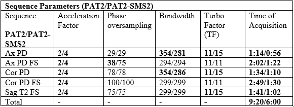
Figure 1. Sequence Parameters. Differences between the PAT2 and PAT2-SMS2 elbow MRI protocols.
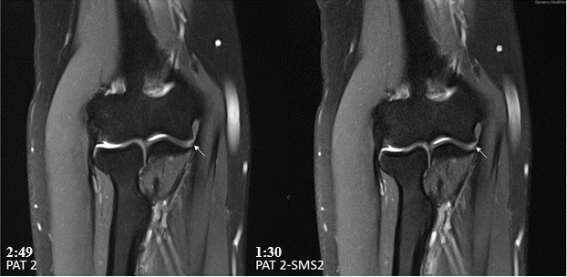
Figure 2. Side-by-side comparison of PAT2-accelerated (left) and PAT2-SMS2-accelerated (right) coronal fat-suppressed proton density-weighted TSE MR images demonstrate excellent visibility of bone, articular cartilage, and the ulnar collateral ligament (arrows), as well as similar good to very good image quality.
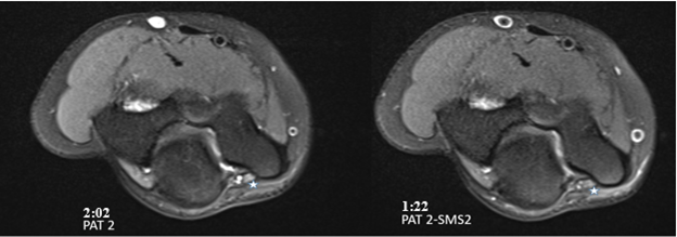
Figure 3. Side-by-side comparison of PAT2-accelerated (left) and PAT2-SMS2-accelerated (right) axial fat-suppressed proton density-weighted TSE MR images demonstrate good visibility of bone, articular cartilage, and the ulnar nerve (asterisk), as well as similar good overall image quality.
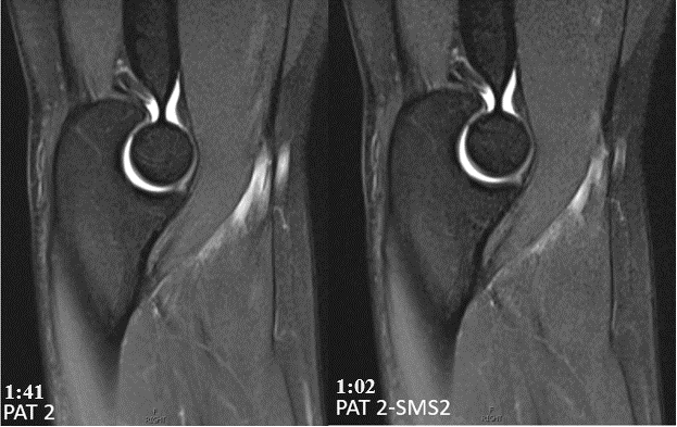
Figure 4. Side-by-side comparison of PAT2-accelerated (left) and PAT2-SMS2-accelerated (right) sagittal fat-suppressed T2-weighted TSE MR images demonstrate excellent visibility of bone, articular cartilage, muscles, and tendons, as well as similar very good overall image quality.
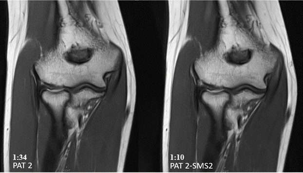
Figure 5. Side-by-side comparison of PAT2-accelerated (left) and PAT2-SMS2-accelerated (right) proton density-weighted TSE MR images demonstrate excellent visibility of bone, articular cartilage, muscles, tendons, and ligaments, as well as similar good to very good image quality.