1682
Influence of breast biopsy clips on diffusion weighted MRI of the breast
Christian Kremser1, Birgit Amort1, and Martin Daniaux1
1Dept. of Radiology, Medical University of Innsbruck, Innsbruck, Austria
1Dept. of Radiology, Medical University of Innsbruck, Innsbruck, Austria
Synopsis
Patients undergoing breast MRI frequently have metallic biopsy clips placed within or adjacent to the lesion, producing metallic artifacts, which could lead to misinterpretation. The aim of our study was to investigate the extent of the artifact of four different clips commonly used in our hospital on diffusion-weighted images obtained with a readout-segmented sequence (RESOLVE) and their influence on ADC maps. It is found that artifacts vary in shape and size between different clips, DWI images and ADC-maps. For the marker with the smallest artifact the minimum size of detectable lesion seems to be approximately 10mm.
Introduction
Diffusion weighted MRI (DWI) of the breast has become an indispensable tool for the detection of breast cancer in clinical practice. Thereby diffusion weighted images together with apparent diffusion coefficients (ADC) can help to increase sensitivity and specificity for the differentiation between benign and malign breast tumors in a multi-parametric approach [1]. Frequently, patients undergoing breast MRI have metallic biopsy clips placed within or adjacent to the lesion [2] producing metallic artifacts, which could lead to misinterpretation. The aim of our study was to investigate the extent of the artifact on diffusion-weighted images and the influence on ADC maps obtained with a readout segmented sequence (RESOLVE) of four different clips commonly used in our hospital.Materials and Methods
According to our clinical routine, experiments were performed on a 1.5T and a 3T whole body MR scanner (Magnetom AvantoFit and Magnetom Skyra, Siemens, Erlangen, Germany) using an 18-channel breast coil. Four different MR conditional biopsy clips were investigated (table 1). For each clip, a cylindrical agar phantom was prepared (500ml isotonic saline with 10g Agar Kobe I). The breast coil was loaded with two of these phantoms at the left and right side. After an empty measurement one clip per phantom was inserted at a depth of 6cm by means of the biopsy needles provided with the clips. For DWI a readout segmented echo-planar imaging (RESOLVE) sequence was used in axial orientation [3, 4] with the same parameters as used for patient exams at our institution: TR=8540ms, TE=53ms, SPAIR fats saturation, FoV: 360mm x 143mm, acquisition matrix: 256 x 102, pixel size: 1.4mm x 1.4mm, number of slices: 42, slice thickness: 4mm, gap: 0, parallel imaging mode: GRAPPA, acceleration factor: 2, b-values: 50 s/mm2 (1 average), 400 s/mm2 (2 averages) and 800 s/mm2 (4 averages), diffusion mode: 3D diagonal, total scanning time: 3min 26sec.The obtained DWI images and ADC maps were analysed using our institution’s picture archiving and communication system (PACS) software Impax EE (R20 XVII SU1, Agfa, Mortsel, Belgium). Thereby the extent of the visible artefact on DWI (b=800 s/mm2) and ADC maps was recorded and ADC maps were inspected for ADC changes.
Results
Fig 1 and 2 show the maximum extent of the obtained artifacts for DWI images and ADC maps, respectively. The size of the observed artifacts was larger and more prominent on DWI images with b=800s/mm2 as compared to ADC maps (table 2 and table 3). The BARD UltraClip Dual Trigger showed the largest artifact, followed by the TUMARK Vision and TUMARK Professional clip. The smallest artifact was observed for HydroMARK Breast Biopsy Site Marker. Whereas most clips showed a clear signal void on the DWI images the HydroMARK Breast Biopsy Site Marker presented with a marked signal increase both at 1.5T and 3T. On ADC maps the observed ADC changes (increased or decreased ADC values) due to the artifact varied between clips and field strength.Discussion
Artifacts produced by biopsy marker clips could render the evaluation of the tissue near the marker impossible or lead to misinterpretations. Size and shape of the artifact depend on the exact design of the respective device. In case of small lesions, the extent of the artifact could completely overlap with the lesion, making a proper assessment of the tissue impossible. Even with the HydroMARK Breast Biopsy Site Marker, which produced the smallest artifact for all investigated clips the minimum size of detectable lesion seems to be approximately 10mm.Conclusions
For the selection of breast biopsy clips the size of the lesion should be taken into account in order to ensure that in subsequent MR examinations the tissue of interest can be properly assessed with diffusion-weighted imaging.Acknowledgements
No acknowledgement found.References
[1] Zhang L, Tang M, Min Z, et al. Accuracy of combined dynamic contrast-enhanced magnetic resonance imaging and diffusion-weighted imaging for breast cancer detection: a meta-analysis. Acta Radiol 2016; 57(6): 651-660.[2] Kapoor MM, Patel MM, Scoggins ME. The Wire and Beyond: Recent Advances in Breast Imaging Preoperative Needle Localization. Radiographics 2019; 39(7): 1886-1906.
[3] Holdsworth SJ, Skare S, Newbould RD, et al. Readout-segmented EPI for rapid high resolution diffusion imaging at 3T. Eur J Radiol 2008; 65(1): 36-46.
[4] Bogner W, Pinker-Domenig K, Bickel H, et al. Readout-segmented Echo-planar Imaging Improves the Diagnostic Performance of Diffusion-weighted MR Breast Examinations at 3.0 T. Radiology 2012; 263(1): 64-76.
Figures
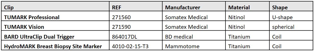
Table 1: List of investigated biopsy markers
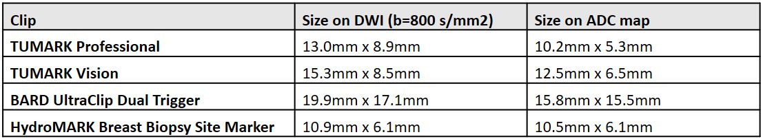
Table 2: Size of artifact @ 1.5T
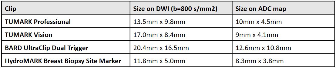
Table 2: Size of artifact @ 3T
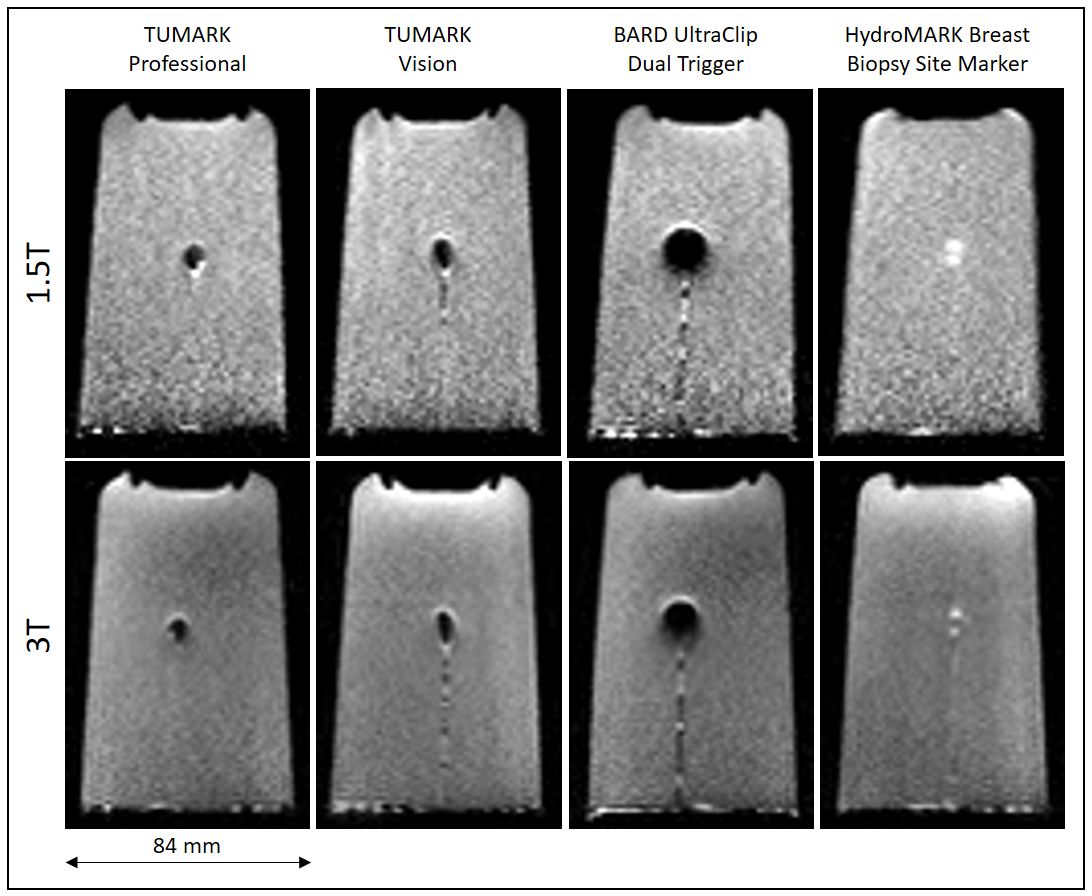
Figure 1: Maximum extend of the obtained artifacts for DWI images (b=800 s/mm2)
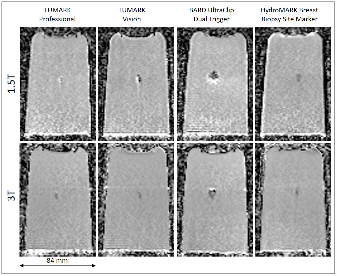
Figure 2: Maximum extend of the obtained artifacts for ADC
maps
DOI: https://doi.org/10.58530/2022/1682