1628
Multishot EPI and a Deep Learning-Based Noise Reduction Strategy for High Resolution Pancreatic DWI1Department of Radiology, Stanford University, Stanford, CA, United States, 2GE Healthcare, Boston, MA, United States
Synopsis
DWI of the pancreas is challenging due to artifacts from physiologic motion, image distortion, and blurring, but has promising applications in pancreatic cancer detection. We conducted a pilot study of pancreatic DWI comparing single-shot and multi-shot EPI protocols as well as multi-shot EPI protocols with and without a new commercially available deep learning (DL) based denoising reconstruction method. Image quality was subjectively scored with key metrics. Multi-shot EPI reduced perceived distortion within the pancreatic bed, while the combination of multi-shot EPI and DL reconstruction subjectively reduced noise.
Introduction
Diffusion Weighted Imaging (DWI) has shown the potential to add clinical value in the detection, diagnosis, and treatment of pancreatic cancer1-6 and has been proposed as a surveillance tool in high-risk patients7-9. The use of DWI for the detection of pancreatic cancer and tumors represents a major challenge due to substantial variability and overlap in quantitative measurements such as apparent diffusion coefficient (ADC)10. Sources of error leading to these ADC inaccuracies may include insufficient spatial resolution, image distortion, and/or insufficient SNR. Multi-shot acquisition approaches have been shown to provide high-resolution DWI while limiting the impact of EPI-induced image distortions11. Deep learning image reconstruction strategies have been shown to reduce noise in DWI while reducing scan time12. Our objective was to evaluate the performance of two-shot multi-shot EPI and a deep learning (DL) based denoising reconstruction method for improving image quality and mitigating artifacts in pancreatic DWI.Methods
This retrospective data acquisition and retrospective review was done with IRB approval and consent waiver. Eleven (N=11) patients underwent MRI of the abdomen, which included both single-shot and multi-shot DWI. All imaging was performed using a 3T SIGNA Premier with a vendor provided respiratory triggered spin echo DWI EPI sequence using a single-shot (SS) and a MUSE11 two-shot multi-shot (MS) sequence with and without a commercially available DL-based denoising reconstruction (AIRTM Recon DL, GE Healthcare, Waukesha, WI). Imaging parameters are summarized in Table 1. To assess the impact of MS and DL-based denoising on image quality, we conducted an observer study performed by two radiologists, each with fellowship training in abdominal MRI and five years of radiology experience (A.S.M. and C.W.H), both of whom were blinded to the clinical data. Pair-wise comparisons for distortion in the pancreas, perceived noise in the pancreatic bed, diagnostic confidence in assessing lesions ≤10mm in size, and diagnostic confidence in assessing lesions >10mm in size were made between 1) SS vs. MS; and 2) MS with and without DL-based denoising. In addition, a score of overall image quality was given for both the liver and the pancreas using a five-point Likert scale, as follows: 1=non-diagnostic; 2=substantial deficits in image quality; 3=moderate image quality; 4=good image quality; and 5=excellent image quality. All reviews were conducted on the high b-value (800s/mm2) diffusion images. Summaries of the pairwise comparisons were tabulated in graphical form for comparison. Likert scores were summarized and compared using two-way ANOVA, and agreement was computed using intra-class correlation coefficients (ICC).Results
Clinical assessment of pancreatic distortion showed that the readers identified reduced distortion on MS relative to SS within the pancreas in over half of cases (55%; Fig. 1A), most commonly in the pancreatic body (45% of all cases). Conversely, the SS images were more commonly assessed to have reduced noise and there was very little difference in diagnostic confidence for either small (≤10mm) or larger (>10mm) lesions (Fig. 1B). As expected, there was very little difference in terms of perceived distortion between MS with and without DL-based denoising (Fig. 1C). MS with DL-based denoising was strongly preferred when it came to image noise and there was a small trend towards increased diagnostic confidence when DL-based denoising was included (Fig. 1D). Analysis of the Likert scores showed that reader 1 preferred MS over SS, in general, while reader 2 had a clear preference for the SS protocol. For both, the addition of DL-based denoising improved image quality in the pancreas, while only reader 2 saw an improvement in the liver (Fig. 2). The combined scores indicate that for both the pancreas and the liver, the addition of DL-based denoising improved perceived image quality while MS with DL-based denoising received the highest overall marks in both anatomic locations. Agreement between the readers was as follows: MS+DL-pancreas (ICC=0.57); MS-pancreas (ICC=0.76); SS-pancreas (ICC=0.02); MS+DL-liver (0.46); MS-liver (0.74); SS-liver (ICC=0.24).Discussion
These data are consistent with prior reports that show that MS can reduce distortion artifacts6, but to our knowledge this is the first demonstration of MS in the pancreas. The tendency to notice reduced distortion in the pancreatic body relative to the head and tail may reflect the impact of gastric air within the body or antrum (Fig. 4), which is immediately adjacent to the pancreatic body. The incorporation of DL-based denoising led to a reduction in perceived noise (Fig. 5), in line with the expected benefits of this tool. The decreased artifact likely contributed to increased reader confidence and the highest overall image quality scores in both the liver and the pancreas for the MS with DL-based denoising images. The current study is limited by a small sample size. The trends suggested here are subtle and not statistically significant. They will need further validation in larger cohorts. Additionally, no quantitative metrics, such as SNR or ADC values, were assessed. Finally, only two readers were used for this pilot study and future studies should include three readers with a range of experiences.Conclusion
Pancreatic DWI with MS with DL-based denoising may offer improved SNR and clinically relevant reductions in distortion, which could improve lesion detection.Acknowledgements
This project was supported, in part, by GE Healthcare.
References
1. Akisik MF, Sandrasegaran K, Jennings SG, Aisen AM, Lin C, Sherman S, Rydberg MP. Assessment of chronic pancreatitis: utility of diffusion-weighted MR imaging with secretin enhancement. Radiology. 2009;250(1):103–109.
2. Lee SS, Byun JH, Park BJ, Park SH, Kim N, Park B, Kim JK, Lee MG. Quantitative analysis of diffusion-weighted magnetic resonance imaging of the pancreas: usefulness in characterizing solid pancreatic masses. J Magn Reson Imaging. 2008;28(4):928–936.
3. Akisik MF, Aisen AM, Sandrasegaran K, Jennings SG, Lin C, Sherman S, Lin JA, Rydberg M. Diagnosis of chronic pancreatitis by using apparent diffusion coefficient measurements at 3.0-T MR following secretin stimulation. Radiology. 2009;252(2):418–425.
4. Zhu M, Zhang C, Yan J, Sun J, Zhao X, Zhang L, Yin L. Accuracy of quantitative diffusion-weighted imaging for differentiating benign and malignant pancreatic lesions: a systematic review and meta-analysis. European Radiology. 2021 Apr 13:1-4.
5. Ren H, Mori N, Hamada S, Takasawa C, Mugikura S, Masamune A, Takase K. Effective apparent diffusion coefficient parameters for differentiation between mass-forming autoimmune pancreatitis and pancreatic ductal adenocarcinoma. Abdominal Radiology. 2021 Apr;46(4):1640-7.
6. Jeon SK, Jang JY, Kwon W, Kim H, Han Y, Kim D, Park D, Kim JH. Diffusion-weighted MR imaging in pancreatic ductal adenocarcinoma: prediction of next-generation sequencing-based tumor cellularity and prognosis after surgical resection. Abdominal Radiology. 2021 Jun 18:1-3.
7. Corrias G, Raeside MC, Agostini A, Huicochea-Castellanos S, Aramburu-Nunez D, Paudyal R, Shukla-Dave A, Smelianskaia O, Capanu M, Zheng J, Fung M, Kelsen DP, Mangino DA, Robson ME, Goldfrank DJ, Carter J, Allen PJ, Conti B, Monti S, Do RKG, Mannelli L. Pilot study of rapid MR pancreas screening for patients with BRCA mutation. Eur Radiol. 2019 Aug;29(8):3976-3985.
8. Kulkarni NM, Mannelli L, Zins M, Bhosale PR, Arif-Tiwari H, Brook OR, Hecht EM, Kastrinos F, Wang ZJ, Soloff EV, Tolat PP. White paper on pancreatic ductal adenocarcinoma from society of abdominal radiology’s disease-focused panel for pancreatic ductal adenocarcinoma: Part II, update on imaging techniques and screening of pancreatic cancer in high-risk individuals. Abdominal Radiology. 2020 Mar;45(3):729-42.
9. Henrikson NB, Bowles EJ, Blasi PR, Morrison CC, Nguyen M, Pillarisetty VG, Lin JS. Screening for Pancreatic Cancer: A Systematic Evidence Review for the US Preventive Services Task Force.
10. Geng R, Zhang Y, Starekova J, Rutkowski DR, Estkowski L, Roldán‐Alzate A, Hernando D. Characterization and correction of cardiovascular motion artifacts in diffusion‐weighted imaging of the pancreas. Magn Reson Med. 2021.
11. Chen NK, Guidon A, Chang HC, Song AW. A robust multi-shot scan strategy for high-resolution diffusion weighted MRI enabled by multiplexed sensitivity-encoding (MUSE). Neuroimage. 2013;72:41-7.
12. Lebel RM. Performance characterization of a novel deep learning-based MR image reconstruction pipeline. arXiv preprint arXiv:2008.06559. 2020.
Figures
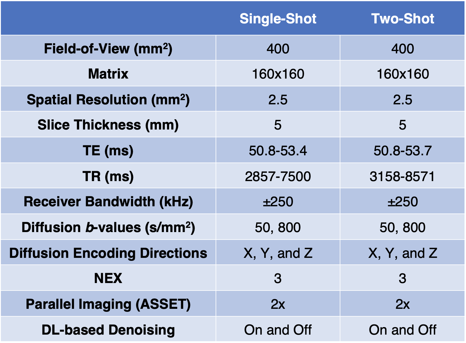
Table 1. Summary of the MRI protocol parameters used for this study.
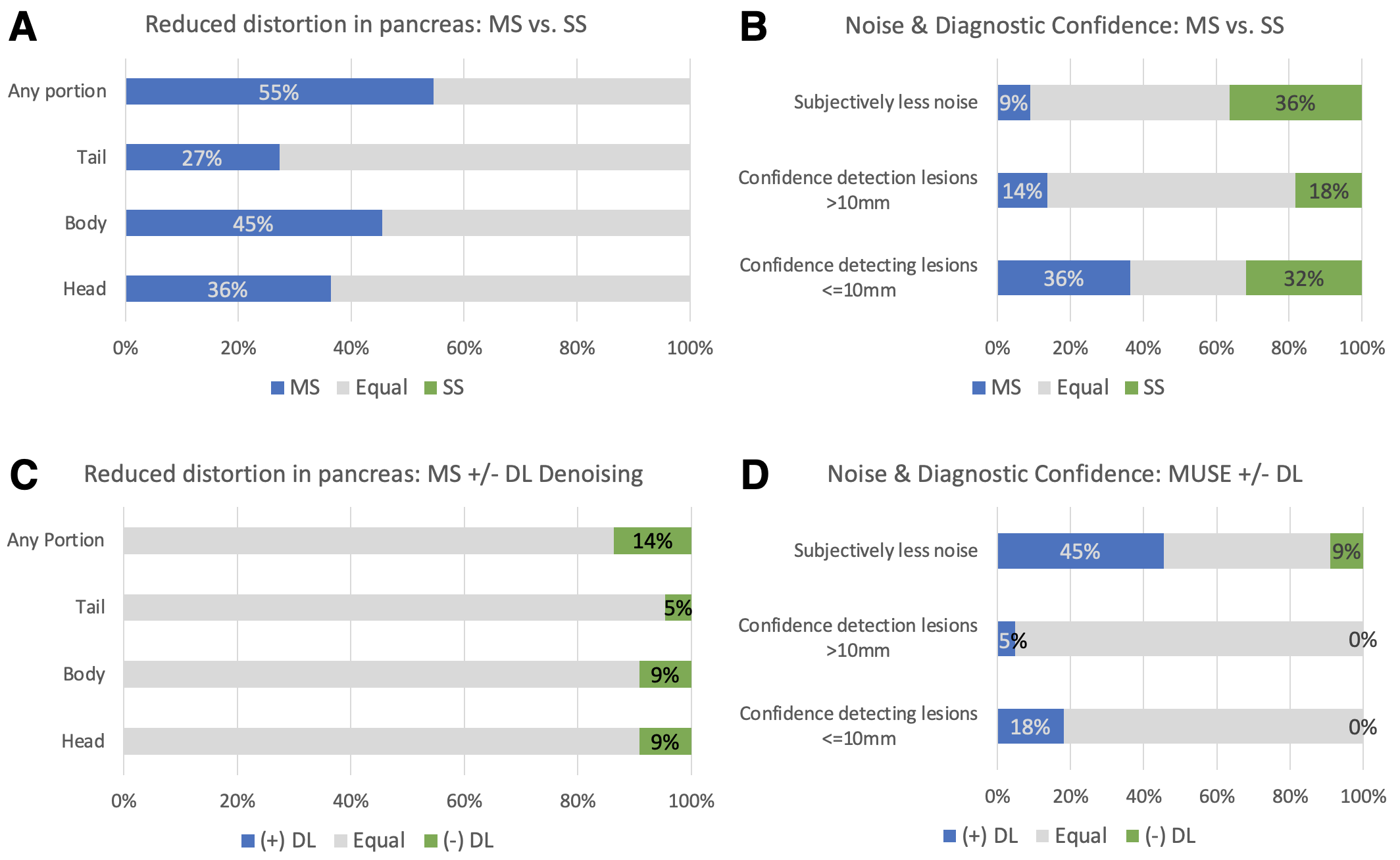
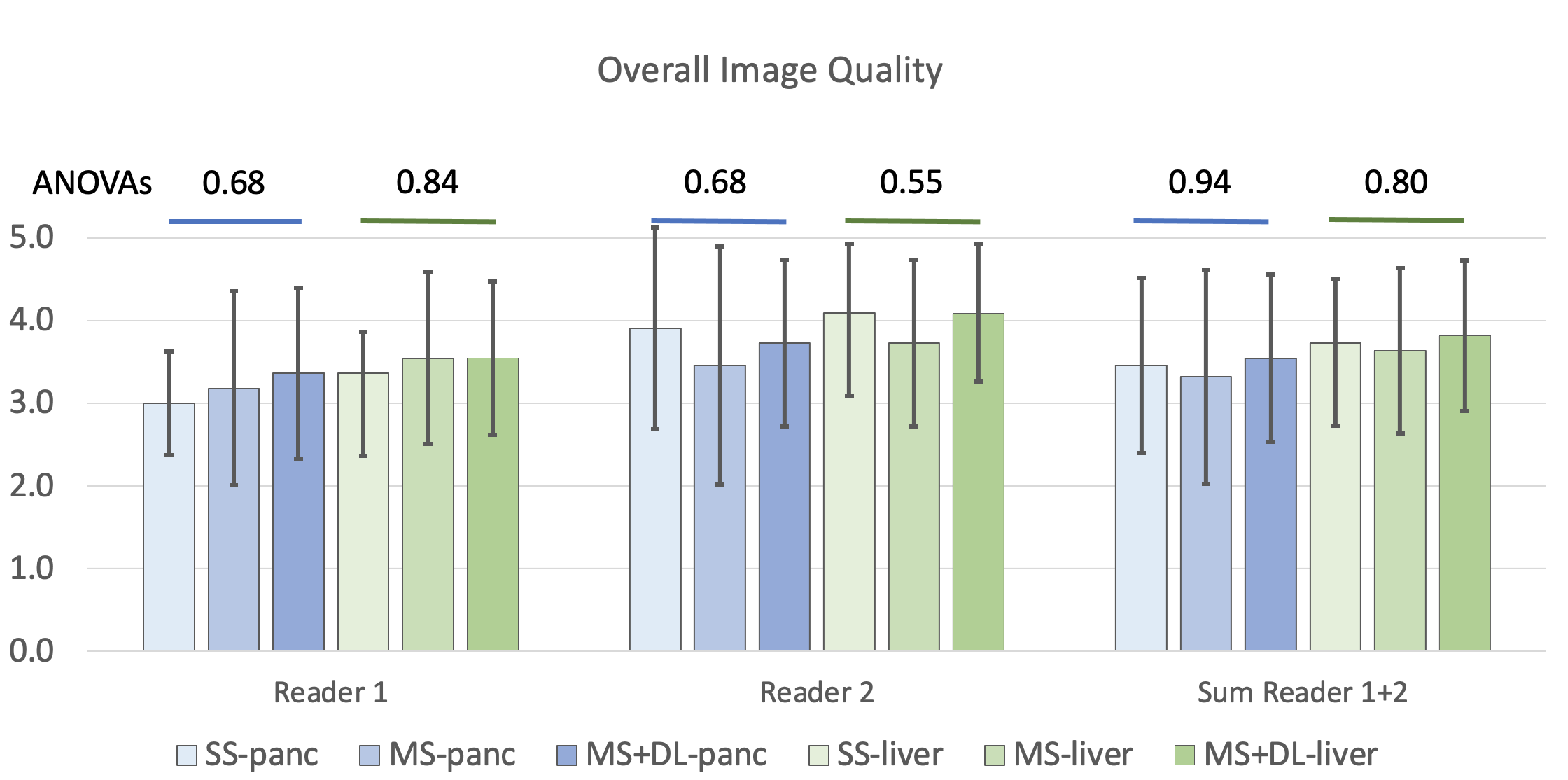
Figure 2. Results of the observer study summarizing the Likert scores for the assessment of overall image quality in the pancreas and the liver. Reader 1 preferred MS over SS while reader 2 showed a preference for SS. The addition of DL-based denoising improved image quality in the pancreas, while only reader 2 saw an improvement in the liver. The combined scores indicate that DL-based denoising improved perceived image quality for both the pancreas and the liver, with MS with DL-based denoising receiving the highest overall scores in both anatomic locations.
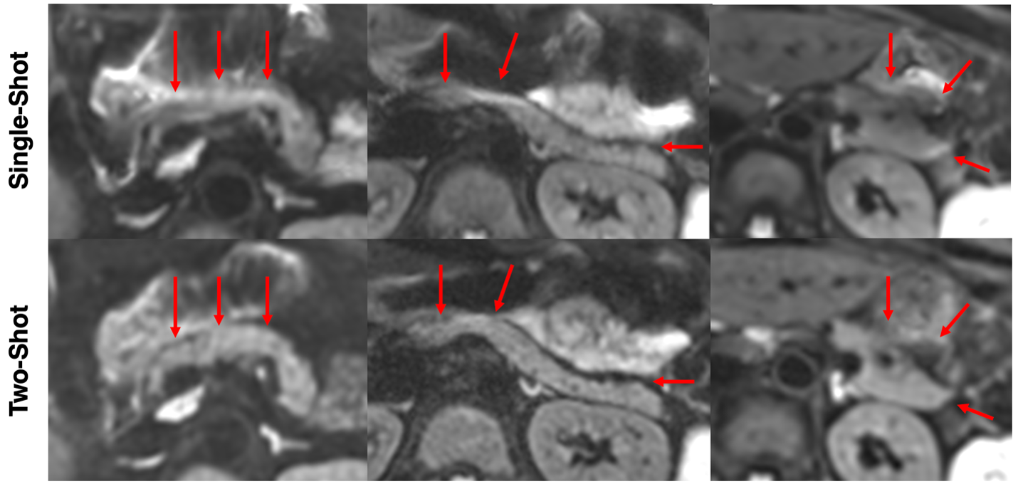
Figure 3. Example images from three patients comparing single-shot (SS, top) and two-shot multi-shot (MS, bottom), which show reduced distortion and image quality in the head, body, and tail of the pancreas for MS. Red arrows indicate areas of the pancreas with notable improvements in distortion and image quality.
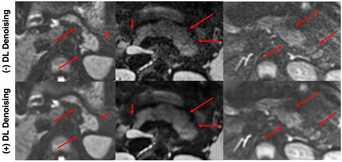
Figure 4. Example images from three patients comparing two-shot multi-shot (MS) without (top) and with (bottom) DL-based denoising, which show lower perceived noise and higher image quality in the head, body, and tail of the pancreas when DL-based denoising is used. Red arrows indicate areas of the pancreas with notable reduction in noise and/or increased margin delineation.