1627
Segmented accelerated multi-shot diffusion imaging combined with reverse polarity gradient (RPG) correction1Department of Radiology, Institute of Clinical Sciences, Sahlgrenska Academy, University of Gothenburg, Gothenburg, Sweden, 2Medical radiation sciences, Institute of Clinical Sciences, Sahlgrenska Academy, University of Gothenburg, Gothenburg, Sweden, 3Department of Medical Physics and Biomedical Engineering, Sahlgrenska University Hospital, Gothenburg, Sweden, 4Department of Radiology, Sahlgrenska University Hospital, Gothenburg, Sweden, 5Department of Radiology, Brigham and Women's Hospital, Boston, MA, United States
Synopsis
The inherent low bandwidth along the phase encoding direction in diffusion-weighted imaging with echo-planar signal readout can cause severe image distortions. These artifacts arise near tissue boundaries and are caused by static magnetic field inhomogeneities. Correction of such artifacts can considerably improve clinical value. We combine a previously presented segmented accelerated multi-shot diffusion imaging method with reverse polarity gradient (RPG) for markedly improved reduction of geometric distortions.
Introduction
Diffusion-weighted imaging, DWI, of the brain can be challenging in areas around the optic nerve, sinuses and the brainstem. These areas exhibit a mixture of tissues ranging from air-filled cavities, bone and brain tissue, which will result in distortion artifacts on diffusion-weighted images. The distortions arise due to susceptibility variations and are pronounced along the direction with the lowest sampling bandwidth. Moreover, higher spatial resolution will further reduce the sampling bandwidth and increase associated distortions. Great work has been done toward utilizing both phase and magnitude information in reconstructions to mitigate artifacts common in DWI1–6. To mitigate susceptibility-related distortions Madore et al.7 introduced a segmented accelerated method, where distortions are reduced according to the segmentation factor. The technique achieves acceleration by exploiting data sparsity that results from processing in x-y-kb-kd space, where kb and kb are the Fourier transform of b and d (b-value and diffusion direction), respectively. The sparsity domain is utilized by displacing aliased data across this domain with a special sub-sampling scheme, allowing non-aliased data to be recovered through regularized reconstruction. Motion induced phase errors are corrected with phase information from a low-resolution navigator echo collected with each shot8–11.Another approach to reduce distortions is the reverse polarity gradient method, RPG12,13. The RPG method relies on the collection of image data with both regular (blip-up) and opposite (blip-down) polarity phase encoding gradients (Fig. 1). The resulting pair of images exhibits distortions of equal magnitude but opposite direction, which is used to estimate distortion for each pixel position. Typically, such data is acquired for only one diffusion weighting level, e.g., for b0, and the estimated distortion is then used to correct all images in a subsequent post-processing step.
The segmented accelerated multi-shot method and RPG are completely independent methods for distortion correction and can thus be combined for further distortion reduction. In the present work the combination of these two methods was tested in phantoms and human brain scans.
Method
The segmented accelerated diffusion technique was extended with the implementation of the reverse polarity gradient method to further reduce distortions in DWI-data. Three datasets were collected with four-, six- and eight-fold segmentation. The b=0 data was fully sampled with forward and reversed phase encoding blips, resulting in pairs of images with distortion shifts in opposite directions. The diffusion-weighted data, b > 0, was accelerated and segmented four-, six-, and eight-fold and collected by traversing k-space in only one phase direction, namely bottom-up. Susceptibility-induced off-resonance fields were estimated from the b=0 image pairs according to a method described by Holland et al.13, as implemented in the Orchestra reconstruction programming suite (GE healthcare, Waukesha, WI). The resulting field maps were then used to correct all the data.Data were collected on a 3 T scanner (MR750w, GE healthcare) with a 48-channel head coil. The imaging parameters where: 8 axial slices, 3.6 mm slice thickness, 0.4 mm slice gap, 192x192 matrix, 240 mm FOV, TR = 3000 ms, TE = 83/71/66 ms, TEnav = 138/111/98 ms, phase encoding bandwidth = 4.1/6.1/8.1 kHz for four-fold/six-fold/eight-fold segmentation, respectively. Eight b-values, evenly spaced between 0 and 1000 s/mm2, along 6 diffusion directions were collected. For comparison, a high resolution T2 propeller data set was also acquired with the same scan coverage.
Result and Discussion
Figure 2 shows the b=0 images with segmentation-related distortion correction only, for which k-space was traversed top-down and bottom-up along with the corrected image, a high resolution T2 anatomical image and ADC maps. The reduction of the distortion in the T2 (b=0) image is evident and can clearly be seen around the eyes and the sinuses in the corrected images. A close-up of the eyes and the optic nerve in the ADC maps and b=1000 s/mm2 images are presented in figure 3.Improved geometric fidelity is demonstrated with increasing segmentation factor, as can been seen around the eyes (Fig.2). Even with an eight-fold accelerated multi-shot acquisition, however, residual distortions are present. Combination with the RPG method results in complete elimination of distortion artifacts in most areas and thus overall improved diagnostic value of the diffusion-weighted images. In the ADC maps, the optic nerve is correctly depicted in the case with eight-fold segmentation, but not for four- or six-fold segmentation (yellow arrows in Fig. 3). This is probably explained by the lower bandwidth in the four- and six-fold case, as this corresponds to higher level of distortions.
It is demonstrated that while the RPG method is efficient for four-fold segmentation, it falls short in areas around the eyes. This is expected in areas where signal pile-up is present, as these cannot be adequately handled by the RPG method and result in both distortion and signal artifacts. By increasing the segmentation factor to eight, and hence the phase-encoding bandwidth, theses signal pile-up is reduced as can been seen for the left eye in figure 2. This helps the RPG methods to work more efficient.
Conclusion
Increasing the segmentation and acceleration factor improves geometric fidelity. Combining the segmented accelerated multi-shot method with RPG can correct distortion artifacts to a greater degree than either method in isolation, and in so doing improves the overall diagnostic value.Acknowledgements
No acknowledgement found.References
Figures
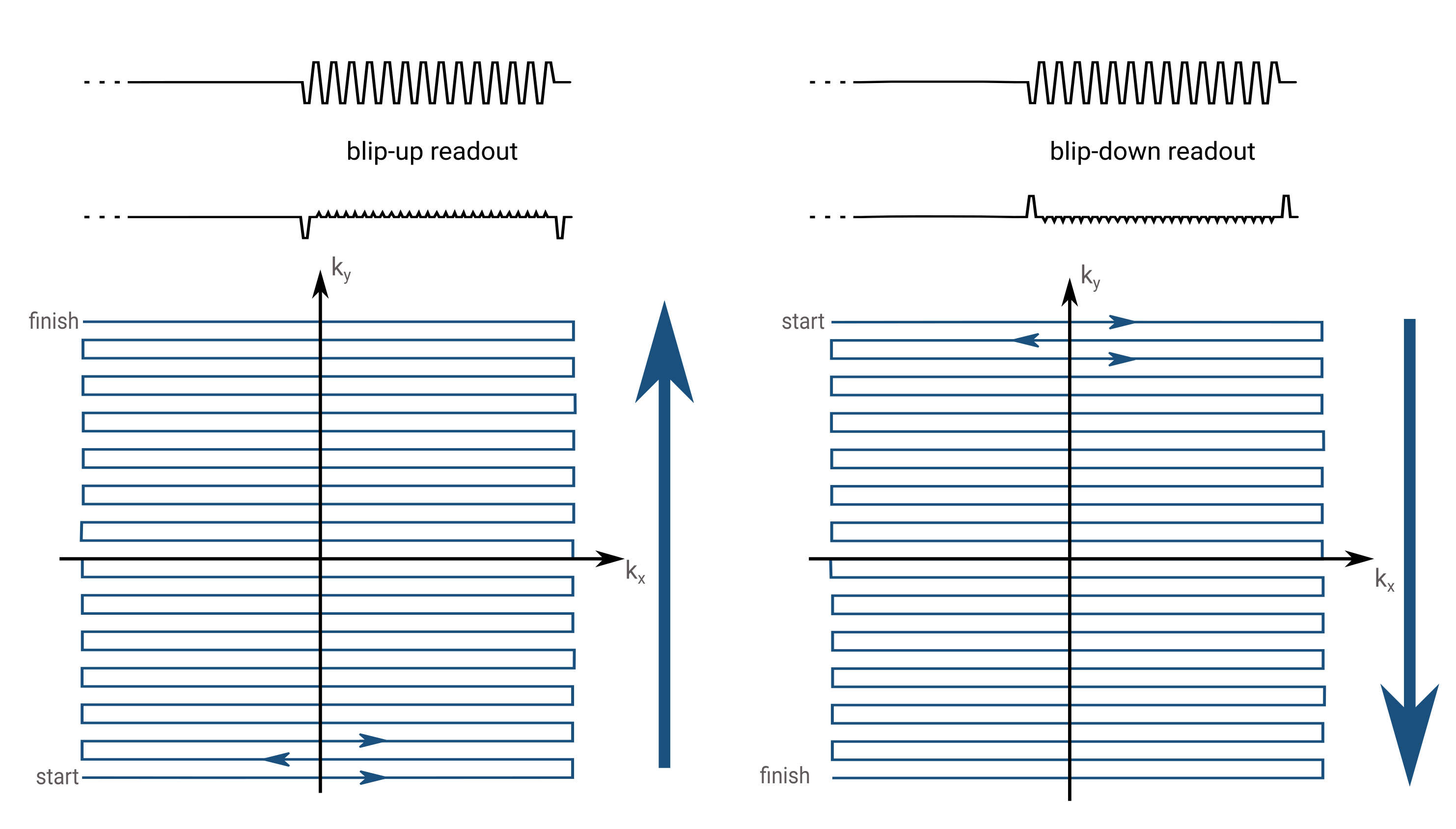
Figure 1: Collection of two identical data set with only a shift in the phase encoding direction between blip-up (collecting the data bottom to top) and blip-down (collecting the data from top to bottom). These data sets are used to create a field-map which is used to correct geometrical distortions in the final image. This method is called the reverse polarity gradient, RPG, method.
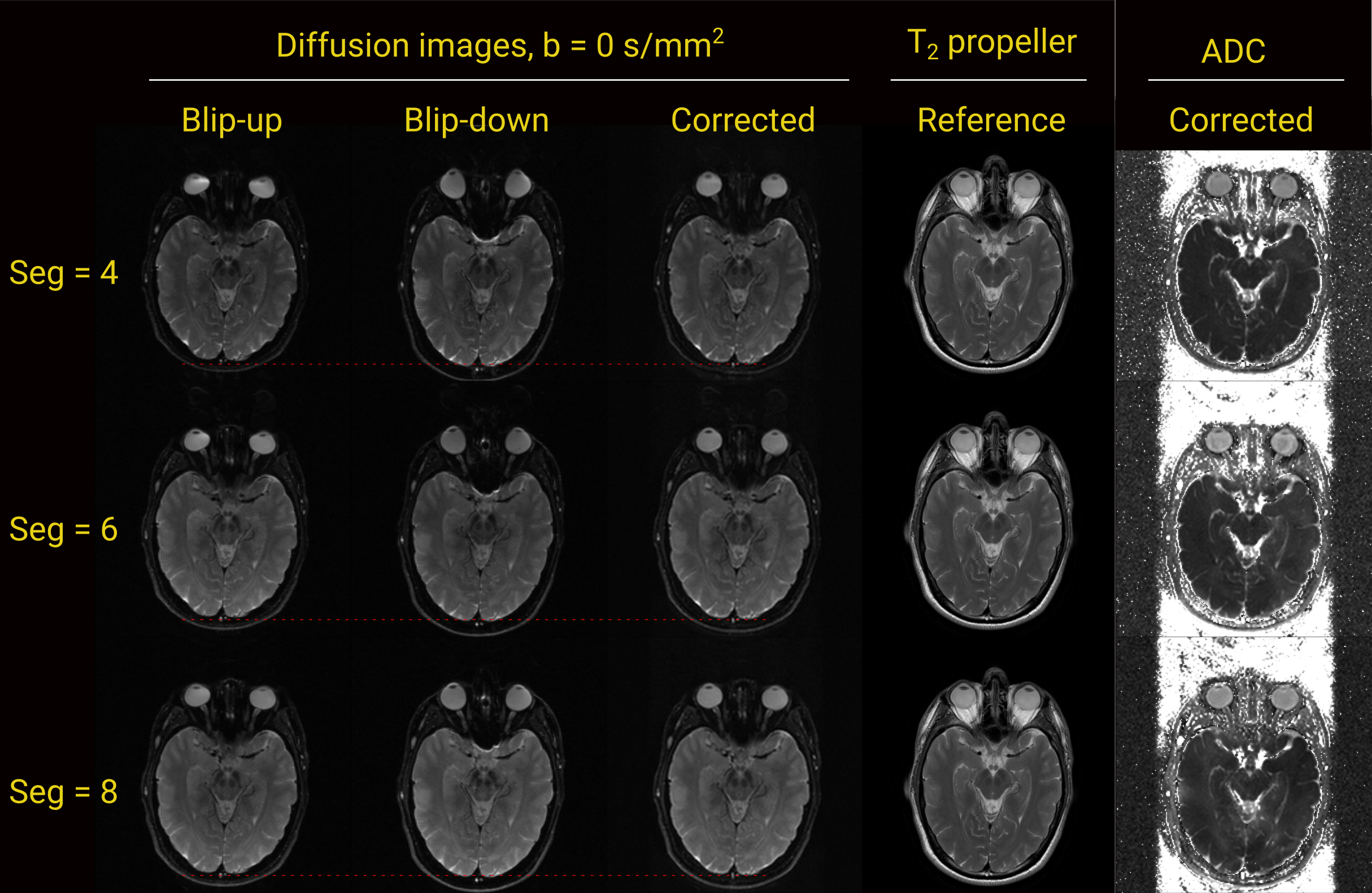
Figure 2: B = 0 s/mm2 images with blip-up read-out and a blip-down read-out along with a corrected image using the RPG method. T2w propeller are shown for reference. Apparent geometrical distortions are visible around the eyes and less obvious distortion including compressing (left) or stretching (middle) of the brain can been seen by comparing the distance from the red marker at the bottom of each image. These distortions reduces with increasing segmentation factor. The eyes and the optic nerve in the corrected image show higher geometric fidelity compared to the uncorrected images.
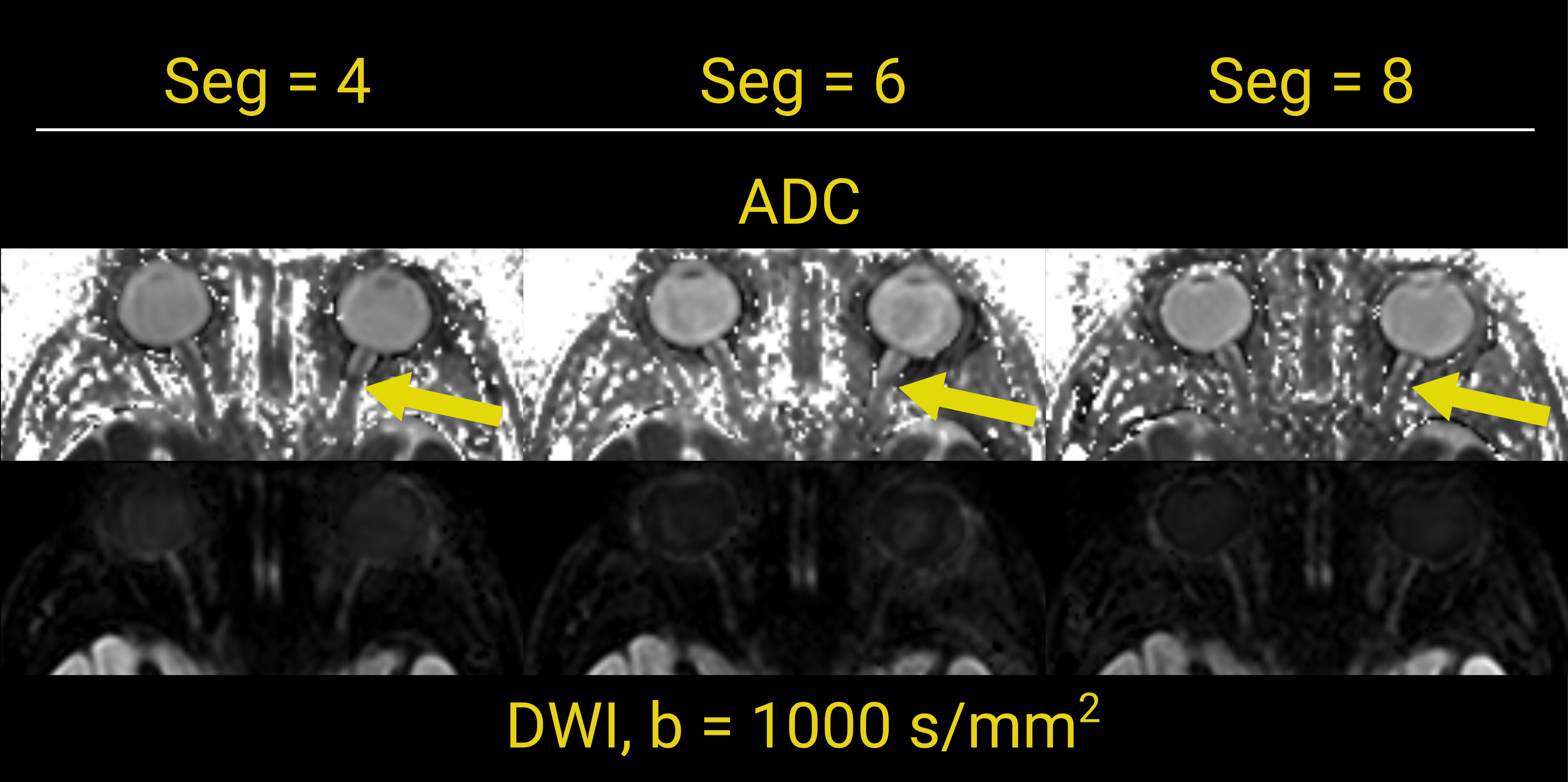
Figure 3: Close up of the ADC maps and b=1000s/mm2 images for four-, six- and eight-fold segmentation and acceleration. Only for the case with eight-fold segmentation is the right optic nerve correctly depicted in the ADC maps (yellow arrows) and b=1000 images. The increase in phase-encoding bandwidth with increasing segmentation factor helps mitigate distortions arising in these areas.