1612
Spiral 3DREAM sequence for fast whole-brain B1 Mapping1Deutsches Zentrum für Neurodegenerative Erkrankungen e.V. (DZNE), Bonn, Germany, 2Department of Physics & Astronomy, University of Bonn, Bonn, Germany
Synopsis
The 3DREAM sequence provides a fast technique for whole-brain B1 mapping, but suffers from different blurring levels in FID and STE* images. We developed a new variant of the 3DREAM sequence with a spiral readout to mitigate this problem. The spiral readout shortens the echo train duration and reduces the number of excitations after the STEAM preparation significantly. This leads to similar blurring levels in FID and STE* images and increases the effective resolution of flip angle maps. As a result, correction strategies for different blurring levels are no longer necessary.
Introduction
Knowledge of the B1 transmit field is a prerequisite for avoiding or correcting inhomogeneities especially at higher field strength. The DREAM sequence1,2 provides fast and accurate B1 mapping for single and multi-channel systems. In the DREAM approach, a set of FID and STE* images is used to calculate flip angle maps. The 3D encoded version of this sequence called 3DREAM3 is able to acquire whole-brain B1 maps in only a few seconds and is therefore well suited as a calibration prescan. However, it suffers from STE* signal saturation due to a relatively long readout, as the STEAM preparation is only applied once in the beginning of the sequence. This leads to significant blurring in the STE* images mostly due to signal saturation at higher flip angles and consequently to artifacts in the flip angle maps. These artifacts are mitigated by applying a blurring filter3 to the FID images, at the cost of losing spatial resolution and accuracy.In this work, the Cartesian 3DREAM readout is replaced by a 3D stack-of-spirals readout. This shortens the single-shot readout and significantly reduces the number of excitations after the STEAM preparation. The spiral version of the 3DREAM sequence is compared to the original Cartesian implementation and an AFI sequence.
Methods
The spiral 3DREAM sequence shown in Figure 1 can be divided into a STEAM preparation block and a readout block. The STEAM preparation consists of two non-selective block pulses α and the gradient Gm2, that dephases the STE* signal.The STEAM block is only applied once, resulting in a single-shot readout of the prepared STE* signal. The readout block starts with two slab-selective binomial water-excitation pulses with flip angle β/2. This is followed by the gradient Gm1, which rephases the STE* signal at TESTE* and dephases the FID signal. The STE* signal is captured with an interleaved 3D stack-of-spirals readout. The spiral readout is accelerated with reduction factor Rint=3 by skipping rotated spiral interleaves. A CAIPIRINHA sampling scheme (shift δ=2) is applied, shifting the set of spiral interleaves in subsequent phase encoding steps. Additionally, the readout is accelerated in phase encoding direction by a factor of Rpe=2. Before reading out the FID signal, it is rephased by the gradient Gmiddle, which dephases the STE* signal at the same time. Afterwards, the FID is captured within the same excitation block as the STE*.
The total duration of the sequence including a parallel imaging calibration prescan and a relaxation delay was about 7s, while the 3DREAM duration was only 470 ms with an echo train length (ETL) of 60. Data was acquired in transversal orientation at 5 mm isotropic resolution with a 200x200x200m3 FOV on a Siemens MAGNETOM Terra 7T scanner with an 1‐channel TX/32‐channel RX head coil (Nova Medical). Both T2 and T2* effects are compensated as effective echo times are equalized such that $$$TS=TE_{FID}-TE_{STE^*}$$$ is fulfilled.
As a reference, B1 maps were acquired using an AFI4 (TA=79s) sequence and the Cartesian version of the 3DREAM sequence3 (TA=10s, TDream=1060ms, ETL=317). FID images of the 3DREAM sequences were filtered with a global filter3. The spiral sequence was implemented with Pulseq5, images were reconstructed with an online pipeline6. The Pulseq sequence file, reconstruction pipeline and raw data will be publicly available.
Results
In Figure 2, STE* images from the Cartesian and the Spiral 3DREAM sequence, as well as filtered and unfiltered FID images of the transversal plane are displayed. Stronger blurring is observed in the Cartesian STE* images compared to the spiral images. This translates to a strong blurring filter for Cartesian FID images, while the spiral filter is much weaker and has almost no effect on FID images. Signal-loss is observed in the frontal and lower parts of the brain especially in spiral images.Figure 3 shows flip angle maps acquired with the AFI and both 3DREAM sequences, with and without FID filtering. The flip angle distribution is similar for all three sequences, though artifacts are reduced in Cartesian 3DREAM maps by filtering. Comparing the flip angles of a small volume between AFI and DREAM maps in Figure 4 indicates good agreement of the three different maps.
Discussion
The spiral readout in the 3DREAM sequence reduces the number of readout excitations by almost a factor of 4.5, leading to significantly reduced blurring in STE* images. This allows much weaker filtering on FID images or even to omit the filtering, which increases the effective resolution of B1 maps. Although the spiral readout is relatively short due to acceleration, typical spiral artifacts such as blurring and signal-loss can be observed in the lower brain and other areas with high B0 inhomogeneity.Increasing the number of spiral interleaves shortens the readout and decreases spiral blurring, but increases STE* blurring due to the higher ETL, while decreasing this number would lead to the opposite effect. Higher acceleration decreases both STE* and spiral blurring, but is limited by the coil sensitivity and decreased SNR.
Conclusion
The presented spiral 3DREAM sequence utilizes a fast readout minimizing the echo train length. This leads to similar blurring levels of FID and STE* images. Therefore, blurring correction is omitted, leading to a higher effective resolution of flip angle maps.Acknowledgements
No acknowledgement found.References
1. Nehrke, K. et. al. DREAM—A Novel Approach for Robust, Ultrafast, Multislice B1 Mapping, MRM, 2012;68:1517–1526
2. Nehrke, K. et. al. Volumetric B1 1 Mapping of the Brain at 7T Using DREAM, MRM, 2014;71:246-256
3. Ehses, P. et. al. Whole-brain B1-mapping using three-dimensional DREAM, MRM, 2019;82:924-934
4. Yarnykh, V. L. Actual Flip-Angle Imaging in the Pulsed Steady State: A Method for Rapid Three-Dimensional Mapping of the Transmitted Radiofrequency Field, MRM, 2007;57:192–200
5. Layton, K. J. et. al. Pulseq: A rapid and hardware-independent pulse sequence prototyping framework, MRM, 2017;77(4):1544-1552
6. Veldmann, M. et. al. Open-Source MR Imaging Workflow, Proceedings of the 2021 ISMRM & SMRT Annual Meeting
Figures
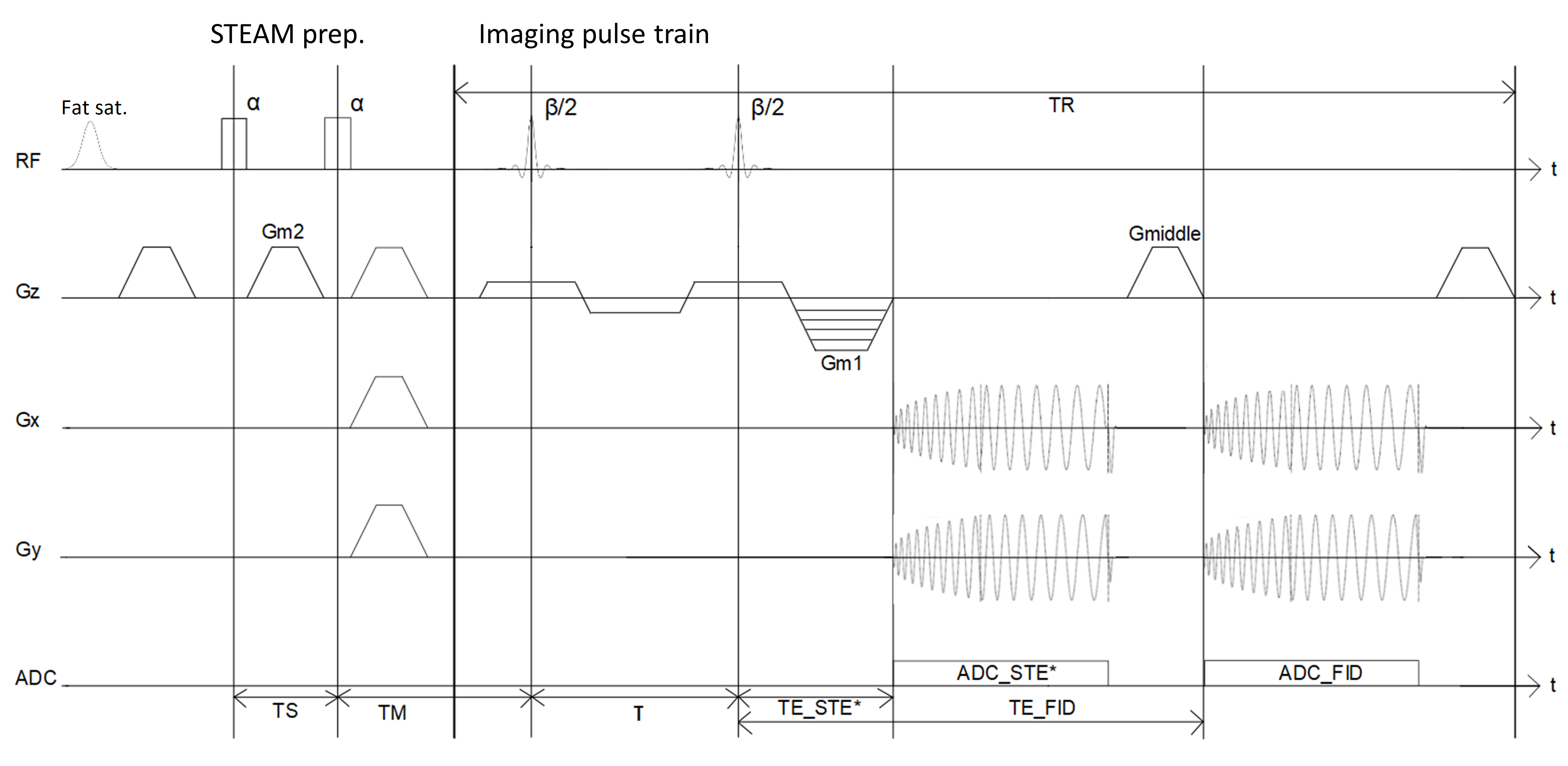
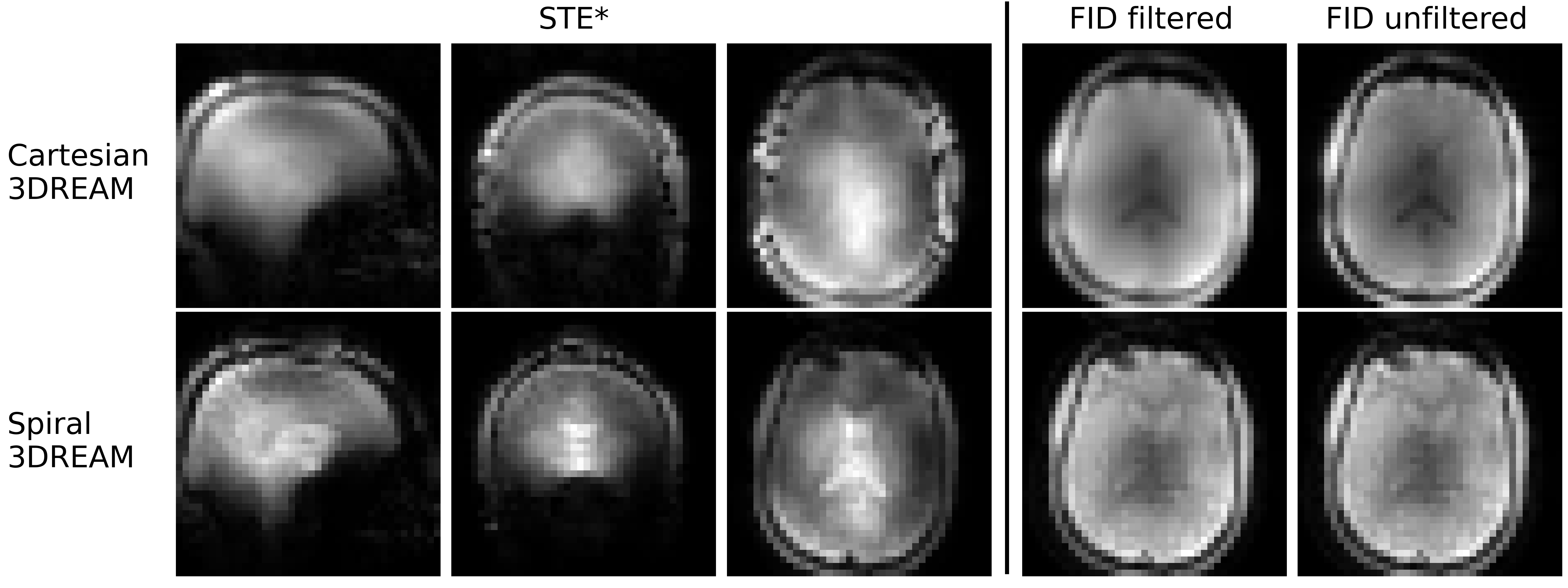
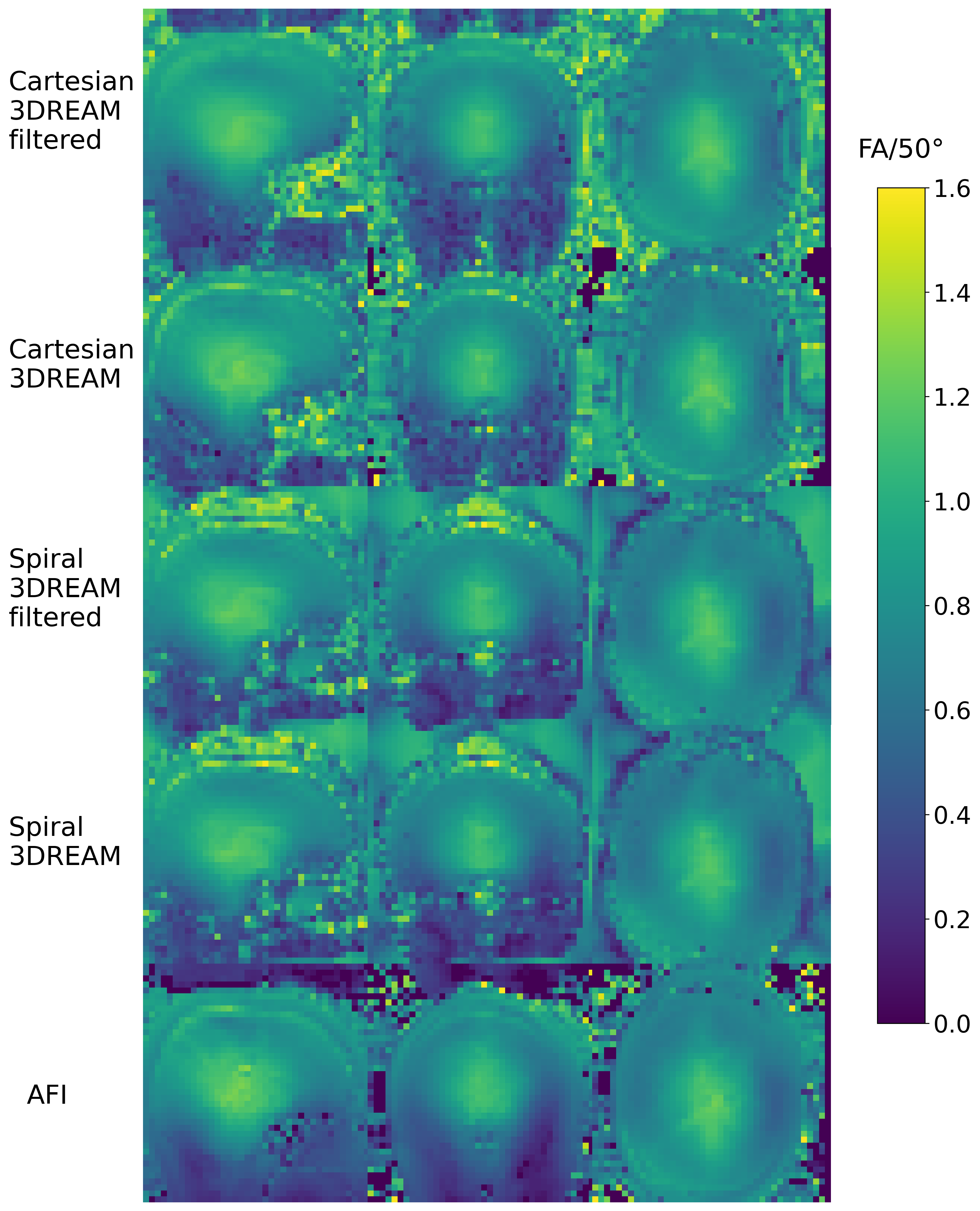
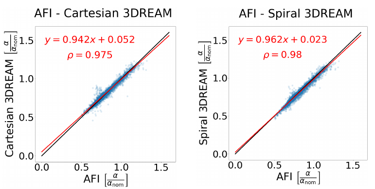
Fig 4: Comparison of flip angle values calculated from a 24x22x7 voxel volume. Left: Comparison between AFI and Cartesian 3DREAM. Right: Comparison between AFI and Spiral 3DREAM. The red line shows a linear fit with Pearson correlation ρ, the black line indicates identity.