1576
Biophysical-Model Based Synthesis of Cine CMR of Healthy and Pathological Left-Ventricular Function1Institute of Biomedical Engineering, ETH Zurich and University of Zurich, Zurich, Switzerland
Synopsis
We propose to augment MRXCAT with left-ventricular anatomies and function, representing realistic population statistics, to enable design and validation of CMR imaging and inference approaches. A variational autoencoder is deployed to identify parametric anatomical representations from a cohort of healthy and diseased anatomies to allow for the generation of new synthetic anatomies. Cardiac function is simulated using a biophysical model also incorporating local tissue defects. The resulting ventricular tissue masks are enriched with background information from XCAT. Synthetic MR images are generated using MRXCAT, making available paired data of CMR images and (patho)physiological parameters including ground-truth ventricular strains and displacements.
Introduction
Anatomical phantoms allow to generate realistic CMR images for which ground truth is available [1,2]. The MRXCAT software [1], for example, leverages the synthetic torso phantoms generated by XCAT [2] to simulate e.g. cine CMR images of the heart. However, while localized scars are available in the implementation [2,3], the XCAT model is primarily focused on healthy cases, which are not a realistic representation of population (patho)physiolgy. Additionally, cardiac function is based on a healthy reference, which is then scaled to match desired output metrics, such as ejection fraction. Such an approach can produce non-physiological displacement and strain fields.We propose to augment MRXCAT with synthetic anatomical cases, generated from a population shape model (SM) defined from a variational autoencoder (VAE) [4] on a cohort of 75 healthy and pathological left-ventricular (LV) geometries [5-8]. We show that the SM can represent healthy, dilated and hypertrophic anatomies and that it is possible to efficiently add local defects such as wall tinning and fibrous tissue. Using a biophysical model [5], realistic cardiac function is simulated for both healthy and pathological cases. The resulting tissue masks, augmented with background information from XCAT, are then used to synthesize realistic CMR cine images. Accordingly, our approach provides a tool to optimize sequence design and inference approaches using paired image data and ground truth physiological parameters and strain fields.
Methods
Left-ventricular meshes from 75 cases of the Multi-Modal Whole Heart dataset [6-8] were parametrized in terms of physiological coordinates as described in [5], including mapping of epicardial and endocardial surfaces onto a unit disk on images of 128x128 pixels. The resulting 6-channel images (one for each of the 3D coordinates of the 2 surfaces) were then used as input to a convolutional VAE [4] to compute a latent space representation of the anatomical features of the dataset. The learned low-dimensional representation was also associated with a normal probability distribution that allows sampling to generate new synthetic anatomies. The VAE was subsequently used to obtain a latent-space parametrization of the Automated Cardiac Diagnosis Challenge (ACDC) dataset [9] which was then input into a k-means clustering algorithm [10] to identify shape clusters for healthy (NOR), dilated (DCM) and hypertrophic (HCM) LV anatomies. Thanks to the parametrization of the shapes, local wall thinning and fibrous tissue properties can easily be added to any shape sampled from the VAE latent space distribution.A biophysical LV model was used to simulate cardiac function of healthy and diseased cases starting from a set of physiological and pathological parameters related to systemic circulation, tissue properties, microstructure, and activation patterns. The model is based on recent work [5] augmented with a pericardium model, to enhance the realism of deformations, and an Eikonal electrophysiological model [11]. The resulting time-dependent tissue masks were utilized as targets for registration of the tissue phantom generated with the XCAT software with 1mm isotropic resolution. Each image slice in the XCAT phantom was warped such that the epicardium contours match the ones of the corresponding slices of the biophysical model. Cardiac views, such as short-axis (SAX) and long-axis (LAX) were then extracted and used in MRXCAT to generate the corresponding images. The approach allows to generate both breathhold and free-breathing MRXCAT data. In case of free-breathing models, the rigid body motion for each cardiac phase is computed from XCAT and superimposed onto the displacement field generated from the biophysical model.
Results
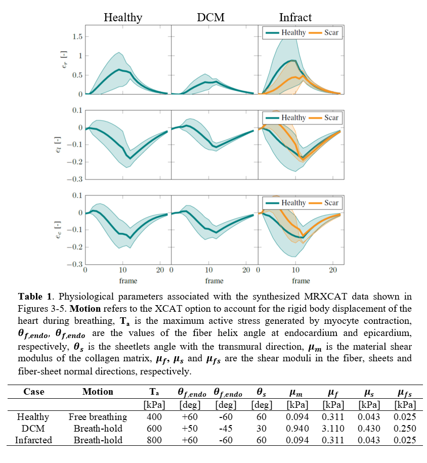
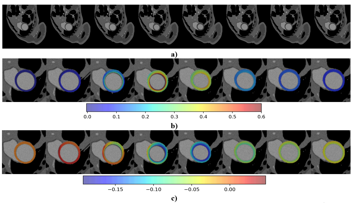
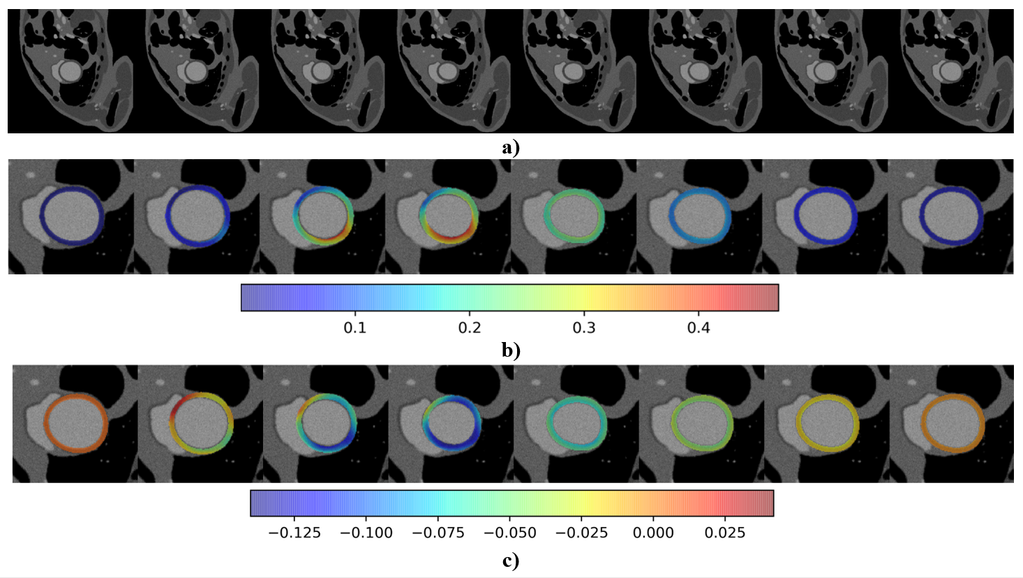
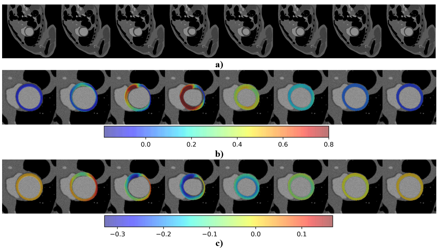
Figure 1 shows SAX and LAX views of the shape clusters obtained. The classification is compared with the clinical annotations available with the ACDC dataset and defines an accuracy of 0.84. Figure 2 summarizes exemplary myocardial strains, defined as in [5], and computed for healthy and dilated anatomical clusters as well as for an infarcted case with preserved ejection fraction. The physiological parameters linked to the three simulations are shown in Table 1 included in Figure 2. The corresponding SAX slices of the cine MRI images of the full cycle are shown in Figures 3 to 5 for healthy, dilated and infracted cases, respectively. Ground truth radial and circumferential strains are superimposed to the myocardium.Discussion
We have demonstrated that by using parametric anatomical and biophysical models augmented with XCAT background information, synthetic CMR images and corresponding ground truth parameters, including strains and displacements, can efficiently be generated using MRXCAT. By sampling from the parameters of a shape model, and by selecting appropriate parameters for cardiac function, it is possible to synthesize cardiac shapes and to derive deformation fields with realistic population variability. These models can then be augmented with torso organs, atria and right ventricle deformations and breathing motion as obtained from the XCAT phantom. The resulting data can then be used for the generation of realistic CMR images in MRXCAT, providing paired ground truth displacement and strain values of the LV function. The possibility of augmenting MRXCAT models with pathophysiological settings allows to study image acquisition sequences, reconstruction and post-processing approaches to assess and improve the accuracy and precision of CMR encoding and inference.Acknowledgements
The authors greatly acknowledge the financial support by the Swiss National Science Foundation (SNF) [grants CR23I3-166485, 325230-197702], and by PHRT SWISSHEART Failure Network of the ETH Domain.References
[1] L. Wissmann, C. Santelli, W. P. Segars and S. Kozerke, ‘Realistic numerical phantoms for cardiovascular magnetic resonance’, Journal of Cardiovascular Magnetic Resonance, vol. 16, no. 1, 2014
[2] W. Segars, S. Sturgeon, S. Mendonca, J. Grimes and B. Tsui, ‘4D XCAT phantom for multimodality imaging research’, Medical physics, vol. 37, no. 9, pp. 4902–4915, 2010
[3] A. I. Veress, W. P. Segars, J. A. Weiss, B. M. W. Tsui and G. T. Gullberg, ‘Normal and pathological NCAT image and phantom data based on physiologically realistic left ventricle finite-element models’, IEEE Transactions on Medical Imaging, vol. 25, no. 12, pp. 1604–1616, 2006.
[4] S. P. Kingma and M. Welling, ‘Auto-encoding variational bayes’, arXiv preprint arXiv:1312.6114, 2013
[5] S. Buoso, T. Joyce, and S. Kozerke, ‘Personalising left-ventricular biophysical models of the heart using parametric physics-informed neural networks’, Medical Image Analysis, p. 102066, 2021
[6] X. Zhuang and J. Shen, ‘Multi-scale patch and multi-modality atlases for whole heart segmentation of MRI’, Medical Image Analysis, vol. 31, pp. 77–87, 2016.
[7] X. Zhuang, ‘Challenges and methodologies of fully automatic whole heart segmentation: A review’, Journal of Healthcare Engineering, vol. 4, no. 3, pp. 371–407, 2013.
[8] X. Zhuang, K. S. Rhode, R. S. Razavi, D. J. Hawkes and S. Ourselin, ‘A registration-based propagation framework for automatic whole heart segmentation of cardiac MRI’, IEEE Transactions on Medical Imaging, vol. 29, no. 9, pp. 1612–1625, 2010
[9] O. Bernard, A. Lalande, C. Zotti, F. Cervenansky, X. Yang, P.-A.Heng, I. Cetin, K. Lekadir, O. Camara, M. A. G. Ballesteret al., ‘Deep learning techniques for automatic MRI cardiac multi-structures segmentation and diagnosis: is the problem solved?’, IEEE Transactions on Medical Imaging, vol. 37, no. 11, pp. 2514–2525, 2018
[10] F. Pedregosa, G. Varoquaux, A. Gramfort, V. Michel, B. Thirion,O. Grisel, M. Blondel, P. Prettenhofer, R. Weiss, V. Dubourg, J. Vander-plas, A. Passos, D. Cournapeau, M. Brucher, M. Perrot and E. Duch-esnay, ‘Scikit-learn: Machine learning in Python’, Journal of Machine Learning Research, vol. 12, pp. 2825–2830, 2011.
[11] A. Neic, F. O. Campos, A. J. Prassl, S. A. Niederer, M. J. Bishop, E. J. Vigmond, and G. Plank, ‘Efficient computation of electrograms and ECGs in human whole heart simulations using a reaction-eikonal model’, Journal of Computational Physics, vol. 346, p. 191–211, 2017
Figures