1574
Interleaved radial linear combination SSFP for improved cine imaging with implanted cardiac devices1Department of Biomedical Engineering, Yale University, New Haven, CT, United States, 2Department of Radiology and Biomedical Imaging, Yale University, New Haven, CT, United States, 3Department of Medicine, Cardiovascular Division, Yale University, New Haven, CT, United States
Synopsis
Interleaved undersampled radial linear-combination balanced SSFP (LC-SSFP) was investigated as a method for improving cardiac cine images, acquired in subjects with metallic implants. Phantoms and healthy volunteer studies were performed, in the presence of metallic artifacts (generated by a stainless steel bolt). We found the proposed method eliminated some artifacts seen using standard Cartesian bSSFP. These findings may lead to improved cardiac cine imaging for patients with a pacemaker or implantable cardioverter defibrillator (ICD).
Introduction
Cardiovascular implantable electronic devices were once assumed to be a contraindication for MRI scanning. However, recent safety studies have demonstrated an acceptable safety profile, and a lack of device or lead failure, in patients with a pacemaker or implantable cardioverter defibrillator (ICD) who underwent nonthoracic and thoracic MRI at 1.5T1-2. However, image quality is another paramount concern, since large B0-field inhomogeneity artifacts are introduced by the implants, especially in the nearby region of the heart. Balanced SSFP (bSSFP) provides high SNR, but recent studies show that device-related artifacts can be present in 94% of the bSSFP images3-4. Linear combination bSSFP (LC-SSFP) is an established way to reduce off-resonance artifacts, approximating a uniform spectral response to off-resonance5. However, scan time is increased 4-fold, making it an impractical solution. Using an undersampled radial LC-SSFP method reduces scan time to that of a feasible breath-hold6. We therefore here explore whether an interleaved radial LC-SSFP method can reduce artifacts (generated by a steel bolt) as a first step toward improvement in the cardiovascula for patients with implanted cardiac devices.Methods
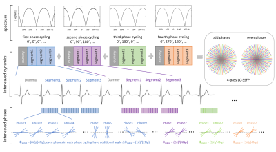
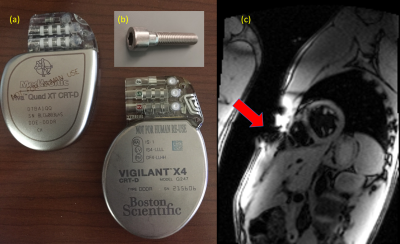
Figure 1 illustrates the undersampled radial sequence we propose. Radial k-space is acquired four times, using four different phase cyclings, with RF phase increment in each TR being 0°, 90°, 180° and 270° respectively. By combining these four acquisitions, off-resonance artifacts from bSSFP banding pattern can be reduced. In order to further improve the sampling pattern, radial views from each dynamic are interleaved by adding a different initial angle offset, where the most complimentary sets of radial views(e.g. initial angle (0π)/(4Np) and (2π)/(4Np), where Np denotes the number of radial views in each dynamic, see Figure 1) are matched to the adjacent phase cycling (e.g. first and second). This rearranged sampling pattern works together with the interleaved projections from cardiac phase to cardiac phase to reduce streak artifacts. Reconstruction was performed with gridding after zero-filling. To reduce artifacts due to bad coils that dominate (off-resonance related) streaks, we used streaking artifact ratio (SR) based k-means clustering, including outlier pruning (OP) and coil removal (CR)7. The UNFOLD8 method was used to take advantage of the interleaved phases in temporal domain, employing a low-pass fermi filter in the temporal Fourier space to reduce radial artifacts. We tested the proposed method on phantoms and healthy subjects with a stainless steel bolt placed on their chest (near the heart, see Figure 2), and compared results to conventional Cartesian bSSFP cine. Imaging was performed on a 3T Siemens.Scan parameters were: 2D Cartesian bSSFP cine, TR/ TE/ q= 3.4ms/1.7ms/35-40°, 36cm FOV, 12 views per segment, 192 matrix, 16 heart-beat breath-hold. For interleaved radial, parameters were identical except using 4 k-space acquisitions, 40-60 projections per k-space, and 20-24 heart-beat breath-hold.
Results
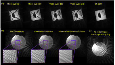
Figure 3A shows the phantom experiment results (Np=63), where intentional B0 shim was added. Images reconstructed from each phase cycle suffer from off-resonance artifacts, that can be removed by linearly combining the four dynamics. Figure 3B shows the same experiment, performed in a uniform phantom using a simple stainless steel bolt to generate metal artifacts representative of an implanted device. For Np=40 radial views in each acquisition, the experiments showed that the sampling pattern with both interleaved dynamics and phases produced the fewest streak artifacts. One striking aspect is how the off-resonant region serves as a source point of streaks especially when odd number of projections is applied (Figure 3C, Np=49).Figure 4 demonstrates the improved cine image quality using LC-SSFP. Compared with the conventional Cartesian acquisition, the proposed interleaved radial LC-SSFP method was able to eliminate the artifacts both on the chest and in the heart chambers.
Discussion
We explored the potential of interleaved radial LC-SSFP to reduce the artifacts caused by implanted cardiac electronic devices. Both phantom and in vivo experiments demonstrated eliminated artifacts compared with the conventional Cartesian method and improved cine image quality compared with the non-interleaved LC-SSFP. Future work will focus on more experiments on artifacts generated by real implants and further optimization for both acquisition and reconstruction, to improve bSSFP cine quality, in patients with cardiac devices.Acknowledgements
The authors acknowledge the support from National Heart Lung and Blood Institute, R01 HL155992.References
1. Russo RJ, Costa HS, Silva PD, Anderson JL, Arshad A, Biederman RWW et al. Assessing the risks associated with MRI in patients with a pacemaker or defibrillator. New England Journal of Medicine. 2017 Feb 23;376(8):755-764.
2. Nazarian S, Hansford R, Roguin A, et al. A prospective evaluation of a protocol for magnetic resonance imaging of patients with implanted cardiac devices. Ann Intern Med. 2011;155(7):415-424.
3. Scheffler K, Lehnhardt S. Principles and applications of balanced SSFP techniques. Eur Radiol. 2003;13(11):2409-2418.
4. Gao X, Abdi M, Auger DA, et al. Cardiac Magnetic Resonance Assessment of Response to Cardiac Resynchronization Therapy and Programming Strategies. JACC Cardiovasc Imaging. 2021;S1936-878X(21)00505-2.
5. Vasanawala SS, Pauly JM, Nishimura DG. Linear combination steady-state free precession MRI. Magn Reson Med 2000;43(1):82-90
6. Robb JS, Hu C, Peters DC. Interleaved, undersampled radial multiple-acquisition steady-state free precession for improved left atrial cine imaging. Magn Reson Med. 2020 May;83(5):1721-1729.
7. Mandava S, Keerthivasan MB, Martin DR, Altbach MI, Bilgin A. Radial streak artifact reduction using phased array beamforming. Magn Reson Med. 2019;81(6):3915-3923.
8. Madore B, Glover GH, Pelc NJ. Unaliasing by fourier-encoding the overlaps using the temporal dimension (UNFOLD), applied to cardiac imaging and fMRI. Magn Reson Med. 1999;42(5):813-828.
Figures


