1556
Accelerated whole-heart mDixon coronary MRA with a deep learning constrained CompressedSENSE: a feasibility study1Department of Radiology, West China Hospital of Sichuan University, Chengdu, China, 2North Sichuan Medical College, Nanchong, China, 3Philips Healthcare, Chengdu, China, 4Philips Healthcare, Shenzhen, China
Synopsis
Currently, modified dixon (mDixon) gradient echo sequence is widely used for respiratory navigated whole-heart coronary magnetic resonance angiography (MRA) for the evaluation of coronary anatomy and abnormalities. However, the main drawback of this approach is that the scan time is longer and prone to interference with motion artifacts. In this study, we investigated the utility of whole-heart coronaryMRA using accelerated mDixonwith compressed sensing (CS) and artificial intelligence(AI) technologies at 3Tesla. The initial results showedtheCS-AI mDixontechnique has potential to be the most viable alternative to enhance the clinical workflow of coronary MRA.
Introduction
Currently, modified dixon (mDixon) gradient echo sequence is widely used for respiratory navigated whole-heart coronary magnetic resonance angiography (MRA) as a noninvasive imaging technique for the evaluation of the anatomy and abnormalities of the coronary artery[1]. However, the main drawback of this approach is the long acquisition time resulting in a greater variability in the depth of respiration, which is prone to create image blurring and motion artifacts. Compressed sensing (CS) could be used to achieve scan time reduction beyond that possible with conventional parallel imaging acceleration. However, using very high acceleration factors with very high resolution can also result in degradation of image quality due to insufficient noise removal.Recently, integrating artificial intelligence (AI) into MRI reconstruction has been proposed to further accelerate MR scans, in which a novel deep neural network was introduced as CS-AI and showed superior performance for reconstructing the image from highly undersampled k-space data[2].Therefore, this study aims to acquire rapid whole-heart coronary MRA images using accelerated mDixon with CS-AI and compare the image quality between images acquired with CS-AI ,CS and Conventional mDixon on a 3T MR scanner.
Methods
This study was approved by the institutional ethics committee. The MR examinations were performed on a 3T MR scanner (Ingenia Elition, Philips Healthcare) using a 16-channel body matrix coil combined with a 12-channel spine matrix coil. Eight volunteers (5 males and 3 females; age range, 19-65 years) were recruited in the study and underwent coronary MRA using three mDixon-based sequences : conventional mDixon, CS mDxion, and CS-AI mDixon in a coronal orientation without contrast material.The details of coronary MRA parameters of Conventional mDixon sequence were listed: FOV=265×301 mm2, TR/TE1/TE2=4.1/1.32/2.4 ms, slice thickness= 0.75mm, flip angle=10°, voxel size=0.75×0.75×0.75mm3, bandwidth =0.355 Hz/Px, In comparison, the CMRA imaging parameters of CS mDxion, and CS-AI mDixon were consistent to conventional mDixon, but with an CS acceleration factor=4. In CS-AI approach, the CS reconstruction chain is merely replaced by a convolution neural network (CNN) reconstruction. Compared to conventional mDixon, the acquisition time of CS & CS-AI decreased (2:55 vs. 4:11 min). Average acquisition efficiency is 58%.
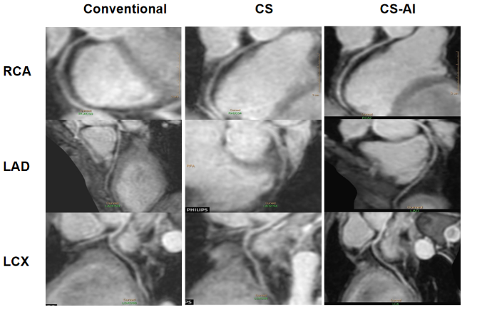
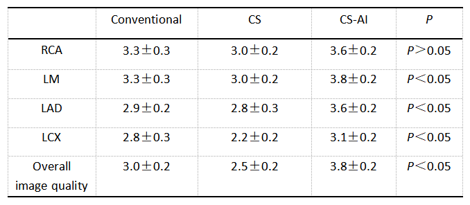
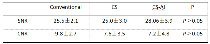
Curved planar reformation (CPR) images of three sequences were used for image display. Subjective image quality was independently scored by two cardiac MR radiologists using a 4-point scale (1=insufficient visualization, 4=excellent visualization) on the assessment of right coronary artery (RCA), left main coronary artery (LM), left anterior descending artery (LAD), left circumflex coronary artery (LCX) and overall image quality on CPR images. For quantitative objective assessment, signal-to-noise ratio (SNR) and contrast-to-noise ratio (CNR) were calculated for comparison between the three techniques. The image scores and quantitative parameters were compared using the Wilcoxon signed rank test. P value less than 0.05 was considered significant.
Results and Discussion
The coronal example and coronary images were shown in Figure 1 and Figure 2,respectively. Overall, the CS-AI images produced visually sharper images, less image blurring and motion artifacts. As shown in table 1, CS-AI demonstrated the excellent image scores in LM, LAD, LCX and overall image quality among the three sequences (P<0.05). The image scores of RCA with CS-AI was also higher than Conventional mDixon and CS mDixon (3.6±0.2 vs. 3.3±0.3 vs. 3.0±0.2, P>0.05). The SNRs and CNRs of the three approach were shown in Table 2, the CS-AI images demonstrated high denoising performance with excellent SNR (28.06±3.9 vs. 25.5±2.1 vs. 25.0±3.0, P>0.05). Despite the promising result, there were no significant difference, maybe in part due to current less subjects included in this study.Conclusion
In this work, we investigated the utility of whole-heart coronary MRA using accelerated mDixon with CS-AI in comparison with CS and Conventional mDixon approach. The results demonstrated that CS-AI can produce excellent image quality and high denoising performance, which could be a viable alternative to enhance the clinical workflow of coronary MRA. A large clinical population is underway to validate the diagnostic value of this technique.Acknowledgements
No acknowledgement found.References
1. Kourtidou S, Jones M R, Moore R A, et al. mDixon ECG-gated 3-dimensional cardiovascular magnetic resonance angiography in patients with congenital cardiovascular disease[J]. Journal of Cardiovascular Magnetic Resonance, 2019,21(1).DOI:10.1186/s12968-019-0554-3.
2. Knoll F, Zbontar J, Sriram A, et al. fastMRI: A Publicly Available Raw k-Space and DICOM Dataset of Knee Images for Accelerated MR ImageReconstruction Using Machine Learning. Radiol Artif Intell. 2020;2(1):e190007.
Figures
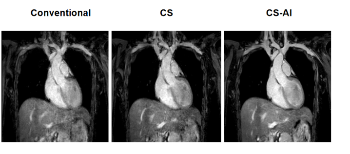
The coronal images acquired on a volunteer using Conventional mDixon, CS mDixon and CS-AI mDixon sequences. The CS-AI coronaryMRA yields best image quality than others.