1549
Sex-differences in CBF changes as a biomarker of preclinical MCI1Physics, Concordia University, Montreal, QC, Canada, 2PERFORM Centre, Concordia University, Montreal, QC, Canada, 3Centre de Recherche de l’Institut Universitaire de G´eriatrie de Montr´eal, Montreal, QC, Canada, 4Centre de Recherche de l’Institut de Cardiologie de Montr´eal, Montreal, QC, Canada, 5StoP-AD, Douglas Mental Health Institute, Montreal, QC, Canada
Synopsis
Alzheimer’s Disease (AD) is the most common form of dementia, preceded by a mild cognitive impairment (MCI) prodromal stage. There are sex differences in the prevalence and severity of MCI and AD. However, the presence of sex differences in cerebral hemodynamics during the transition from normal cognition to MCI is unknown. This is the first study to assess whether sex differences exist in cerebral blood flow (CBF) in the years prior to MCI diagnosis in patients with a familial history of AD.
Introduction
AD is a type of dementia, with a prodromal stage associated with mild cognitive impairment (MCI) and a higher rate of conversion to AD than in cognitively normal individuals. This neurodegenerative disorder results in cerebrovascular and structural changes in the brain1. Importantly, longitudinal studies on cerebrovascular changes, including CBF, have demonstrated that CBF changes occur years before AD2,3,4. Evidence suggests that CBF is an important biomarker to predict who is likely to convert to MCI and AD5. AD appears with different prevalence and severity in males compared to females, where females have increased severity and prevalence over time6 . Yet, to date, sex differences in CBF prior to the onset of MCI have not been investigated. Since the presence of these sex difference could partially account for the increased severity and prevalence of AD in females, it is imperative to investigate CBF longitudinally in males and female patients who develop MCI compared to those who do not convert. This will help determine whether CBF can be used as an early biomarker of MCI conversion and the efficacy of this biomarker in males compared to females.Methods
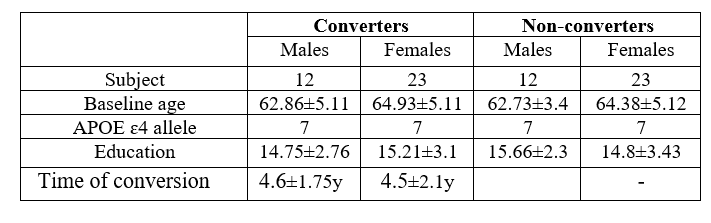
Data acquisition was completed in 385 participants with an increased risk of developing AD based on familial and genetic information. Pseudo-continuous arterial spin labelling (pCASL) at rest and structural data were collected annually. All participants also completed an extensive neuropsychological testing battery at baseline and all other time points7. All participants had no evidence of cognitive impairment at baseline. Over time, some of the participants were diagnosed with MCI based on the neuropsychological assessment. Specifically, within the approximate 4-year period of this study, 35 participants (females = 23, mean age at baseline= 64.3 ± 5.2, Table. 1 for demographics) were diagnosed with MCI. A control group of 35 individuals with normal cognition at the final time point were selected, matched to the MCI converters for age, sex, education and APOE ɛ4 allele status. Preprocessing of pCASL data included brain extraction and motion correction in FSL and MATLAB 8. Resting CBF was quantified9 using a CSF M0 mask for each participant. All CBF maps were registered to native T1 images and then to MNI space using ANTS 10. CBF was compared in 12 ROIs related to MCI and AD pathology and symptomology 3 with age, education, and APOE ɛ4 status as covariates.Results
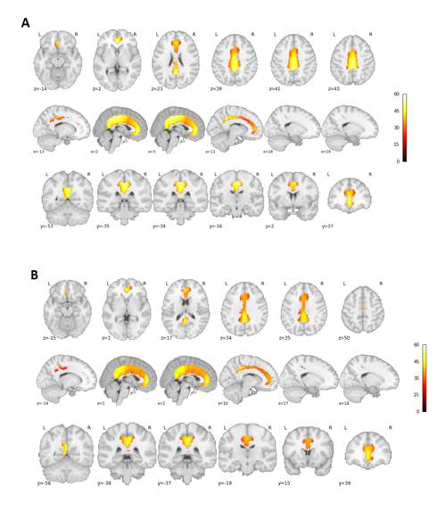
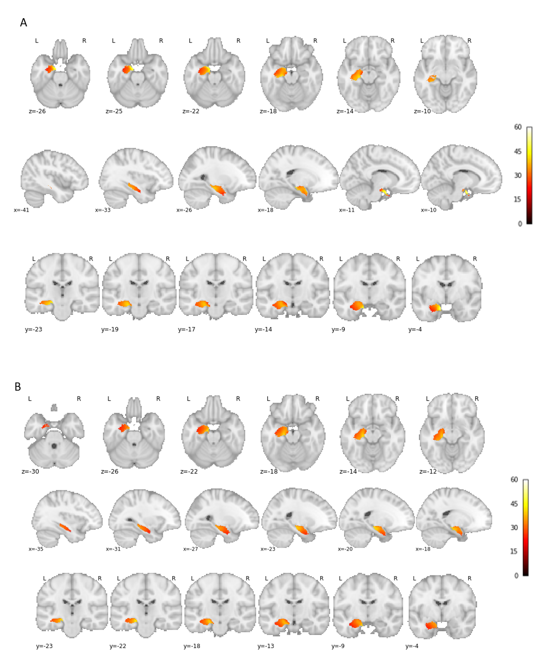
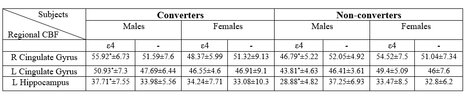
A Mann-Whitney U test revealed significant differences only in males at baseline between MCI-converters and non-converters. Higher CBF was observed in the right (p = 0.039) and left (p = 0.014 cingulate gyrus and the left hippocampus (p=0.01) in MCI-converters compared to non-converters group. This significant difference maybe driven by the APOE ɛ4 carriers in the sample, as a comparison of converter and non-converter APOE ɛ4 carriers revealed the same hyperperfusion (Fig .1and fig. 2), which was however not present in the same comparison for non-carriers. No significant differences were observed in females for any of the ROIs (p > 0.05, see Table 2). It should be noted that females were two years older than males at the baseline in this database (see Table 1) and both groups were diagnosed with MCI after approximately 4 years.Discussion
Our results revealed hyperperfused regions in male MCI-converters compared to the control group at baseline. Similar patterns of hyperperfusion in the years prior to MCI development have previously been observed and have been interpreted as a compensatory process for metabolic changes4. Moreover, these regions have been shown to be associated with cognitive decline in structural and functional outcomes in those who are transitioning from normal cognition to MCI, highlighting their importance in MCI4,11 . Some of the effects observed may be specific to the APOE ε4 genotype, as hyperperfusion was only observed in male APOE ε4 carriers. Similar hyperperfusions have been observed in APOE ε4 carriers and have been similarly interpreted as an early compensatory mechanism5. To our knowledge, this is the first study to investigate sex-specific differences in CBF in those who convert to MCI and non-converters. Previous findings of perfusion differences in MCI or the interaction between APOE ε4 and CBF could be driven by a greater portion of males in specific sample14,15. Future studies should validate these findings in a larger sample of MCI converters. Future studies could also use upstream cerebrovascular markers, such as cerebrovascular reactivity (CVR) to better understand the development of cerebrovascular impairments in the development of MCI and AD. Future studies could also use upstream cerebrovascular markers, such as cerebrovascular reactivity (CVR) to better understand the development of cerebrovascular impairments in the development of MCI and AD.Acknowledgements
Data used in preparation of this article were obtained from the Pre-symptomatic Evaluation of Novel or Experimental Treatments for Alzheimer’s Disease (PREVENT-AD) program (https://douglas.research.mcgill.ca/stop-ad-centre), data release 5.0 (November 30, 2017). A complete listing of PREVENT-AD Research Group can be found in the PREVENT-AD database: https://preventad.loris.ca/acknowledgements/acknowledgements.php?date=[2019-06-03]. The investigators of the PREVENT-AD program contributed to the design and implementation of PREVENT-AD and/or provided data but did not participate in analysis or writing of this report. The authors would also like to thank Heart and Stroke Foundation New Investigator Award and J.M. Barnett fellowship, and the Michal and Renata Hornstein Chair in Cardiovascular Imaging.References
1. Dai, Weiying, et al. "Mild cognitive impairment and alzheimer disease: patterns of altered cerebral blood flow at MR imaging." Radiology 250.3 (2009): 856-866.
2. Hays, Chelsea C., Zvinka Z. Zlatar, and Christina E. Wierenga. "The utility of cerebral blood flow as a biomarker of preclinical Alzheimer’s disease." Cellular and molecular neurobiology 36.2 (2016): 167-179.
3. Duan, Wenna, et al. "Cerebral Blood Flow Predicts Conversion of Mild Cognitive Impairment into Alzheimer’s Disease and Cognitive Decline: An Arterial Spin Labeling Follow-up Study." Journal of Alzheimer's Disease Preprint (2021): 1-13.
4. Beason-Held, Lori L., et al. "Changes in brain function occur years before the onset of cognitive impairment." Journal of Neuroscience 33.46 (2013): 18008-18014.
5. Hays, Chelsea C., Zvinka Z. Zlatar, and Christina E. Wierenga. "The utility of cerebral blood flow as a biomarker of preclinical Alzheimer’s disease." Cellular and molecular neurobiology 36.2 (2016): 167-179.
6. Laws, Keith R., Karen Irvine, and Tim M. Gale. "Sex differences in Alzheimer's disease." Current opinion in psychiatry 31.2 (2018): 133-139.
7. Tremblay-Mercier, Jennifer, et al. "Open Science Datasets from PREVENT-AD, a Longitudinal Cohort of Pre-symptomatic Alzheimer’s Disease." NeuroImage: Clinical (2021): 102733.
8. https://www.mathworks.com/
9. Alsop, David C., et al. "Recommended implementation of arterial spin‐labeled perfusion MRI for clinical applications: A consensus of the ISMRM perfusion study group and the European consortium for ASL in dementia." Magnetic resonance in medicine 73.1 (2015): 102-116.
10. http://stnava.github.io/ANTs/
11. Sambuchi, Nathalie, Yonas Endale Geda, and Bernard François Michel. "Cingulate cortex in pre-MCI cognition." Handbook of clinical neurology 166 (2019): 281-295.
12. Barron, Anna M., and Christian J. Pike. "Sex hormones, aging, and Alzheimer’s disease." Frontiers in bioscience (Elite edition) 4 (2012): 976.
13. Krause, Diana N., Sue P. Duckles, and Dale A. Pelligrino. "Influence of sex steroid hormones on cerebrovascular function." Journal of applied physiology 101.4 (2006): 1252-1261.
14. Thambisetty, Madhav, et al. "APOE ε4 genotype and longitudinal changes in cerebral blood flow in normal aging." Archives of neurology 67.1 (2010): 93-98.
15. Sun, Z‐W., et al. "Decreased cerebral blood flow velocity in apolipoprotein E ɛ4 allele carriers with mild cognitive impairment." European journal of neurology 14.2 (2007): 150-155.
Figures