1532
Effect of post mortem interval and body temperature on tissue susceptibility1InBrain, Departamento de Física, Faculdade de Filosofia, Ciências e Letras de Ribeirão Preto, Universidade de São Paulo, Ribeirão Preto, Brazil, 2Departamento de Radiologia e Oncologia, Faculdade de Medicina, Universidade de Sao Paulo, São Paulo, Brazil
Synopsis
On this study, the impact of temperature and postmortem interval (PMI) on QSM were evaluated for a total of 21 subjects. Temperature and PMI were correlated with values from CSF, white matter (WM) and gray matter (GM). Only the GM presented a significant correlation with both temperature and PMI.
Introduction
Quantitative Susceptibility Mapping (QSM) is a magnetic resonance imaging (MRI) technique to non-invasively quantify tissue magnetic susceptibility, and as such it represents a very important tool to assess many pathological processes, in particular the ones related to iron and other metals deposition. The correlation of postmortem QSM-MRI to histopathological studies is vital in the elucidation of the role of iron in neurodegeneration, and for establishing such associations the access to post-mortem in situ brain MRI presents an excellent opportunity for histological validation of MRI biomarkers. However, it is still not fully evaluated how variables such as post-mortem interval (PMI) or temperature can affect QSM susceptibility values. A recent study [1] compared an ante mortem to post-mortem MRI (with post-mortem interval of only 9 hours) and they were able to detect significant changes in brain structures volumes, diffusivity and fractional anisotropy values. The aim of this study is to investigate the possible effect of body temperature and PMI on measured susceptibility values.Methods:
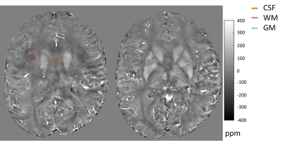
A total of 21 cases were recruited from the City Death Verification Service after the family signed informed consent. Post-mortem in situ brain MRI examination was performed with a Magnetom 7T scanner (Siemens, Erlangen, Germany) and a 32ch head coil (Nova Medical) just before the autopsy. QSM was based on a 3D GRE multiecho acquisition, with five echoes (first echo 5ms, ΔTE of 4ms), 1mm thick axial slices and 0.5mm in plane resolution. PMI was calculated by the difference between the death time declared by the subject’s relatives and the starting time of the MRI acquisition. Body temperature was measured with an infrared thermometer pointed at the forehead right before and after MRI examination. A mean temperature was considered for statistical analysis.Phase images were unwrapped using the Laplacian method and the background field was removed using the PDF algorithm. Susceptibility maps were calculated using the MEDI-L1 algorithm, assuming the mean whole brain as the reference.Three spherical ROIs were centered in anatomical regions and considered representative of Cerebrospinal Fluid (CSF), white (WM) and gray (GM) matter, respectively. CSF in the right ventricle, GM in the right caudate nucleus and WM near the right frontal region (Figure 1).For statistical analysis, correlations were assessmented between temperature and PMI with mean susceptibility values obtained on CSF, WM and GM ROIs, respectively. All correlations were controlled by the age factor.Results:
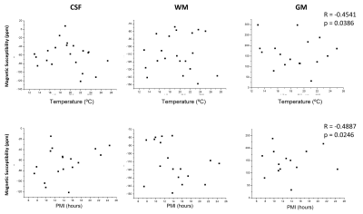
Mean age was 66±16 years, and 6 were female. Mean susceptibility values were -5.67±3.15, -702.7±52.2 and 133.2±91.7 ppm for CSF, WM and GM respectively. A significant negative correlation was obtained between susceptibility values in GM and body temperature (Figure 2). Susceptibility was also negatively correlated with the PMI in GM. No other correlations were found.Discussion:
According to Curie’s law, the paramagnetic part of the magnetic susceptibility shows an inverse temperature dependency [2]. Even in a small temperature range, it was possible to detect this tendency in GM, suggesting a significant contribution of the paramagnetic ions in this tissue. On the other hand, WM and CSF did not show any correlation with body temperature, as expected for tissues mostly composed of water (diamagnetic). A recent study confirmed the difference in temperature dependence of the paramagnetic and diamagnetic components within brain tissue [3].The PMI range we were able to study spanned from 7-24 hours, and within this range we observed for GM a small negative correlation between susceptibility and PMI. This decrease in susceptibility might be caused by an increase in water, since we know that after 9 hours it is possible to observe a brain swelling of about 7% [1]. For WM we expect similar or even a higher increase in volume compared to subcortical GM [1], but the negative correlation between WM susceptibility and PMI was not significant. This might be due to a counteracting effect of membrane decomposition, which would lead to an increase in susceptibility.An important limitation of the study is that the cases included were not of hospital’s inpatients, therefore the declared time of death might not always be very accurate, especially in cases of death during sleep or while being home alone. This might account for the high dispersion in Figure 2. The ideal experimental setup would be a 24hrs longitudinal study with the same body, however such a study is rarely possible given the urge of the family to proceed with burial service. Another limitation is that some of the cases included were kept for some hours in the refrigerator, which slows down decomposition processes relative to room temperature. But analysis excluding the cases with T<20C (not shown here) did not alter the results as a function of PMI.Conclusion
Temperature affected mostly subcortical GM, while CSF and WM were temperature invariant.Within a PMI range of 7-24 hours only subtle changes for subcortical GM, probably related to water accumulation, were observed. But we acknowledge that this relation should be further investigated, accounting for confounding factors such as temperature. When evaluating post-mortem brain in situ susceptibility values it is important to keep PMI and T as constant as possible, specially for subcortical GM regions.Acknowledgements
No acknowledgement found.References
[1] Boon BDC, Pouwels PJW, Jonkman LE, Keijzer MJ, Preziosa P, van de Berg WDJ, Geurts JJG, Scheltens P, Barkhof F, Rozemuller AJM, Bouwman FH, Steenwijk MD. Can post-mortem MRI be used as a proxy for in vivo? A case study. Brain Commun. 2019; 24: 1(1):fcz030. doi: 10.1093/braincomms/fcz030.
[2] Birkl C, Langkammer C, Krenn H, Goessler W, Ernst C, Haybaeck J, Stollberger R, Fazekas F, Ropele S. Iron mapping using the temperature dependency of the magnetic susceptibility. Magn Reson Med. 2015 Mar;73(3):1282-8. doi: 10.1002/mrm.25236.
[3] Chen J, Gong NJ, Chaim KT, Otaduy MCG, Liu C. Decompose quantitative susceptibility mapping (QSM) to sub-voxel diamagnetic and paramagnetic components based on gradient-echo MRI data. Neuroimage. 2021; 15: 242:118477. doi: 10.1016/j.neuroimage.2021.118477.
[4] Schweser F, Deistung A, Lehr B W, Reichenbach J R. Quantitative imaging of intrinsic magnetic tissue properties using MRI signal phase: An approach to in vivo brain iron metabolism? NeuroImage. 2011; 54: 2789-2807. doi: 10.1016/j.neuroimage.2010.10.070
[5] Liu J, Liu T, de Rochefort L, Ledoux J, Khalildov I, Chen W, Tsiouris A J, Wisnieff C, Spincemaille P, Prince M R, Wang Y. Morphology enabled dipole inversion for quantitative susceptibility mapping using structural consistency between the magnitude image and the susceptibility map. NeuroImage. 2012; 59: 2560-2568. doi: 10.1016/j.neuroimage.2011.08.082
Figures

