1530
Deep learning based image reconstruction for improved 3D multiparameter mapping and synthetic MR imaging1Department of Imaging Physics, The University of Texas M.D. Anderson Cancer Center, Houston, TX, United States, 2Department of Neuroradiology, The University of Texas M.D. Anderson Cancer Center, Houston, TX, United States, 3GE Healthcare, Hino, Japan, 4Global MR Applications and Workflow, GE Healthcare, Calgary, AB, Canada, 5SyntheticMR, AB, Linkoping, Sweden, 6GE Healthcare, Menlo Park, CA, United States, 7GE Healthcare, Houston, TX, United States
Synopsis
Brain data with high grade glioma were acquired with a 3D multiparameter mapping sequence and reconstructed with a novel deep learning based method (DL Recon). Synthetic images created from maps fitted to the reconstructed images were rated by experienced radiologists for image quality and diagnostic utility. Images from the DL Recon workflow consistently rated equal to or better than those created from conventional reconstruction. We find that the SNR and resolution benefits of 3D DL Recon extend to improve the resulting relaxation maps and subsequent synthetic images.
Introduction
Deep learning based image reconstruction (DL Recon) offers the potential for increased SNR, reduced artifacts, and enhanced resolution1. It has previously been applied to 2D quantitative multiparameter mapping in synthetic MR imaging methods2, by which multiple images are acquired with varying sequence parameters and then fitted or matched to spin models to produce maps of T1 and T2 relaxation times and proton density, PD. More recent developments have extended both techniques to 3D imaging3,4. In this work, we investigate the effect of a prototype 3D DL Recon on 3D multiparameter mapping and synthetic MR Imaging of high grade gliomas in the brain.Methods
The 3D DL Recon used in our study (AIR Recon DL, GE Healthcare)1 is a vendor-supplied deep convolutional residual encoder network trained to reconstruct images from MR data with reduced noise, reduced Gibbs ringing, and enhanced resolution. Ringing reduction and improved sharpness occur in all three dimensions. The convolutional network is embedded in the image reconstruction, operating on raw complex image volumes and has an adjustable parameter that ranges between 0 and 100% to control denoising strength of the final reconstructed images.Four subjects were prospectively acquired with 3D QALAS sequence with the following parameters: FOV = 24cm, acquired matrix = 256x240 slice thickness = 1.4mm (reconstructed to 0.7mm), number of slices = 120-136, ARC acceleration factor = 2x1, overall scan time = 7:57-8:55. Raw data were reconstructed offline with conventional parallel imaging reconstruction and with 3D DL recon at 75% noise reduction level to produce two sets of 3D image data, each set containing 5 stacks of images from the 5 timepoints on the QALAS signal model (fig 1). The conventional set of base images was processed offline with a dedicated mapping and image synthesis application (SyMRI, SyntheticMR, Linkoping, Sweden) to produce T1, T2, and PD parameter maps, as well as synthetic T1w, T2w, FLAIR, and PSIR images (SynMRI). The post processing was also applied to the set of base images generated from the DL reconstruction to generate a second set of synthetic images (DL-SynMRI) for comparison.
Two neuroradiologists performed a consensus review of both SynMRI and DL-SynMRI. Anatomical and pathological delineation were both scored on a 5-point Likert scale. Four anatomical structures (optic chiasm, red nucleus, central sulcus, gray-white matter differentiation including visibility of the caudate nucleus and deep gray nuclei) were each rated for visibility sufficient for accurate diagnosis. Any presence of four image quality deficiencies (low SNR, flow artifact, motion artifact, and bright cortical interface artifact) was also noted if they were suspected of hindering diagnostic ability.
Results
Of the 16 series of synthetic images (4 subjects, 4 contrasts), DL-SynMRI scored equal to or higher than SynMRI for all images for anatomical delineation, and for all images except for one PSIR image on one subject for pathological delineation. Overall, DL-SynMRI scored higher than SynMRI for all image contrasts, though with relatively few number of cases, only T2 and T2 FLAIR scores demonstrated statistical significance at p<0.05. DL-SynMRI had improved visualization of the optic chiasm in two images and gray-white differentiation in two images. Low SNR was not noted on any of the DL-SynMRI, but was noted on 9 out of 16 of the SynMRI, including all four T2w images. DL-SynMRI exhibited less flow artifact in one image; all other cases of flow or motion artifact were unchanged between SynMRI and DL MRI. No cortical interface artifact was observed in any images.Discussion
Our study showed that 3D DL Recon resulted in the expected benefits of reduced noise and image enhancement when applied to the raw data acquired by the 3D QALAS sequence. DL Recon also had little negative effects on the resulting SynMRI images. We therefore conclude that DL recon can be advantageously applied to 3D synthetic imaging techniques such as 3D QALAS. Our method of scoring was designed to separate gains in diagnostic ability from general impressions of image quality. While none of the SynMRI images were deemed undiagnostic, confidence in most T2w and FLAIR SynMRI images was degraded by low SNR, which was corrected in the corresponding DL-SynMRI dataset. It was noted that none of the images exhibited much cortical brightening artifact seen in many 2D synthetic FLAIR images. While this cannot be attributed to DL Recon, it is presumably due to the smaller voxel sizes of the 3D acquisition as well as certain improvements in the SyMRI software.3D QALAS acquires 5 stacks of varying contrast images, and 3D DL Recon was applied to each stack independently. While it is possible that some improvement may be gained from training a network on the entire signal model, the current generalized reconstruction can be applied to most individual 3D Cartesian data sets. Since the 3D DL recon acts on the reconstructed base images before parameter fitting, improvements in synthetic T2w and FLAIR imaging would be the result of improved fits of the 5 base images to the signal model, rather than the denoising of any spin parameter maps.
In conclusion, 3D DL Recon improved SNR and resolution of 3D SynMRI without negatively affecting the artifacts, thus demonstrating a positive impact on diagnostic ability.
Acknowledgements
No acknowledgement found.References
1. Lebel RM. Performance characterization of a novel deep learning-based MR image reconstruction pipeline. 2020. arXiv preprint, arXiv:2008.06559.
2. Hwang KP, Wang X, Lebel M, Johansson P, Petersen C, Warntjes M, Bayram E, Banerjee S, Ma J, Johnson J. Deep learning based image reconstruction for improved multiparameter mapping and synthetic MRI. ISMRM Scientific Meeting and Exhibition, 2020.
3. Kvernby S, Warntjes MJ, Haraldsson H, Carlhäll CJ, Engvall J, Ebbers T. Simultaneous three-dimensional myocardial T1 and T2 mapping in one breath hold with 3D-QALAS. J Cardiovasc Magn Reson. 2014;16(1):102.
4. Fujita S, Hagiwara A, Takei N, Hwang KP, Fukunaga I, Kato S, Andica C, Kamagata K, Yokoyama K, Hattori N, Abe O, Aoki S. Accelerated Isotropic Multiparametric Imaging by High Spatial Resolution 3D-QALAS With Compressed Sensing: A Phantom, Volunteer, and Patient Study. Invest Radiol. 2021;56(5):292-300.
Figures
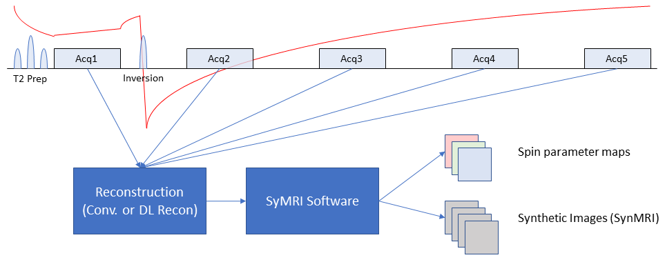
Figure 1. Signal and data flow of the 3D QALAS sequence. Images from 5 timepoints of the QALAS signal model are acquired and reconstructed with both the conventional and 3D DL Recon. These are each processed with SyMRI software to produce 3D spin parameter maps and synthetic images.
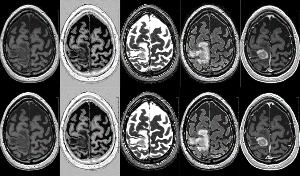
Figure 3. (Left to right) Synthetic T1W, PSIR, T2W, FLAIR, and contrast enhanced T1W images produced from 3D QALAS data reconstructed with conventional (top) and DL Recon (bottom).
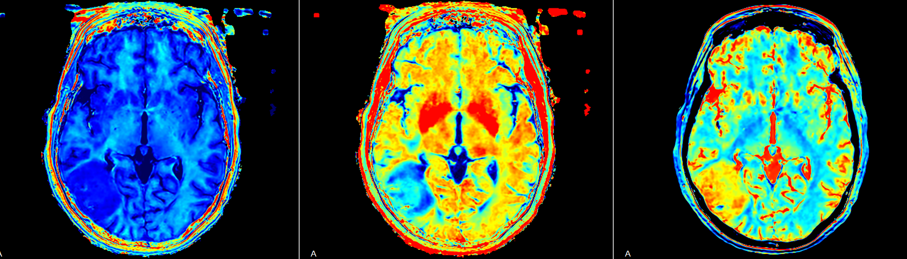
Figure 5. Axial slice of (left to right) 3D R1, R2, and proton density parameter maps produced from 3D QALAS data reconstructed with 3D DL Recon.