1509
Characterization of relationship between MR microstructural parameters and memory in older subjects1Biomedical Engineering, University of Michigan, Ann Arbor, MI, United States, 2Functional MRI Laboratory, University of Michigan, Ann Arbor, MI, United States, 3Neurology, University of Michigan, Ann Arbor, MI, United States, 4Psychiatry, University of Michigan, Ann Arbor, MI, United States, 5Mental Health Service, VA Ann Arbor Healthcare System, Ann Arbor, MI, United States
Synopsis
This work examined the relationship between white matter microstructure parameters calculated by NODDI and a composite measure of memory function. Connectome predictive modeling was done on fractional anisotropy, orientation dispersion, and neurite density maps. Significant relationships were found for all parameters, with orientation dispersion having the highest correlation between predicted and observed memory scores.
Introduction
Mild Cognitive Impairment (MCI) and Alzheimer's disease (AD) are associated with neurodegenerative effects upon the white matter structure of the brain1. Multi-shell DTI analyses such as NODDI (Neurite Orientation Direction and Density Imaging) have increased sensitivity over regular single tensor DTI computed metrics2. In this work, we examine the relationship between white matter microstructure metrics calculated by NODDI and a composite measure of memory function.Methods
Subjects: Data from 79 participants were included in this study; 45 participants met criteria as cognitively intact, 24 as amnestic MCI with a presumed AD etiology, and 10 with a diagnosis of dementia of the Alzheimer’s Type (DAT). All study procedures took place under approval of the local IRBMED. Consensus diagnoses follow those set forth by the National Alzheimer's Coordinating Center (NACC). A composite measure of memory was created using the mean z-scores of the delayed recall portions of the Craft story, Hopkins Verbal Learning Test, and Benson Complex Figure.MRI data acquisition and analysis: DTI data were acquired on a 3T GE MR750. Multi-shell DTI was acquired using a multiband EPI sequence (TR=3900, Min TE, Flip angle=77, FOV 24 cm, 1.7 mm slice thickness, MB factor=3, 102 total directions: 12 @ b=0, 30@b=711, 60@b=2000). DTI data was preprocessed for eddy-current and distortions in FSL, with calculation of fractional anisotropy (FA) maps. NODDI parameters, Neurite Density (ND) and Orientation Dispersion (OD), were calculated using the AMICO toolbox3. Tract-based spatial statistics (TBSS) operations were then computed using FSL, including FA normalization, mean FA skeletonization, and projection of the NODDI parameter maps to the FA skeleton.
Predictive Modeling: CPM (connectome predictive modelling)4 in MATLAB was adapted to examine the predictive relationship of each of the NODDI parameters with the composite memory score. Using a Leave-One-Out prediction framework, each skeletonized parameter map were correlated across training subjects. Feature reduction was done by selecting the voxels that met 0.01 significance. Then, the raw values for all features were summed to give a composite brain score for each subject, which was then fit to a linear model to predict composite memory; this was done separately for positive and negative correlations. The fit model was then applied to the left-out subject to predict their value, and repeated over all leave-one-out folds. Goodness of fit was evaluated by correlating the predicted versus actual metrics.
Results
Significant relationships between DTI measures and the composite memory measure were found for all calculated parameters (Table I). Results for both the positive and negative correlation of Orientation Dispersion were observed to be stronger than the FA results; while the results for the negative correlations of Neurite Density were not significant. Figure 1 plots predicted vs. actual composite memory scores for the OD positive correlations. Figure 2 shows the voxels used for each predictive analysis, overlaid on the average FA map.Discussion and Conclusions
Significant relationships were found between NODDI parameters and a composite memory index in older subjects. Compared to the FA positive edges, ND and OD corresponded well to the voxels used in the FA predictive analysis, while also showing sensitivity to additional areas. For the FA negative edges, the OD positive edges correspond well, while the ND negative edges do not (and do not have a significant relationship with the composite memory score). This indicates that the underlying microstructural effect driving the FA negative relationship is OD, not ND. Of note, the tracts involved (uncinate and fronto-occipital fasciculi) have been implicated in memory function in MCI/AD5,6.Acknowledgements
This project was supported in part by the National Institute on Aging via Michigan Alzheimer’s Disease Research Center (MADRC; 5P30 AG053760) and R01 AG058724 (to BMH) as well as the Department of Veterans Affairs (I01RX001534 to BMH), and the University of Michigan Precision Health Investigators Award.References
1. Doan NT, Engvig A, Zaske K, Persson K, Lund MJ, Kaufmann T, Cordova-Palomera A, Alnæs D, Moberget T, Brækhus A, Barca ML. Distinguishing early and late brain aging from the Alzheimer's disease spectrum: consistent morphological patterns across independent samples. Neuroimage. 2017, 158:282-95.
2. Venkatesh A, Stark SM, Stark CE, Bennett IJ. Age-and memory-related differences in hippocampal gray matter integrity are better captured by NODDI compared to single-tensor diffusion imaging. Neurobiology of Aging. 2020, 96:12-21.
3. Daducci A, Canales-Rodríguez EJ, Zhang H, Dyrby TB, Alexander DC, Thiran JP. Accelerated microstructure imaging via convex optimization (AMICO) from diffusion MRI data. NeuroImage. 2015, 105:32-44.
4. Shen X, Finn ES, Scheinost D, Rosenberg MD, Chun MM, Papademetris X, Constable RT. Using connectome-based predictive modeling to predict individual behavior from brain connectivity. nature protocols. 2017 Mar;12(3):506-18.
5. Fujie S, Namiki C, Nishi H, Yamada M, Miyata J, Sakata D, Sawamoto N, Fukuyama H, Hayashi T, Murai T. The role of the uncinate fasciculus in memory and emotional recognition in amnestic mild cognitive impairment. Dementia and geriatric cognitive disorders. 2008, 26(5):432-9.
6. Li X, Wang H, Tian Y, Zhou S, Li X, Wang K, Yu Y. Impaired white matter connections of the limbic system networks associated with impaired emotional memory in Alzheimer's disease. Frontiers in aging neuroscience. 2016, 8:250.
Figures
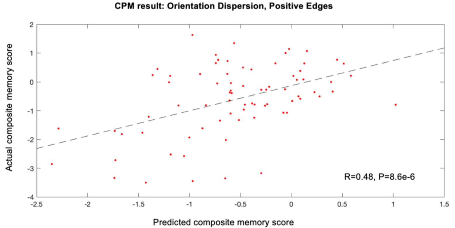
Figure 1. Plot of predicted vs. actual composite memory scores for Orientation Dispersion positive edges. Dashed line is least-squares fit line

Red: voxels positively correlated with composite memory score, Blue: voxels negatively correlated with composite memory score
