1488
Longitudinal comparison of medial and lateral meniscal T2* values within elite basketball players and swimmers1Radiology and Imaging, Hospital for Special Surgery, New York, NY, United States, 2Stanford University, Stanford, CA, United States, 3University of California, San Francisco, San Francisco, CA, United States
Synopsis
Basketball players represent a population with an inherently high risk of sustaining meniscal injuries. Evaluation of quantitative MRI (qMRI) metrics of the meniscus within these athletes may improve clinical insight. To date, no studies have longitudinally evaluated meniscal T2* values in high performance athletes. Therefore, the purpose of this study was to utilize ultra-short TE (UTE) MRI to longitudinally evaluate medial and lateral meniscal T2* values within elite weight-bearing (basketball) and non-weight bearing (swimmers) athletes. Significant differences of T2* values were found between the medial and lateral menisci. No significant difference of meniscal T2* values were found between basketball players and swimmers.
INTRODUCTION:
Acute meniscal injuries often result from high compressive forces with concomitant flexion or rotation of the knee joint and are most common in cutting and jumping athletes such as basketball players. 1,2,3 As the presence of meniscal pathology is highly correlated with the early onset and progression of degenerative joint disease,4 and elite basketball players have an inherently high risk of sustaining meniscal injuries, evaluation of quantitative MRI (qMRI) metrics of the meniscus within these athletes may improve clinical insight. Ultra-short echo (UTE) MRI sequences can capture the rapid transverse relaxation times associated with the menisci,5,6 and allow for quantitative evaluation of tissue microstructure with T2* mapping. While studies have demonstrated that T2* metrics are correlated to meniscal degeneration,7,8 no studies have evaluated meniscal T2* metrics within elite athletes over time, and no previous studies have attempted to evaluate the differences in T2* metrics between weight-bearing (WB) and non-weight bearing (NWB) athletes. As such, the objective of this study was to evaluate meniscal T2* metrics within and between collegiate basketball players (WB) and swimmers (NWB) and determine if T2* metrics were impacted by time.METHODS:
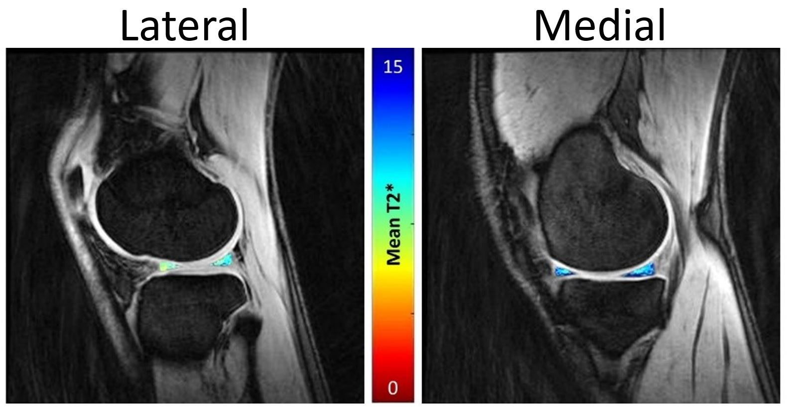
This was an IRB approved longitudinal multi-site study that included 32 collegiate athletes (16 swimmers and 16 basketball players). Longitudinal data were collected at 2 pre-season study visits (TP#1 and TP#2: spaced 12 months apart) for 11 of 32 subjects (7 swimmers, 4 basketball players) while all other subjects participated in a single pre-season study visit (TP#1). MRI: Bilateral 3-Tesla MRI examinations were obtained on a clinical scanner (GE Healthcare, Waukesha, WI) using an 8-channel phased array knee coil (Invivo). Three-dimensional, Cones UTE sequences were acquired for evaluation of T2* metrics (TEs: 5 echoes between 0.03-24 ms, TR: 188 ms, slice thickness: 3 mm, field of view: 16 cm, acquisition matrix: 256x256mm, RBW: ±83.3kHz, Flip-Angle: 16o). All menisci were manually segmented using MeVisLab software, and T2* values were calculated (Matlab, Natick, MA) using a mono-exponential fit of signal intensity to corresponding echo time. Statistical Analysis: A three-way repeated measures ANOVA was performed to evaluate the effects of sport (swimming/basketball) and knee compartment (medial/lateral meniscus) and timepoint (TP#1 and TP#2) on T2* values (SAS, V9.3, Cary, NC). Based on the ANOVA results, an unpaired t-test was performed to evaluate differences of meniscal T2* values between basketball players and swimmers for each compartment. Significance was set to p<0.05.RESULTS:
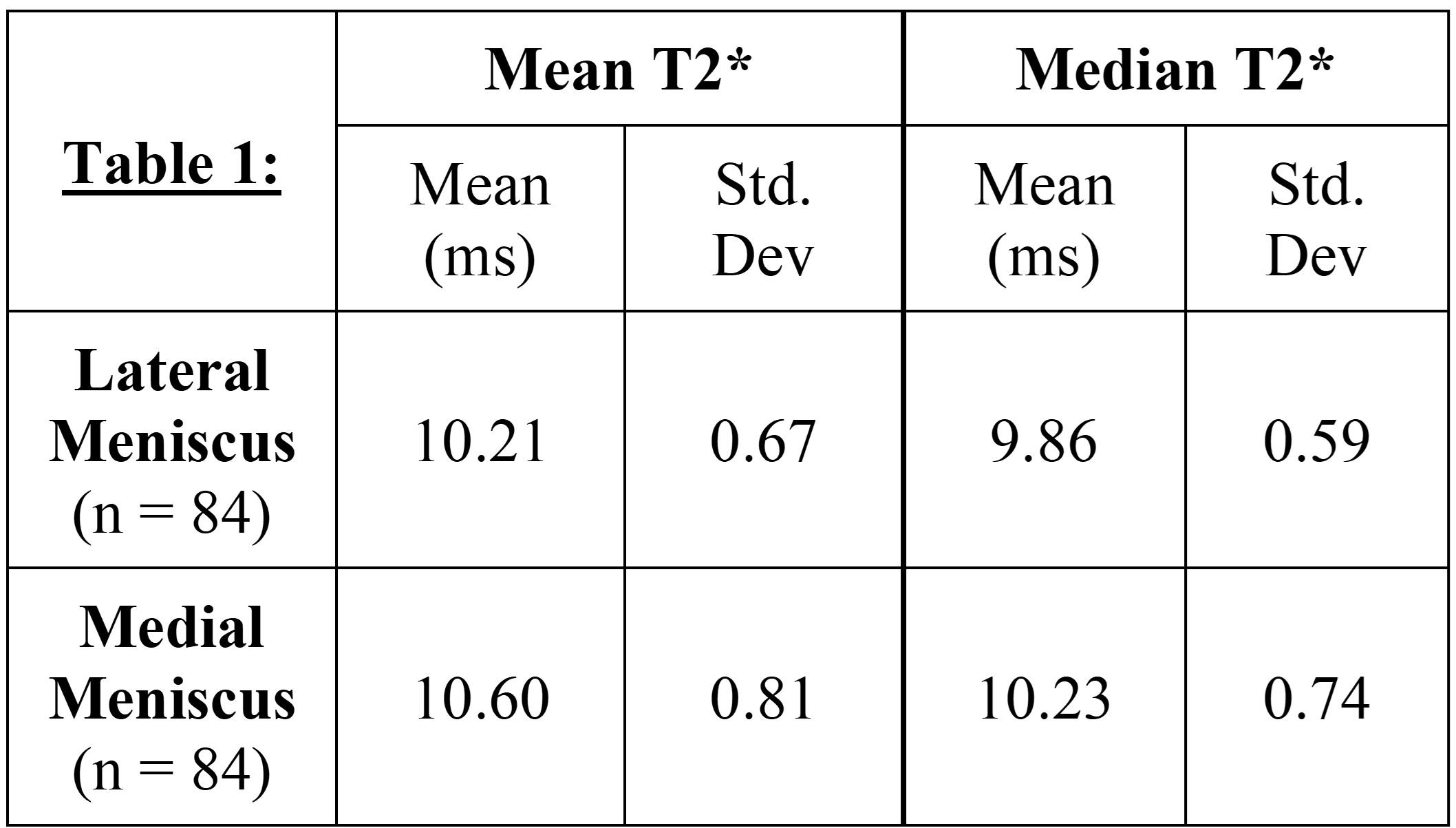
No significant differences in meniscal T2* values were found between weight-bearing and non-weightbearing athletes and no significant differences in meniscal T2* were found between timepoints (p>0.05). However, significant differences in in both mean and median T2* values were found between the medial and lateral menisci (p=<0.0005, Table 1).DISCUSSION:
No significant differences in meniscal T2* values were found between timepoints. Though basketball players are exposed to higher compression and shear forces in their knee during play, no significant differences in meniscal T2* metrics were evident between WB and NWB athletes. The lack of differences of T2* between the basketball players and swimmers may be attributable to limited enrollment, and the need for further evaluation of meniscal subregions (anterior horn, meniscal body, posterior horn) to localize differences that may not be evident with a global analysis. As TP#1 and TP#2 data were collected 12 months apart, all athletes were in the same phase of training (pre-season) for both visits; this may explain the consistency of medial and lateral T2* values over time. Significant differences in T2* metrics detected between the medial and lateral menisci likely reflect the differences in structure and function between the medial and lateral menisci9,10 and are likely the result of the disparate loading environments between the medial and lateral compartments.10CONCLUSION:
These initial findings demonstrate differences in medial and lateral meniscal T2* values and will benefit from a subregional analysis with concomitant assessment of the adjacent femoral and tibial articular surfaces. Meniscal T2* values may provide a non-invasive means to assess response of the tissue specific to the loading environment and identify the early onset meniscal degeneration. A better understanding of the longitudinal changes in meniscal T2* values may help define the progression of meniscal degeneration in elite athletes.Acknowledgements
The authors acknowledge funding from the GE/NBA Research Consortium. The authors would also like to thank the HSS MRI staff and technologists for their help and support on this study.References
1. Zedde 2014
2. Yeh 2012
3. Ripani 2012
4. Lohmander 2007
5. Gold 1995
6. Gatehouse 2003
7. Koff 2014
8. Williams 2012
9. Bloecker 2012
10. Stoller 2007
Figures