1487
Bi-Component Cartilage T2 Analysis in Subjects With Anterior Cruciate Ligament Reconstruction
Richard Kijowski1, Robert Moskwa2, and Fang Liu3
1Radiology, New York University School of Medicine, New York, NY, United States, 2Medical Physics, University of Wisconsin, Madison, WI, United States, 3Radiology, Massachusetts General Hospital, Boston, MA, United States
1Radiology, New York University School of Medicine, New York, NY, United States, 2Medical Physics, University of Wisconsin, Madison, WI, United States, 3Radiology, Massachusetts General Hospital, Boston, MA, United States
Synopsis
mcDESPOT was used to measure single-component T2 relaxation time (T2Single) and the bi-component T2 parameter fraction of the fast relaxing macromolecular bound water component (FF) of the cartilage of the knee at 3.0T in 10 subjects with ACL reconstruction and 10 control subjects. There were significant differences (p<0.05) between ACL reconstruction and control subjects on 5 of 6 articular surfaces of the knee for FF compared with 2 of 6 surfaces for T2Single with FF having higher absolute effect size than T2Single for all surfaces. The results demonstrate the superiority of bi-component over single-component T2 analysis for detecting cartilage degeneration.
Introduction: Changes in cartilage T2 relaxation time may be
difficult to interpret and often challenging to detect due to the multiple
competing factors influencing the measurement including water and
macromolecular content, tissue anisotropy, and magic angle effect (1, 2, 3). Bi-component
T2 mapping may improve the specificity of cartilage T2 analysis by evaluating the
individual water components of cartilage (4, 5, 6). Multi-component-Driven Equilibrium Single
Pulse Observation of T1 and T2 (mcDESPOT) is a clinically feasible MRI method for
bi-component T2 mapping of cartilage at 3.0T (7, 8). This study was performed to compare single-component
and bi-component cartilage T2 analysis using mcDESPOT for detecting early cartilage
degeneration within the knee in subjects with anterior cruciate ligament (ACL) reconstruction
surgery.
Methods: The study group consisted of 10 subjects with ACL reconstruction and no associated meniscus tear (mean time 1.7 years after surgery) and 10 age, gender, ethnicity, and body mass index matched healthy control subjects. All subjects underwent an MRI examination of the knee on a 3.0T scanner (Discovery MR750, GE Healthcare, Waukesha, WI) using an 8-channel extremity coil. A 3D fast spin-echo (3D-FSE) sequence was acquired with TR/TE=2216/23.6ms, 16cm field of view, 384 x 384 matrix, 1.0mm slice thickness, and 31.2kHz bandwidth. mcDESPOT was performed using a series of 3D spoiled gradient echo (SPGR) and 3D balanced steady-state free precession (bSSFP) scans. A SPGR scan was acquired with TR/TE=5.5/2.6ms over a range of flip angles (α =3, 4, 5, 6, 7, 9, 13, 18°). Two bSSFP scans with radiofrequency phase cycling on and off were acquired with TR/TE=7.1/3.6ms over a range of flip angles (α=2, 5, 10, 15, 20, 30, 40, 50°). An inversion recovery IR-SPGR scan with TR/TE=5.5/2.6ms, TI=450ms, and α=5° was obtained to estimate the transmit B1 field. All scans were performed using 16cm field of view, 256 x 256 matrix, 3mm slice thickness, 83.3kHz bandwidth, 32 slices, and one excitation with total scan time of 17 minutes. Single-component T2 relaxation time maps (T2Single) were created using DESPOT T2 Full Modeling (DESPOT2-FM) reconstruction (9). Bi-component T2 relaxation time maps for the fast relaxing macromolecular bound water component (T2F) and slow relaxing bulk water component (T2S) and water fraction maps for the fast relaxing water component (FF) of cartilage were created using two-pool mcDESPOT reconstruction (Figure 1) (7, 8). Single-component and bi-component cartilage T2 analysis was performed on 6 articular surfaces of the knee (Figure 2) (8). A musculoskeletal radiologist reviewed the 3D-FSE images to detect morphologic cartilage degeneration within the knee. Wilcoxon-Mann-Whitney tests were used to compare T2 parameters between ACL reconstruction subjects and control subjects with Cohen tests used to calculate absolute effect sizes between subject groups.
Results: No ACL reconstruction subject or control subject showed morphologic cartilage degeneration on 3D-FSE images. ACL reconstruction subjects had significantly higher cartilage T2Single than control subjects on the trochlea (p=0.001) and medial femur (p=0.011). ACL reconstruction subjects had significantly lower cartilage FF on the patella (p=0.010), trochlea (p<0.001), medial femur (p<0.001), lateral femur (p=0.023), and lateral tibia (p=0.029) are marginally lower cartilage FF on the media tibia (p=0.064) than control subjects. There was no significant differences (p=0.384-0.912) between ACL reconstruction subjects and control subjects in cartilage T2F and T2S on any articular surface of the knee (Figure 3). Absolute effect sizes between ACL reconstruction subjects and control subjects were higher for cartilage FF than cartilage T2Single, T2F, and T2S for all articular surfaces of the knee (Figure 4).
Discussion: Our study showed that T2Single can detect early cartilage degeneration in the knee of subjects with ACL reconstruction, which is similar to the findings of previous studies (10, 11, 12, 13). However, our study also showed that the bi-component T2 parameter FF was more sensitive than T2Single for detecting early cartilage degeneration in our subject population. Previous studies have also shown that FF measured using mcDESPOT was superior to T2Single for distinguishing between subjects with and without knee osteoarthritis and cartilage regions with and without morphologic degeneration (14). Experiments performed using Carr-Purcell-Meiboom-Gill sequences at high field strength NMR (5, 6) and mcDESPOT at 3.0T (15) have shown a significant (p<0.05) direct correlation between FF and the proteoglycan content of ex-vivo cartilage specimens. The lower cartilage FF in subjects with ACL reconstruction in our study likely reflects a combination of decreased proteoglycan content and increased bulk water content of cartilage, compositional changes that concomitantly occur during the earliest stages of cartilage degeneration. In contrast, cartilage T2Single is a nonspecific parameter influenced by multiple potentially competing biological changes that occur with cartilage degeneration. The lower sensitivity of T2F, and T2S compared to FF for detecting cartilage degeneration in our study is similar to the findings of previous studies (14, 15), which further indicates that FF is the most important bi-component T2 parameter for evaluation cartilage.
Conclusion: Bi-component cartilage T2 analysis is superior to single-component T2 analysis for detecting early cartilage degeneration within the knee in subjects with ACL reconstruction. Attempts should be made in future osteoarthritis research studies to use bi-component T2 analysis to maximize sensitivity for detecting cartilage degeneration within the knee.
Acknowledgements
We gratefully acknowledge funding from National Institute of Health R01AR068373 grant.References
(1) Nishioka, H, Hirose J, Nakamura E, et. al. Journ Magn Reson Imaging. 2012;35:147. (2) Liess C, Lusse S, Karger N, et al. Osteoarthritis Cartilage. 2002;10:907. (3) Xia Y, Moody JB, Burton-Wurster N, et al. Osteoarthritis Cartilage. 2001;9:393. (4) Reiter D, Lin P, Fishbein K, et al. Magn Reson Med. 2009;61:803. (5) Reiter D, Roque R, Lin P, et al. Magn Reson Med. 2011;65:377. (6) Reiter D, Roque R, Lin P, et al. NMR Biomed. 2011;24:1286. (7) Deoni S, Rutt B, Arun T, et al. Magn Reson Med. 2008;60:1372. (8) Liu F, Chaudhary R, Hurley S, et al. Journ Magn Reson Imaging. 2014;39:1191. (9) Deoni S. Journ Magn Reson Imaging. 2009;30:411. (10) Theologis A, Kuo D, Cheng J, et al. Arthroscopy. 2011;27:65. (11) Li X, Kuo D, Theologis A, et al. Radiology. 2011;258:505. (12) Su F, Hilton J, Nardo L, et al. Osteoarthritis Cartilage. 2013;21:1058. (13) Van Ginckel A, Verdonk P, Victor J, et al. Am J Sports Med. 2013;41:550. (14) Liu F, Choi K, Samsonov, et al. Radiology. 2015;277:477. (15) Grondin, M, Liu F, Vignos M, et al. J Biomech. Epub 2020 Dec 31.Figures
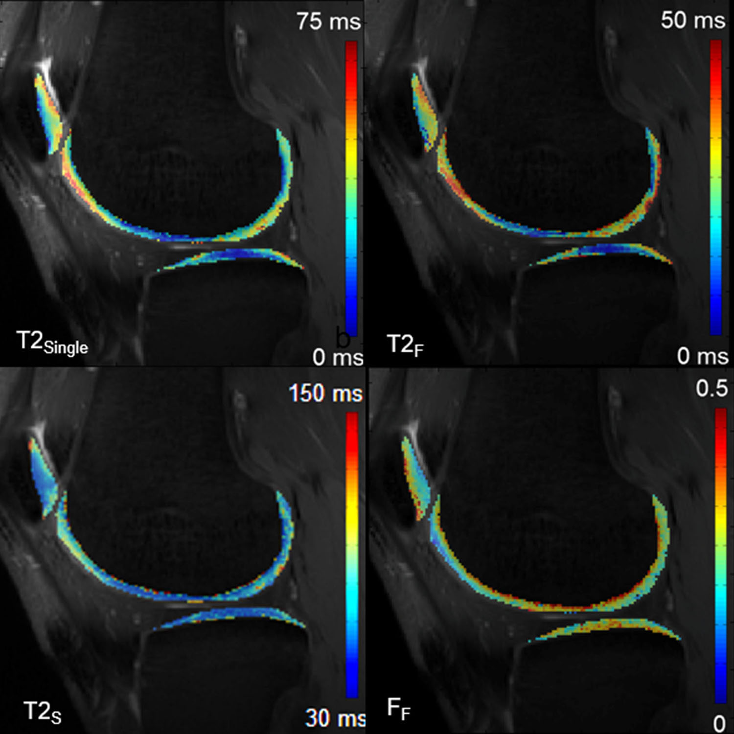
Figure 1: Single-component T2 relaxation time map
(T2Single), T2 relaxation time maps for the fast relaxing
macromolecular bound water component (T2F) and slow relaxing bulk
water component (T2S), and water fraction maps for the fast relaxing
water component (FF) of cartilage obtained using mcDESPOT.
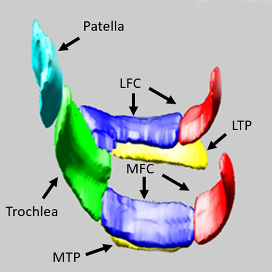
Figure
2: Single-component and bi-component T2
parameters of cartilage obtained using mcDESPOT were measured on the patella,
trochlea, medial femur (medial femoral condyle or MFC), medial tibia (medial
tibia plateau or MTP), lateral femur (lateral femoral condyle or LFC), and
lateral tibia (lateral tibia plateau or LTP).
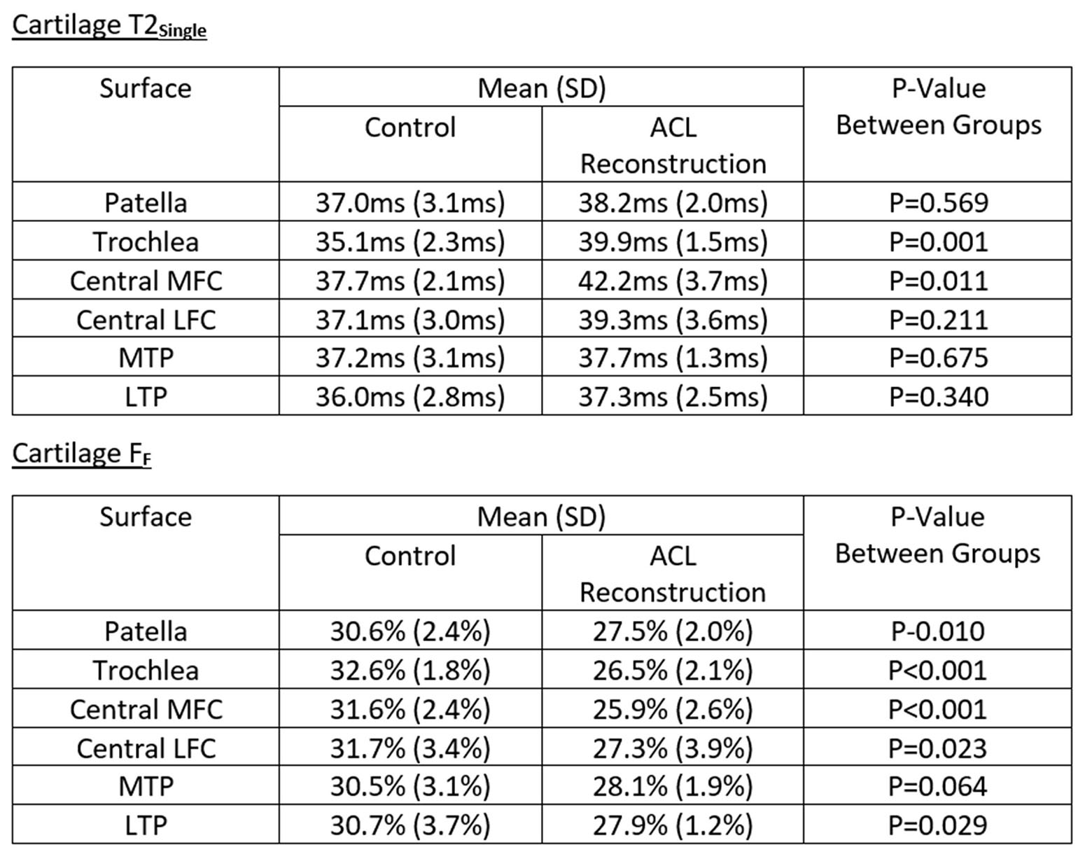
Figure 3: Mean and standard deviation (SD)
of cartilage T2Single and cartilage FF obtained using
mcDESPOT on each articular surface of the knee in ACL reconstruction subjects
and control subjects.
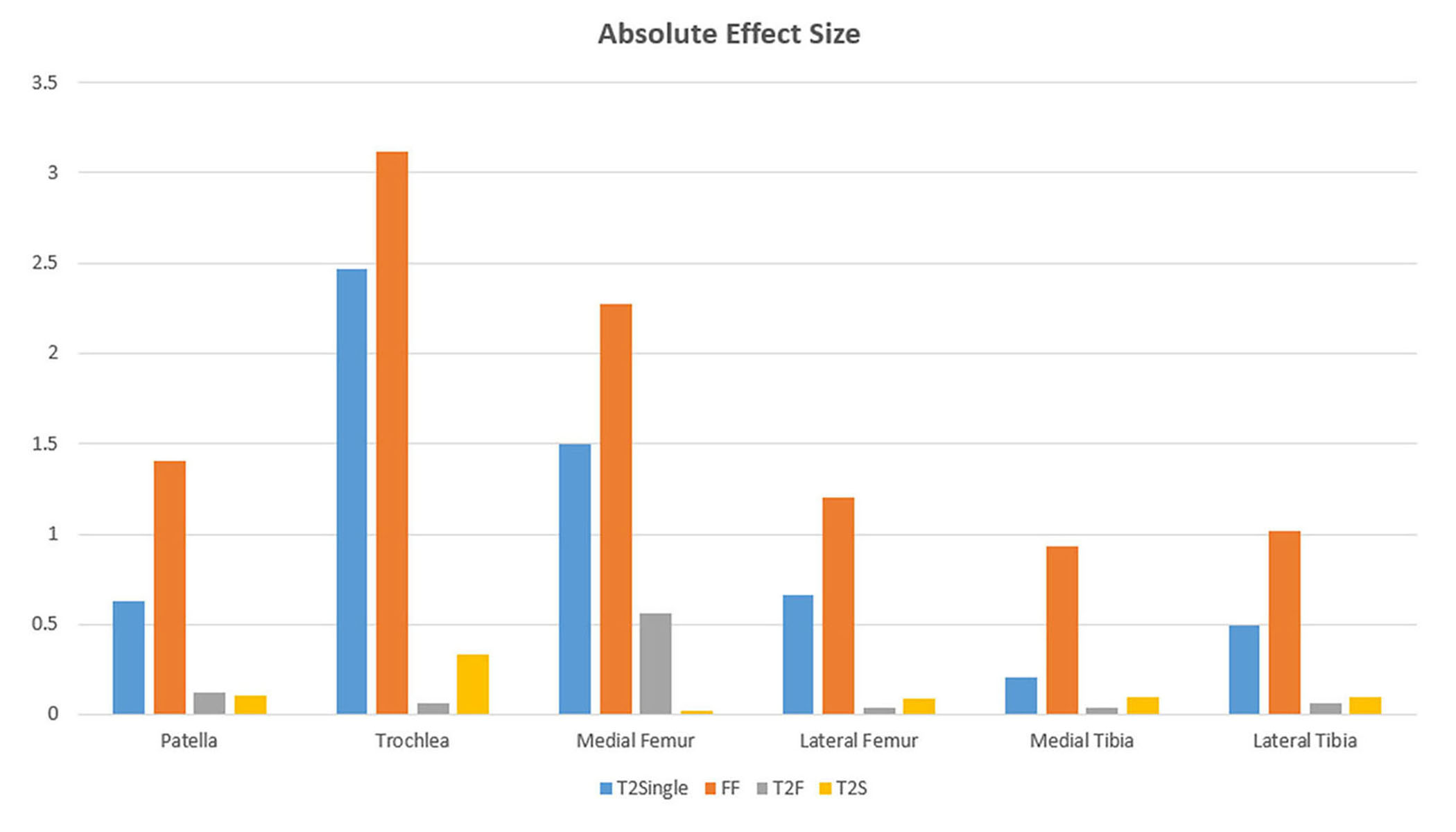
Figure 4: Comparison of absolute
effect size for single-component and bi-component T2 parameters obtained using
mcDESPOT for detecting early cartilage degeneration of the knee in subjects
with ACL reconstruction. Note that FF
had the highest absolute effect size for all articular surfaces.
DOI: https://doi.org/10.58530/2022/1487