1408
Simultaneous morphologic and quantitative imaging of the knee joint using pseudo-3D MIXTURE sequences: from deliberation to implementation1Diagnostic and Interventional Radiology, University Hospital Aachen, Aachen, Germany, 2Philips GmbH Market DACH, Hamburg, Germany, 3Philips Japan, Tokyo, Japan, 4Clinic for Diagnostic and Interventional Radiology, Uniklinik RWTH Aachen University, Aachen, Germany
Synopsis
This work aimed to propose a clinical sequence platform for simultaneous morphologic and quantitative imaging of joints and identified TSE-based MIXTURE (Multi-Interleaved X-prepared Turbo-Spin Echo with Intuitive Relaxometry) sequences in terms of PDFS-T2 and T1-T1ρFS combinations for clinical implementation. Even though capable of isotropic resolution, MIXTURE sequences were acquired as pseudo-3D (thicker slices, higher in-plane resolution) to match standard-clinical 2D TSE sequences. After identification of clinical demands, MIXTURE sequences were systematically optimized, while maintaining clinically feasible acquisition times of 5:00 min (PDFS-T2 for T2-mapping) and 6:40 min (T1-T1ρFS for T1ρ-mapping), and evaluated in a cadaveric human knee cartilage defect model.
Introduction
Quantitative MRI techniques such as T2 and T1ρ mapping are diagnostically beneficial in the comprehensive evaluation of various pathologies of the joint, yet require long acquisition times which limits their clinical adoption. MIXTURE (Multi-Interleaved X-prepared Turbo-Spin Echo with Intuitive Relaxometry) sequences have recently been proposed for simultaneous morphologic and quantitative MRI of joints1. Quantitative data are calculated based on two or more interleaved turbo-spin echo (TSE) images with different preparation modules and hence adjustable contrast. Standard 2D TSE sequences (proton density (PD)-weighted, T2-weighted with or without fat-saturation (FS) and T1-weighted) are the diagnostic mainstay of joint imaging in our department and beyond. Previously, isotropic MIXTURE sequences were suggested, e.g., a PD-T2FS MIXTURE sequence with T2 preparation pulses of different lengths2, and a PD-T1ρFS MIXTURE sequence with T1ρ preparation pulses of different spin lock times (TSL)3. Pseudo-3D MIXTURE variants with higher in-plane resolution and thicker slices resemble the clinical-standard 2D TSE sequences more closely and may hence be used for comparative evaluation of the MIXTURE sequence platform. Hence, we present optimized pseudo-3D MIXTURE sequences of human cadaveric knee joint specimens with standardized traumatic cartilage defects and visually evaluate diagnostic aspects in comparison to clinical-standard 2D TSE sequences.Methods
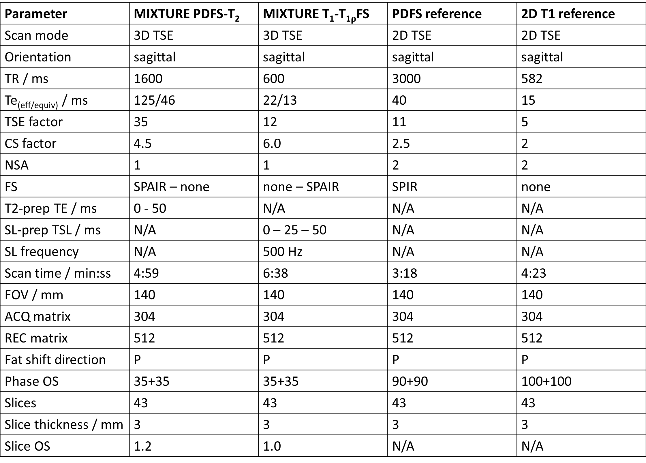
Measurements were performed on human cadaveric knee joint specimens following approval by the local Ethical Committee on a clinical 3T system (Philips Elition X, Best, The Netherlands with a 16-channel clinical T/R knee coil (Philips). Clinical needs and optimization criteria for morphologic MIXTURE images were discussed with a clinical radiologist (8 years of experience in musculoskeletal imaging) and implemented accordingly. Guided by the our institution’s clinical standard imaging protocol for the knee that consists of PDFS sequences (axial, coronal, sagittal) and a T1-weighted sequence (sagittal), two sagittal MIXTURE sequences, i.e., PDFS-T2 MIXTURE for T2 mapping and T1- T1ρFS MIXTURE for T1ρ mapping were acquired in pseudo-3D configuration and optimized to provide similar diagnostic quality as the 2D scans while optimizing scan time (sequence parameters of reference sequences and optimized MIXTURE protocols c.f. Figure 1). To evaluate the different sequences in close-to-clinical conditions, the MIXTURE and clinical reference sequences were acquired before (i.e., intact joint) and after creating standardized defects of 3, 5, and 8 mm in diameter at the weight-bearing area of the lateral femoral condyle of n=3 knee joint specimens.Results
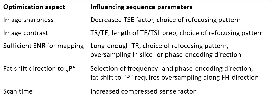
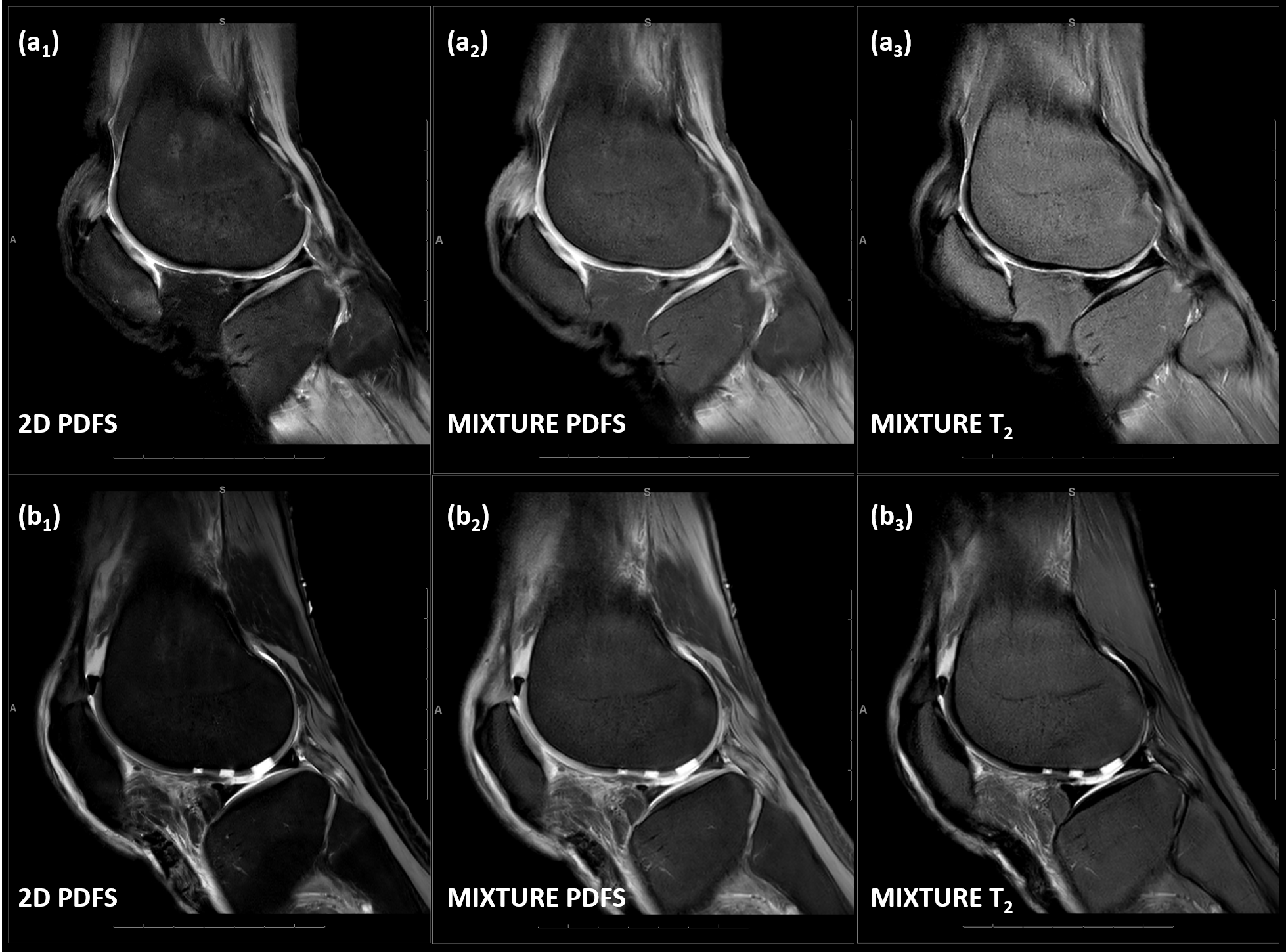
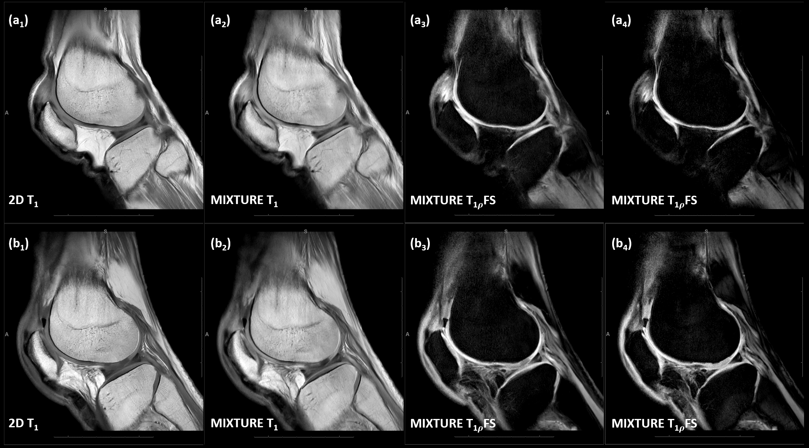
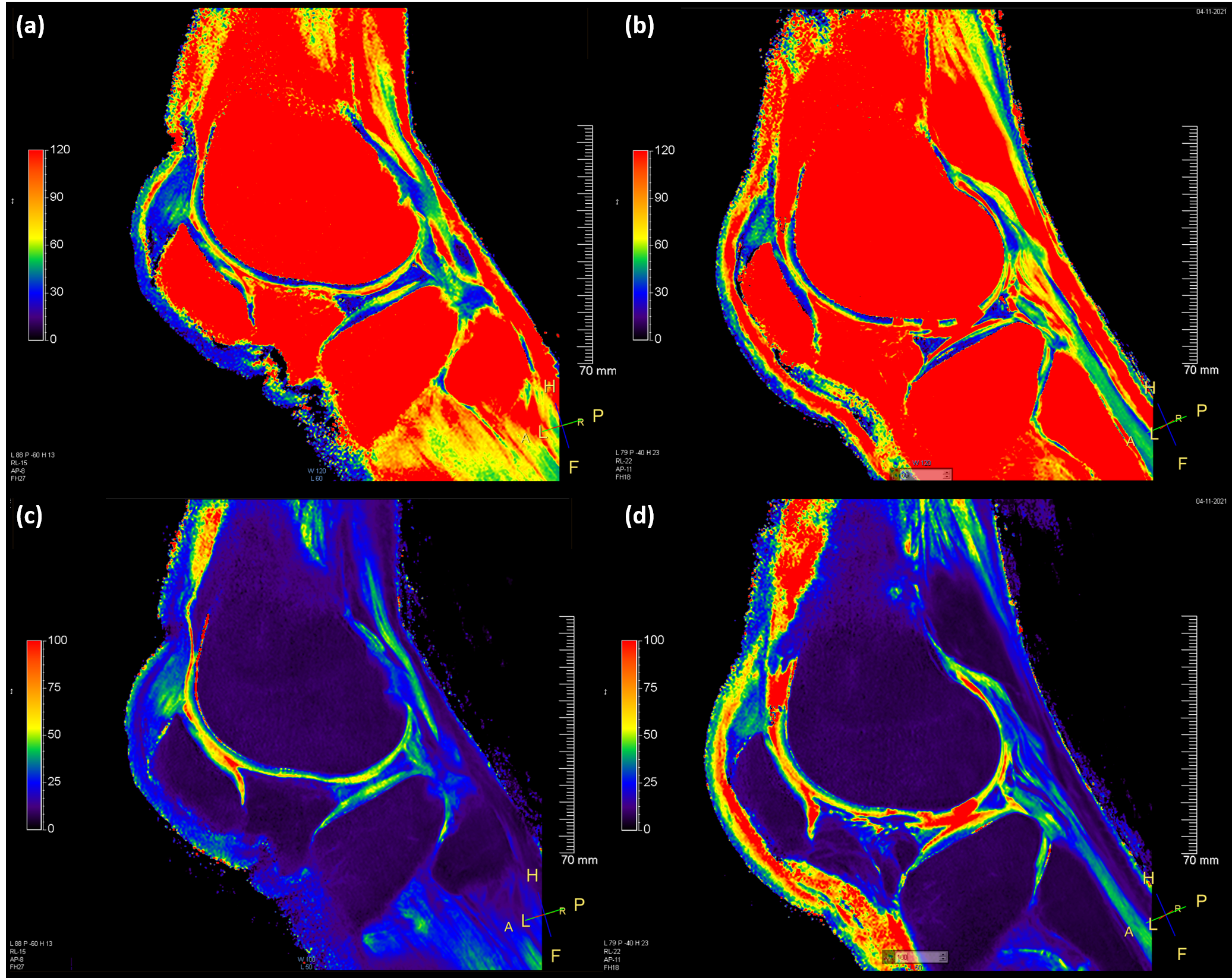
Figure 2 shows the optimization aspects with regard to clinical needs and relevant sequence parameters. Image blurring in MIXTURE sequences associated with the underlying 3D technique and long TSE readouts were largely reduced by using shorter TSE factors and adapting refocusing patterns4. Figure 3 shows the 2D PD-weighted FS TSE (reference) sequence and the morphologic images of the optimized MIXTURE PDFS-T2 sequence for the knee without and with standardized cartilage defects, while Figure 4 indicates the corresponding 2D T1-weighted reference sequence and the morphologic images of the optimized MIXTURE T1-T1ρFS sequence. After optimization, the morphologic PDFS MIXTURE images allow clear visualization and delineation of the cartilage tissue and their defects with comparable overall image quality, contrast, and signal strength. The morphologic T1 MIXTURE image depicts the same anatomic features, in particular regarding bone texture and structure, as the clinical reference sequence. Figure 5 visualizes the MIXTURE-based T2 and T1ρ maps of the respective sagittal slices, which demonstrate the layered structure of cartilage as well as the defects.Discussion
T2 and T1ρ MIXTURE sequences, acquired in pseudo-3D configuration in human cadaveric knee joint specimens, enabled a voxel-wise, motion-free comparison with the clinical 2D protocols which is a step towards additional validation and clinical implementation of the MIXTURE sequence platform. After optimization, morphologic MIXTURE images are diagnostically sufficient in terms of image quality, contrast, and signal strength. Thus, MIXTURE sequences acquired as pseudo-3D sequences may be useful candidates for diagnostic morphologic joint imaging with adjunct mapping of distinct biophysical properties that allow inferences on tissue ultrastructure and composition beyond mere morphology. This study is the first to propose pseudo-3D PDFS-T2 and T1-T1ρFS variants of MIXTURE in a human model of cartilage pathology.Conclusion
MIXTURE sequences are promising candidates for combined whole-joint morphologic and quantitative assessment in the future at scan times of around 5-6 minutes. Future research should validate the MIXTURE-based relaxation times against reference T2 and T1ρ mapping sequences in dedicated phantoms and collect MIXTURE data in volunteers and patients.Acknowledgements
No acknowledgement found.References
1Yoneyama M, Sakai T, Zhang S, et al. MIXTURE: a novel sequence for simultaenous morphological and quantitative imaging based on multi-interleaved 3D turbo-spin echo MRI. Proc. Intl. Soc. Mag. Reson. Med. 29 (2021), 4203.
2Sakai T, Yoneyama M, Watanabe A, et al. Simultaneous anatomical, pathological and T2 quantitative knee imaging with 3D submillimeter isotropic resolution using MIXTURE. Proc. Intl. Soc. Mag. Reson. Med. 29 (2021), 0845.
3Saruya S, Suzuki M, Yoneyama M, et al. 3D sub-millimeter isotropic knee cartilage T1rho mapping using multi-interleaved fluid-attenuated TSE acquisition (MIXTURE). Proc. Intl. Soc. Mag. Reson. Med. 29 (2021), 2977.
4Mugler JP. Optimized Three-Dimensional Fast-Spin-Echo MRI. J Magn. Reson. Med. (2014) 39:745–767.
Figures