1383
Surface-Based Morphometry Analysis Shows Increased Cortical Sulcus Depth after Sound Therapy in Patients with Tinnitus1Department of Radiology, Beijing Friendship Hospital, Capital Medical University, Beijing, China, 2MR Collaboration, Siemens Healthcare Ltd., Beijing, China
Synopsis
In this study, we performed brain surface-based morphometry to evaluate changes in sulcal depth after sound therapy in patients with idiopathic tinnitus. Our results showed that sulcal depth was significantly reduced in the left medial temporal cortex (MTC) and right somatosensory and motor cortex (SMC) of patients with tinnitus compared to the healthy controls, but increased significantly at 24 weeks after sound therapy. Therefore, sulcal depth in the auditory sensory regions of the brain is a potential neuroimaging biomarker for evaluating treatment efficacy in tinnitus patients.
Introduction
Tinnitus is a conscious awareness of a sound in the absence of any external acoustic stimulation, and it is a major health issue in society worldwide [1-3]. Narrow-band noise sound therapy has been one of the commonly used methods for the treatment of tinnitus in recent years [4]. Tinnitus and hyperacusis can lead to significant brain structural changes. Sulcal depth is a quantitative measure of the cerebral cortex. It is widely used as an important marker in brain morphology research. In this study, we performed brain surface-based morphometry using magnetic resonance imaging (MRI) data to evaluate changes in sulcal depth after sound therapy in patients with idiopathic tinnitus.Materials and Methods
We enrolled 33 patients with tinnitus and 26 age- and sex-matched healthy control (HC) individuals for this study. We then acquired high-resolution 3D T1-weighted brain structural images of all tinnitus patients at baseline and 24 weeks after sound therapy. We also obtained MR images of healthy controls at baseline and at 24 weeks. We used the 3D magnetization-prepared rapid gradient echo (MPRAGE) sequence in a MAGNETOM Prisma 3T MRI system (Siemens Healthcare, Erlangen, Germany) equipped with a 64-channel phase-array head coil. The 3D MPRAGE sequence parameters were as follows: repetition time (TR): 2530 ms, echo time (TE): 2.98 ms, inversion time (TI): 1100 ms, FA: 7°, bandwidth: 240 Hz/Px, field of view (FOV): 256 mm × 224 mm, slice thickness: 1 mm, number of slices: 192, matrix: 256 × 224, isotropic voxel size: 1 mm × 1 mm × 1 mm, and acquisition time: 5min58sec. The brain structural image data was preprocessed using the DPABISurf toolbox. The severity of tinnitus was assessed before and after sound therapy using Tinnitus Handicap Inventory (THI) scores. Statistical data was evaluated using two-way mixed model analysis of variance (ANOVA) and Pearson’s correlation analysis. Student-Newman-Keuls (SNK) method was used for the post hoc analysis.Results
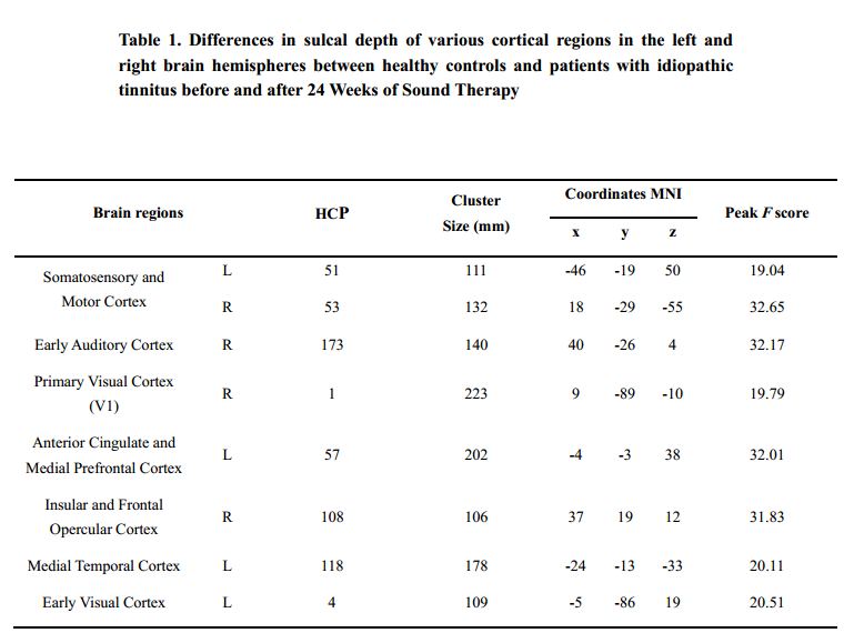
Group differences in sulcal depthAs shown in Figure 1, Figure 2 and Table1, there were significant differences in sulcal depth among the tinnitus patients before sound therapy (baseline), tinnitus patients after sound therapy (24 weeks), HC individuals at baseline and HC individuals after 24 weeks. The relevant brain regions included both somatosensory and motor cortexes (SMCs) and the right early auditory cortex (EAC), right primary visual cortex (PVC), left anterior cingulate and medial prefrontal cortex (ACC&MPC), right insular and frontal opercular cortex (IFOC), left medial temporal cortex (MTC) and left early visual cortex (EVC) (P<0.05 (P<0.025 for each hemisphere) corrected by Monte Carlo simulation; L, left; R, right).
Post hoc analysis showed significant changes in sulcal depth of left MTC and right SMC in tinnitus patients before and after sound therapy
Sulcal depth was significantly decreased in the left MTC and right SMC of the participants in the tinnitus baseline group compared to participants in the HC baseline group and HC 24-week group, and there were no significant differences between the two HC groups (Figure 3). The 24-week sound therapy tinnitus group demonstrated a significantly higher sulcal depth in the left MTC and right SMC than the tinnitus baseline group (Figure 3). Compared with the HC baseline group and the HC 24-week group, the 24-week sound therapy tinnitus group demonstrated slightly lower or similar sulcal depths in the left MTC and right SMC, and there were no significant differences between the 24-week sound therapy tinnitus group and the HC groups. Compared with the HC baseline group, the HC 24-week group did not show a difference in sulcal depth that reached statistical significance in the left MTC or right SMC (Figure 3).
Discussion
Our study demonstrated that sulcal depth was significantly reduced in the left MTC and right EAC of patients with tinnitus compared to the healthy controls. However, sulcal depth increased significantly in the left MTC of patients with tinnitus at 24 weeks after sound therapy and was comparable to the two HC groups. Both left MTC and right EAC are involved in auditory sensing as well as language-related activities and processes [5]. MTC is also involved in the processing of complex sounds [6] and integration of auditory-related actions. The auditory cortex is subdivided into early auditory cortex (EAC), secondary auditory cortex (SAC), and auditory association cortex (AAC) regions [7]. Lv et al reported that regional brain activity increased in the EAC after sound therapy [8]. Moreover, Han et al reported increased functional connectivity between the left parahippocampal gyrus and the cingulate cortex as well as other brain regions that regulate auditory sensing in tinnitus patients after sound therapy [9]. We also observed increased sulcal depth in the right SMC of tinnitus patients after sound therapy, thereby suggesting recovery of sensorimotor functions. Sulcal depth has not been reported as a quantitative surface-based morphometry measure of cortical thickness in tinnitus previously. Our study suggested that reduced sulcal depth and disrupted connectivity of the medial frontal cortex may be associated with negative emotions experienced by individuals with tinnitus [10].Acknowledgements
Our study demonstrated that sound therapy resulted in significant recovery of sulcal depth in the cortical regions that control auditory and sensorimotor functions in tinnitus patients. Therefore, sulcal depth in the auditory sensory regions of the brain is a potential neuroimaging biomarker for evaluating treatment efficacy in tinnitus patients.References
(1) Esmaili AA, Renton J. A review of tinnitus. Aust J Gen Pract, 2018, 47(4): 205-208.
(2) Bauer CA. Tinnitus. N Engl J Med, 2018, 378(13): 1224-1231.
(3) Conlon B, Langguth B, Hamilton C, Hughes S, Meade E, Connor CO, Schecklmann M, Hall DA, Vanneste S, Leong SL, Subramaniam T, D'Arcy S, Lim HH. Bimodal neuromodulation combining sound and tongue stimulation reduces tinnitus symptoms in a large randomized clinical study. Sci Transl Med, 2020, 12(564).
(4) Henry JA, Schechter MA, Nagler SM, Fausti SA. Comparison of tinnitus masking and tinnitus retraining therapy. J Am Acad Audiol, 2002, 13(10): 559-581.
(5) Hickok, G. (2012). The cortical organization of speech processing: feedback control and predictive coding the context of a dual-stream model. J Commun Disord. 45(6): 393-402.
(6) Mirz, F., Ovesen, T., Ishizu, K., Johannsen, P., Madsen, S., Gjedde, A., et al. (1999). Stimulus-dependent central processing of auditory stimuli: a PET study. Scand Audiol. 28 (3): 161-169.
(7) Glasser, M.F., Coalson, T.S., Robinson, E.C., Hacker, C.D., Harwell, J., Yacoub, E., et al. (2016). A multi-modal parcellation of human cerebral cortex. Nature. 536 (7615): 171-178.
(8) Lv, H., Liu, C., Wang, Z., Zhao, P., Cheng, X., Yang, Z., et al. (2020). Altered functional connectivity of the thalamus in tinnitus patients is correlated with symptom alleviation after sound therapy. Brain Imaging Behav 14 (6): 2668-2678.
(9) Han, L., Pengfei, Z., Chunli, L., Zhaodi, W., Xindi, W., Qian, C., et al. (2020). The effects of sound therapy in tinnitus are characterized by altered limbic and auditory networks. Brain Commun. 2 (2): fcaa131.
(10) Aldhafeeri, F.M., Mackenzie, I., Kay, T., Alghamdi, J., and Sluming, V. (2012). Neuroanatomical correlates of tinnitus revealed by cortical thickness analysis and diffusion tensor imaging. Neuroradiology. 54 (8): 883-892.
Figures
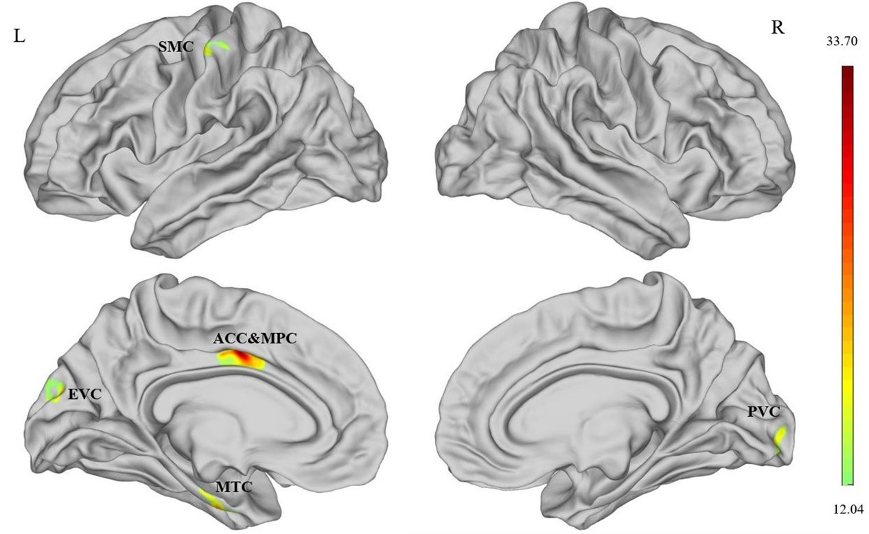
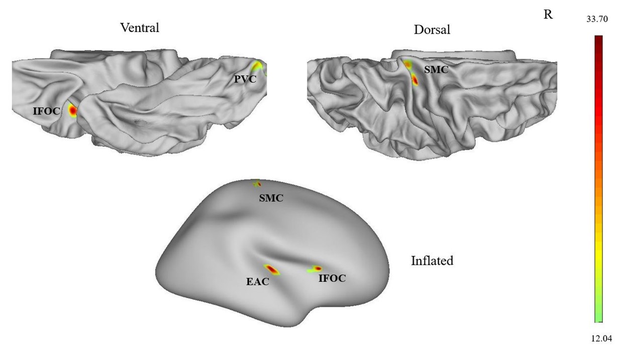
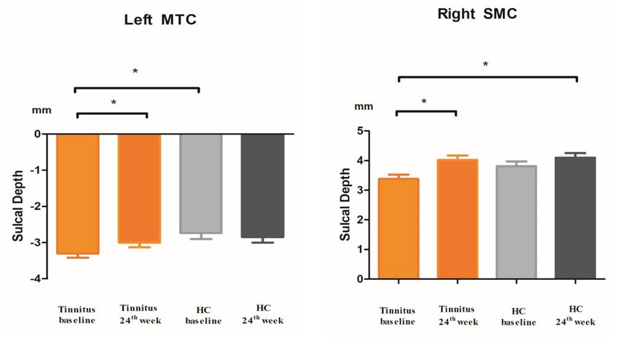
Fig.3 Post hoc analyses showed that the sulcal depth changed among the tinnitus baseline, 24-week sound treatment, HC baseline and HC 24-week groups in both hemispheres. *P < 0.05. Abbreviations: ANOVA: analysis of variance; HC: healthy control; SMC: somatosensory and motor cortex; MTC: medial temporal cortex.