1322
Difference of stiffness between the fetal and the maternal part of the placenta by virtual magnetic resonance elastography
Ting Liu1, Jiaojiao Lu1, Junjun Li1, and Jian Yang2
1Department of Radiology, the first Affiliated Hospital of Xi'an Jiaotong University, Xi'an, China, 2the first Affiliated Hospital of Xi'an Jiaotong University, Xi'an, China
1Department of Radiology, the first Affiliated Hospital of Xi'an Jiaotong University, Xi'an, China, 2the first Affiliated Hospital of Xi'an Jiaotong University, Xi'an, China
Synopsis
Virtual Magnetic Resonance Elastography (vMRE) is based on IVIM, which does not require any mechanical vibration, can assess the elastography of your organization. dysfunctional placenta had higher stiffness than normal placenta has been found by ultrasound shear‑wave elastography. This study intends to use this vMRE method to study the difference in elasticity between the fetus and the maternal compartment.
Introduction
Ultrasound shear‑wave elastography has confirmed that dysfunctional placenta, such as FGR, preeclampsia, had higher stiffness than normal placenta[1, 2]. However, there is no similar report on MRI. Because MRI elasticity imaging needs to provide an additional any mechanical vibration[3], it may have potential risks to pregnant women and fetuses. Recently discovered a non-invasive method to assess the elasticity of tissues. This method, referred to as virtual Magnetic Resonance Elastography (vMRE), is based on IVIM. It does not require any mechanical vibration, but based on information of two b-values in IVIM for simulation calculation, the hardness value similar to elastography can be obtained[4]. This method has been validated in liver fibrosis and some tumors[3, 5, 6], confirming that it has a very high agreement with the real elasticity. Therefore, this study intends to use this vMRE method to study the difference in elasticity between the fetus and the maternal compartment.Materials and Methods
This study was approved by ethical committee of First Affiliated Hospital of Xi’an Jiaotong University. Written parental informed consent was obtained for all pregnant women. 83 pregnant women were recruited from January 2017 to October 2021 in First Affiliated Hospital of Xi’an Jiaotong University. No sedatives were used for MRI. The scanning was stopped immediately once the pregnant woman has chest discomfort or other discomfort. Placental MR examination for each gestational woman was performed on a 3T 750 MRI system (GE Healthcare, Milwaukee, Wisconsin) with an 8-channel body flex coil. Single shot fast spin echo (SSFE) T2-weighted images were acquired using: TR minimum; TE 85 ms, matrix 228×256, FOV 360mm, section-thickness 4 mm without gap. Intra Voxel Incoherent Motion (IVIM) images were collected with each of the following b values: 0, 20, 50, 80, 100, 150, 200, 400, 600,800 sec/mm2. The scanning parameters were as follows: TR 2000ms; TE 63 ms, matrix 128×128, FOV 380mm, section-thickness 4mm with 1mm gap. A total examination time was less than 10 min. Diffusion weighted images of the lower b-value (Slow, b value = 200 s/mm2) and those of the higher b-value (Shigh, b value = 800 s/mm2) were used to estimate the virtual shear stiffness[3, 6]: virtual shear stiffness = a·ln (Slow/Shigh) + b. The scaling (a) and the shift (b) factors were separately set to −9.8 and 14 according to the previous calibration studies[3, 6]. The drawing diagram of the regions of interest (ROI) is shown in Figure 1. For each placenta, select the middle 3 layers to draw ROI, and take the average of the measured values as the final stiffness value.Results
A total of 83 pregnant women undergoing MRI examination were included in this study. After screening, 28 subjects were left, as shown in Figure 2. The basic information of such subjects is shown in Table 1. Analyzing the relationship between the stiffness value of the fetus and the maternal part and the gestational age, it was found that the stiffness value of the maternal compartment was higher than that of the fetal part (P<0.05). In addition, the stiffness value of the compartment was the lowest at 26 weeks, and showed an upward trend from 26 weeks to 36 weeks. The stiffness changes of the fetal compartment are basically the same. Further analysis of the relationship between the stiffness difference between the fetal and the maternal compartment that the stiffness of the maternal compartment of the placenta is less than that of the fetus only in the 32-34 week period. In addition, we also found that the stiffness of the fetal and maternal compartment of the placenta was significantly correlated with the thickness of the placenta after the gestational age> 26 weeks (P<0.05).Discussion
This study found, for the first time, the difference in elasticity between the fetal and the maternal compartment during the development of the placenta. First, it was found that the stiffness value of the fetal compartment was lower than the maternal compartment (except for 32-34 weeks), and secondly, it was found that the stiffness value of the placenta was related to the thickness of the placenta. In general tissues, the higher the stiffness, the larger the proportion of fibrous tissue. In the maternal compartment of the placenta, the villous structure goes deep into the decidua and even the muscle layer of the uterus, requiring more fibrous tissue support. In the fetal compartment of the placenta, there are more places for blood exchange[7, 8], so the stiffness may be slightly lower than that of the maternal compartment. With the development of the placenta, the function of the placenta in the first and second trimester of pregnancy gradually changes from immature to mature, and its thickness will increase. In the third trimester, placental function may deteriorate and may show increased stiffness. At present, few studies have reported on the relationship between placental stiffness and placental development. Some ultrasound reports show that placental stiffness was not closely related to gestational age, which may be related to insufficient number of cases and individual differences.Conclusion
Using vMRE can discover the difference in elasticity of the fetus and maternal compartment of the placenta at different developmental stages.Acknowledgements
This work was supported by Shaanxi Natural Science Basic Research Program Fund (2017JQ8034), the first Affiliated Hospital of Xi'an Jiaotong University Fund(XJTU1AF-3D-2018-005).References
[1] M. Spiliopoulos, C. Kuo, A. Eranki, M. Jacobs, C. Rossi, S. Iqbal, J. Fisher, M. Fries, P. Kim, Characterizing placental stiffness using ultrasound shear-wave elastography in healthy and preeclamptic pregnancies, Archives of gynecology and obstetrics 302(5) (2020) 1103-1112. [2] H. Arioz Habibi, E. Alici Davutoglu, S. Kandemirli, M. Aslan, A. Ozel, A. Kalyoncu Ucar, P. Zeytun, R. Madazli, I. Adaletli, In vivo assessment of placental elasticity in intrauterine growth restriction by shear-wave elastography, European journal of radiology 97 (2017) 16-20. [3] K. Lagerstrand, N. Gaedes, S. Eriksson, D. Farahmand, E. De Coursey, G. Johansson, L. Jönsson, T. Skoglund, Virtual magnetic resonance elastography has the feasibility to evaluate preoperative pituitary adenoma consistency, Pituitary 24(4) (2021) 530-541. [4] D. Le Bihan, What can we see with IVIM MRI?, NeuroImage 187 (2019) 56-67. [5] M. Kromrey, D. Le Bihan, S. Ichikawa, U. Motosugi, Diffusion-weighted MRI-based Virtual Elastography for the Assessment of Liver Fibrosis, Radiology 295(1) (2020) 127-135. [6] D. Le Bihan, S. Ichikawa, U. Motosugi, Diffusion and Intravoxel Incoherent Motion MR Imaging-based Virtual Elastography: A Hypothesis-generating Study in the Liver, Radiology 285(2) (2017) 609-619. [7] I. Carrasco-Wong, A. Moller, F. Giachini, V. Lima, F. Toledo, J. Stojanova, L. Sobrevia, S. San Martín, Placental structure in gestational diabetes mellitus, Biochimica et biophysica acta. Molecular basis of disease 1866(2) (2020) 165535. [8] P. Grigsby, Animal Models to Study Placental Development and Function throughout Normal and Dysfunctional Human Pregnancy, Seminars in reproductive medicine 34(1) (2016) 11-6.Figures
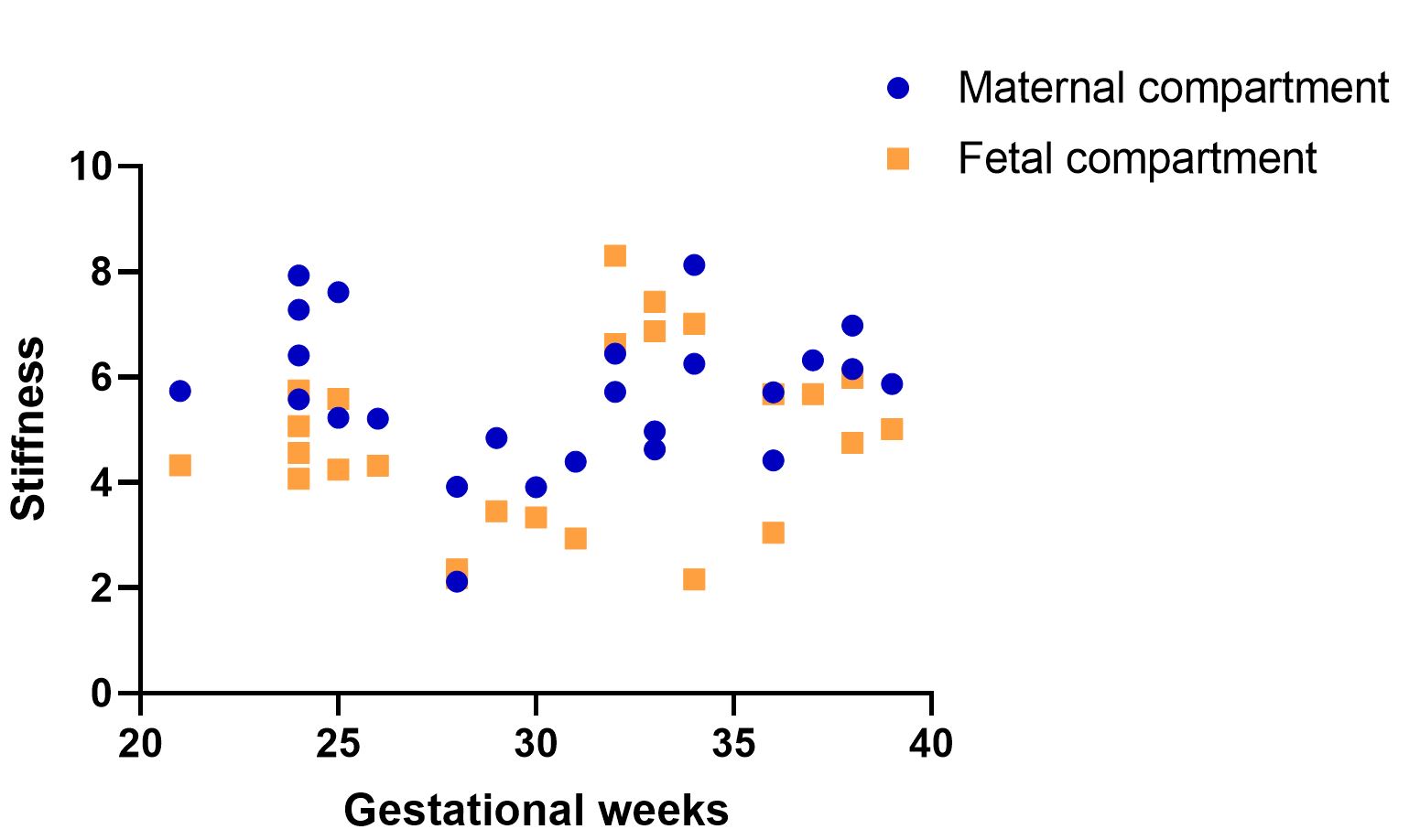
Figure: 3. The relationship between the stiffness value of the fetus and the maternal
part and the gestational age.
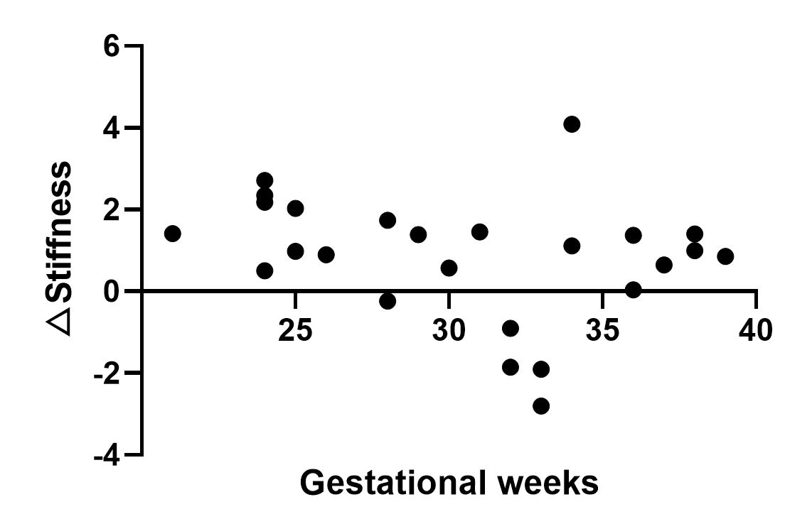
Figure: 4. Relationship between △ Stiffness(maternal- fetal)and gestational age.
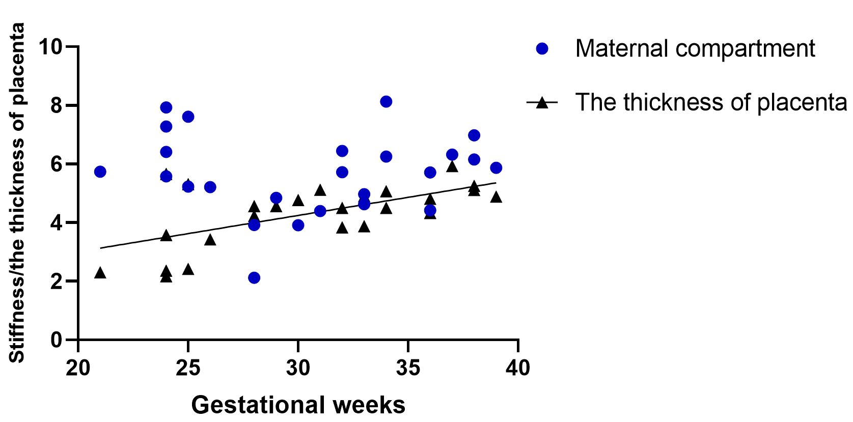
Figure: 5. The relationship between the stiffness value of the the maternal part and thickness of the placenta.
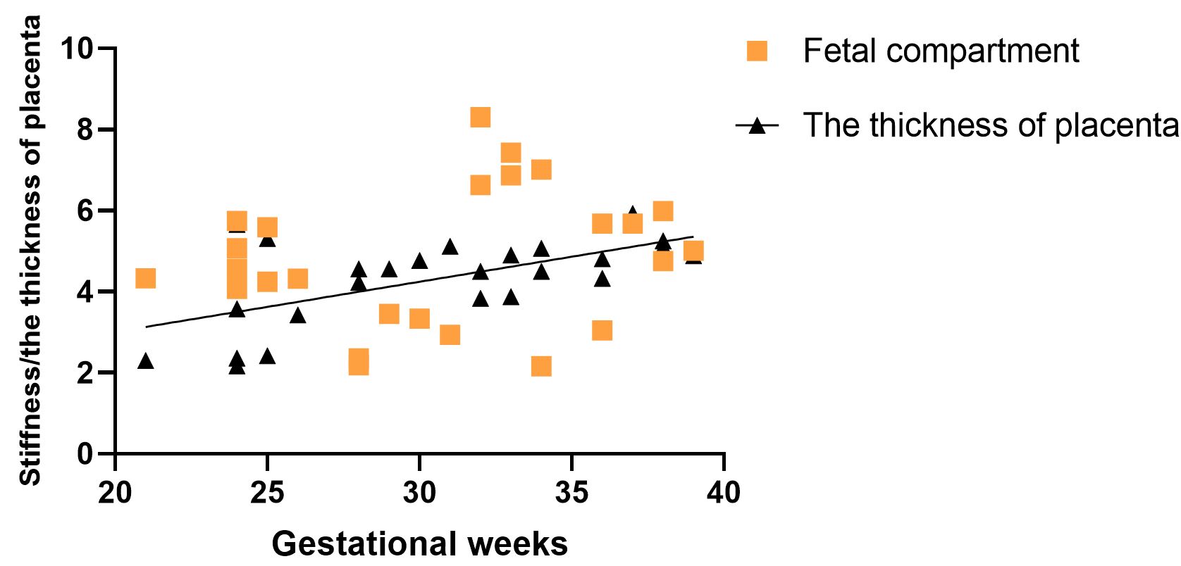
Figure: 6. The relationship between the stiffness value of the the fetus part and thickness of the placenta.
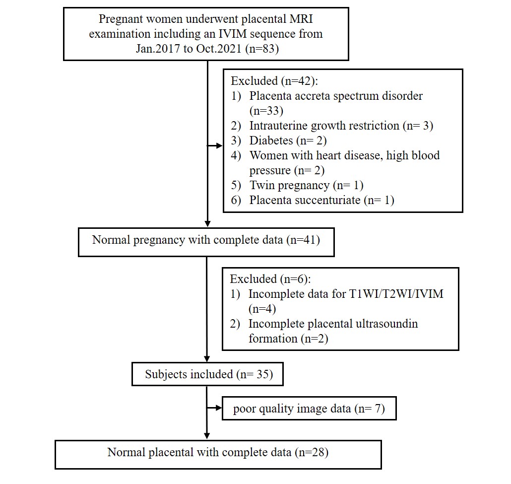
Figure: 2. Subject enrollment process
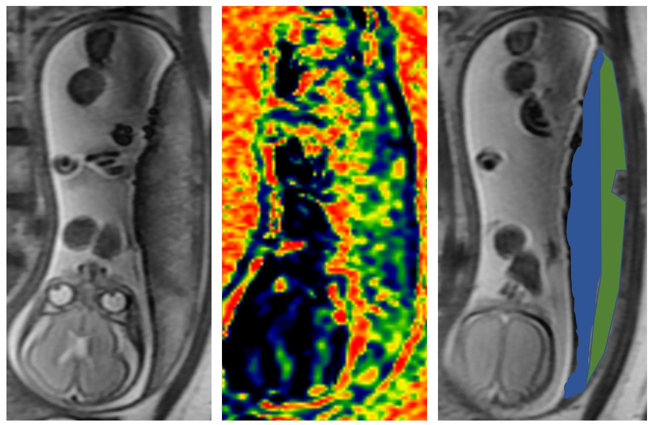
Figure: 1. ROI diagram and vMRE diagram
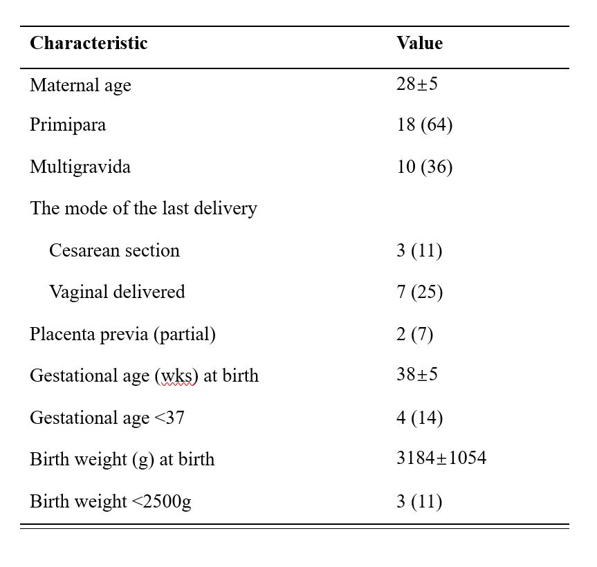
Table
1 Maternal and fetal characteristics of 28 pregnancies that underwent placental
IVIM examination.
DOI: https://doi.org/10.58530/2022/1322