1314
Quantification of regional cerebral blood flow using diffusion imaging with phase-contrast (DIP)1Division of Health Sciences, Graduate School of Medical Sciences, Kanazawa University, Kanazawa, Ishikawa, Japan, 2Department of Radiology, Kanazawa University Hospital, Kanazawa, Ishikawa, Japan, 3Department of Radiology, University of Miami, Miami, FL, United States
Synopsis
Perfusion-related diffusion coefficient (D*) in intravoxel incoherent motion analysis is closely correlated with the regional cerebral blood flow (rCBF). However, the D* is only a semiquantitative relative value of rCBF, which makes absolute rCBF quantification challenging. To solve this problem, we developed a novel method of diffusion imaging with phase contrast (DIP), in which the total CBF (tCBF) from phase-contrast (PC)-MRI was used to convert the perfusion-related parameters in the brain to absolute rCBF. Using this method, we measured rCBF in gray matter and white matter. Each value was consistent with literature values assessed using [15O]-water positron emission tomography.
INTRODUCTION
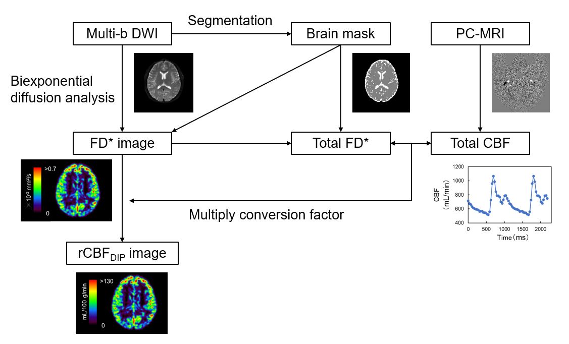
Perfusion-related diffusion coefficient (D*) in intravoxel incoherent motion analysis is closely correlated with the regional cerebral blood flow (rCBF) assessed by arterial spin labeling (ASL).1 However, the D* is only a semiquantitative relative value of rCBF, which makes absolute rCBF quantification challenging. To solve this problem, we developed a novel method of diffusion imaging with phase contrast (DIP), in which the total cerebral blood flow (tCBF) from phase-contrast (PC)-magnetic resonance imaging (PC-MRI) was used to convert perfusion-related diffusion parameters in the brain to absolute rCBF.MATERIALS AND METHODS
Eleven healthy volunteers (nine men and two women; mean age, 23.9 years) participated in this study. On a 3.0-T MRI, diffusion-weighted images of the whole brain were acquired using single-shot diffusion echo-planar imaging with multiple b-values (0, 50, 200, and 1000 s/mm2). Next, we performed voxel-wise estimations of the D*, perfusion fraction (F), multiplication of D*and F (FD*),2 and restricted diffusion coefficient using biexponential function. Moreover, PC-MRI was performed to obtain tCBF from the volumetric flow rate at the main feeding arteries into the cranium. Using tCBF obtained from PC-MRI, we converted the FD* in the brain into absolute rCBF (Fig. 1). We measured rCBF using DIP and ASL and their correlations in gray and white matter (GM and WM, respectively) in healthy volunteers and assessed the relationship between the two methods.RESULTS AND DISCUSSION
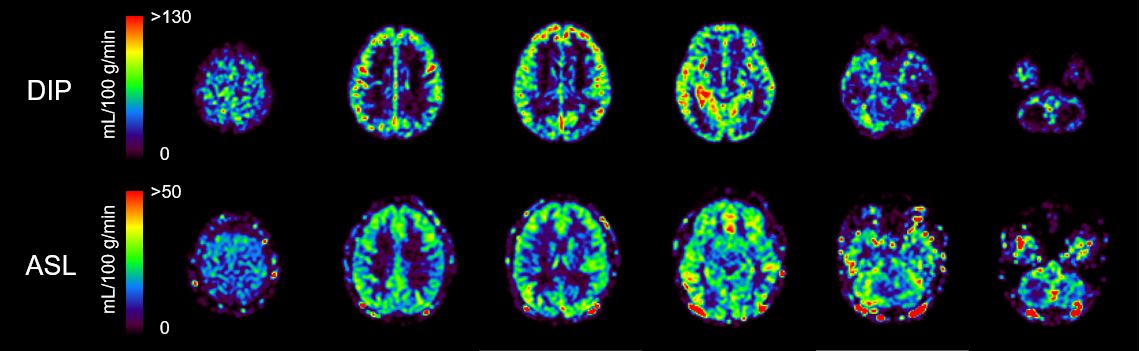
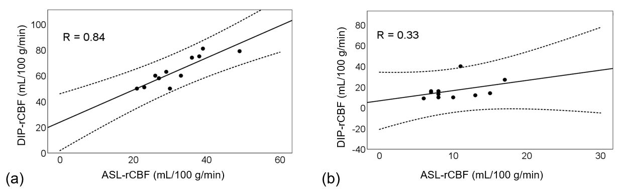
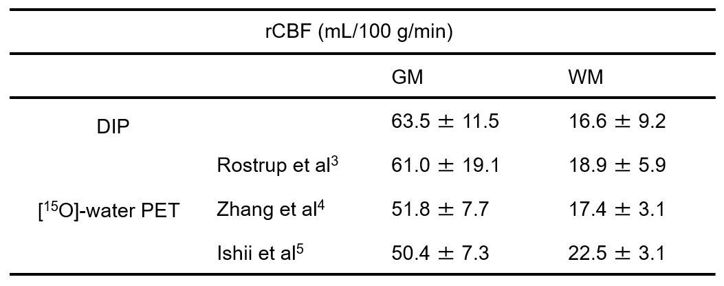
Figure 2 shows representative rCBF images obtained using DIP and ASL. A highly significant positive correlation between DIP and ASL in terms of rCBF was observed in GM (R = 0.84), whereas the correlation between the methods was weak in WM (R = 0.33) (Fig. 3). The rCBF values in GM (63.5 ± 11.5 mL/100 g/min) and WM (16.6 ± 9.2 mL/100 g/min) obtained using DIP were consistent with the values assessed using [15O]-water positron emission tomography reported in literature (Table 1).3-5 These results indicate the ability of DPC to quantitatively evaluate rCBF.CONCLUSION
DIP makes it possible to quantitatively evaluate rCBF as well as standard diffusion parameters.Acknowledgements
No acknowledgement found.References
1. Ohno N, et al., Modified triexponential analysis of intravoxel incoherent motion for brain perfusion and diffusion. J Magn Reson Imaging. 2016; 43: 818-823.
2. Le Bihan D, et al., The Capillary Network: A Link between IVIM and Classical Perfusion. Magnetic Resonance in Medicine. 1992; 27: 171-178.
3. Rostrup E, et al., The relationship between cerebral blood flow and volume in humans. Neuroimage. 2005; 24: 1-11.
4. Zhang K, et al., Comparison of cerebral blood flow acquired by simultaneous [15O]water positron emission tomography and arterial spin labeling magnetic resonance imaging. J Cereb Blood Flow Metab. 2014; 34: 1373-1380.
5. Ishii Y, et al., Simultaneous Phase-Contrast MRI and PET for Noninvasive Quantification of Cerebral Blood Flow and Reactivity in Healthy Subjects and Patient With Cerebrovascular Disease. J Magn Reason Imaging. 2020; 51: 183-194.
Figures