1308
Clinical evaluation of accelerated wideband cardiac perfusion pulse sequence in stress testing in patients with a CIED1Department of Radiology, Northwestern University Feinberg School of Medicine, Chicago, IL, United States, 2Department of Radiology, Mayo Clinic, Rochester, MN, United States, 3Department of Medicine, Division of Cardiology, Northwestern University Feinberg School of Medicine, Chicago, IL, United States, 4Department of Radiology, University of Chicago, Chicago, IL, United States
Synopsis
This study evaluates visual scores of image quality (conspicuity, artifact, noise; Likert scale 1[worst]-5[best], 3 acceptable) produced by a wideband cardiac perfusion sequence in patients with CIED and a standard sequence in matching non-device patients in the setting of vasodilated stress perfusion imaging. The median conspicuity scores were not significantly different between wideband (4) and standard (4). While the median artifact score was significantly worse for wideband (4) than standard (5), it was above the acceptable cutoff. The median noise score was significantly better for wideband than standard. Our wideband perfusion sequence produces diagnostically acceptable image quality in CIED patients.
Introduction
Over 3 million patients in the United States have a cardiac implantable electronic device (CIED); there is an increasing rate of implantation secondary to an aging population and increasing evidence of symptomatic and mortality benefits from CIEDs [1-2]. CIED patients often require diagnostic imaging to investigate the cause of new cardiac symptoms. Due to underlying and often complex heart disease CIED patients are likely to benefit from stress perfusion MRI which offers a comprehensive examination of function, myocardial ischemia, and scarring. Previously, image quality was significantly hindered by CIEDs. However, a recent study demonstrated that a wideband cardiac perfusion pulse sequence effectively suppresses image artifacts associated with a CIED at rest [3]. The purpose of this study was to evaluate the image quality of a wideband cardiac perfusion sequence in CIED patients undergoing vasodilated stress perfusion imaging in comparison to a standard cardiac perfusion pulse sequence in matching non-device patients.Methods
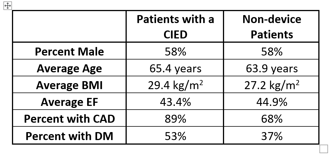
We retrospectively identified 19 consecutive patients with a CIED who had undergone cardiac stress perfusion imaging with the previously described accelerated wideband pulse sequence [3]. We identified 19 matching controls from a database of non-device patients who had underwent clinical cardiac stress perfusion MRI. We chose controls based on gender, age, BMI, LVEF, and when possible, CAD and diabetes (Figure 1). Both wideband and standard stress perfusion MRI scans were done on a 1.5 T scanner (Siemens, Avanto) using identical adenosine and gadolinium dose protocols. Two clinical raters (4-20 years of experience) independently evaluated the image quality in three categories on a 5-point Likert scale (1: worst, 3: acceptable, 5: best): conspicuity of the myocardial wall enhancement, image artifact of the heart region, and noise throughout the image. The two raters were given training datasets to calibrate their scores together prior to independent reads. Each rater provided a composite score for all slices. Inter-rater reproducibility was calculated using Cohen’s kappa coefficient [4]. For statistical analysis, the rater scores for patients with a CIED and non-device patients were compared using the Mann-Whitney’s test.Description of Results
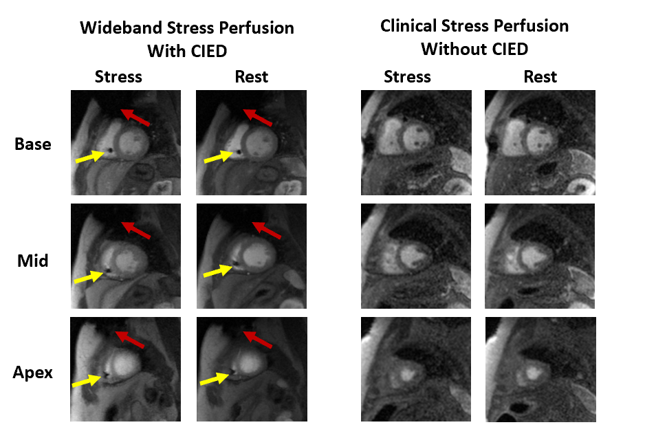
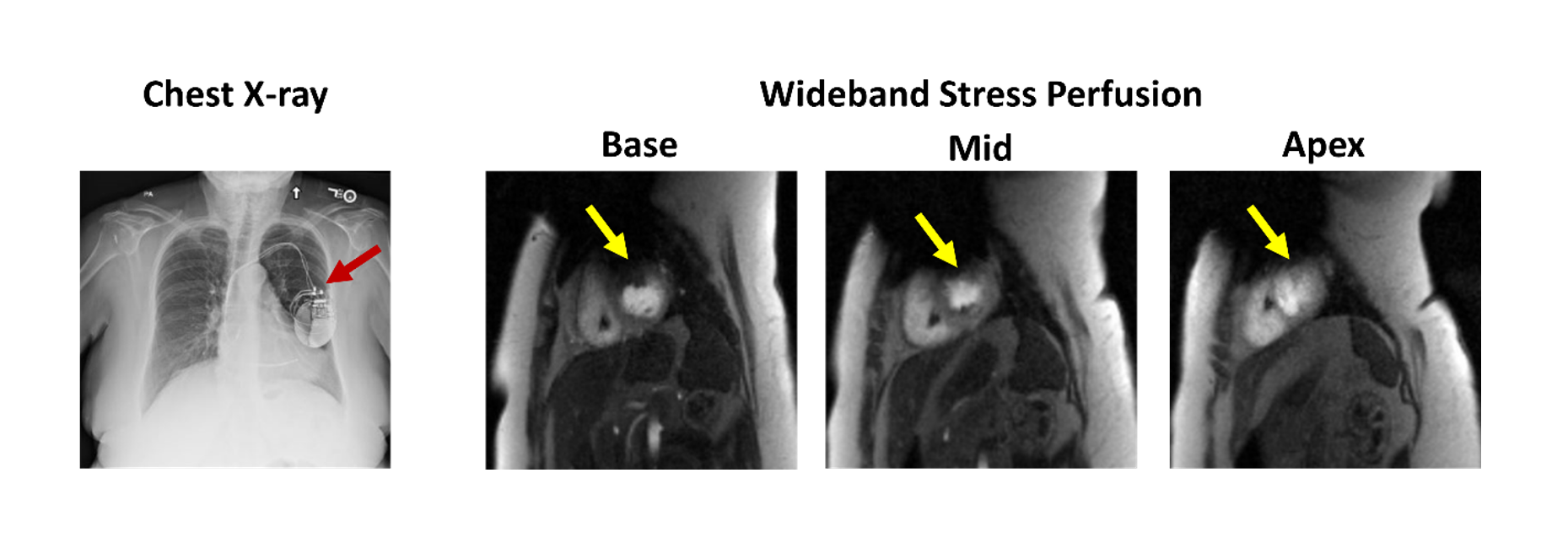
Figure 2 shows a comparison of image quality between wideband and standard pulse sequences at stress and rest. As summarized in Figure 3, the median conspicuity, artifact, and noise scores were 4 for wideband at stress and rest; median conspicuity, artifact, and noise scores ranged from 3 to 5 for standard at stress and rest. In two CIED patients, artifact scores were less than 3 (non-diagnostic) at both stress and rest. As shown in Figure 4, one CIED patient with artifact scores below 3 had large signal voids in the anterior wall due to the proximity of CIED to the heart (see chest X-ray).Using the Mann-Whitney’s test, the median conspicuity score was not significantly different between wideband and standard at stress or rest (p = 0.269 and 0.238, respectively). The median artifact score was significantly worse for wideband than standard at stress and rest (p < 0.001, p < 0.001, respectively), but both were above the acceptable cutoff. The median noise score was significantly better for wideband than standard at stress and rest (p = 0.01 and 0.018, respectively).
Using Cohan’s kappa coefficient, there was moderate inter-rater agreement (k=0.595, 95% CI, 0.507 to 0.6832).
Conclusions
This study demonstrates that an accelerated wideband cardiac perfusion sequence produces diagnostically acceptable image quality in CIED patients in the context of stress testing. It shows that image quality of an accelerated wideband cardiac perfusion sequence in patients with a CIED is similar to a standard cardiac perfusion sequence in non-device patients. In all image quality categories, the median score was 4 for wideband, supporting its clinical use in patients with a CIED. Future possible studies include quantification of myocardial blood flow, regional analysis of image quality, and additional strategies to eliminate residual image artifacts.Acknowledgements
This work was partially supported by the following grants: National Institutes of Health (R01HL116895, R01HL138578, R21EB024315, R21AG055954, R01HL151079, R21EB030806) and American Heart Association (19IPLOI34760317).References
[1] Greenspon, A.J., et al., Trends in permanent pacemaker implantation in the United States from 1993 to 2009: increasing complexity of patients and procedures. J Am Coll Cardiol, 2012. 60(16): p. 1540-5.
[2] Bradshaw PJ, Stobie P, Knuiman MW, Briffa TG, Hobbs MS. Trends in the incidence and prevalence of cardiac pacemaker insertions in an ageing population. Open Heart. 2014 Dec 10;1(1):e000177. doi: 10.1136/openhrt-2014-000177. PMID: 25512875; PMCID: PMC4265147.
[3] Hong K, Collins JD, Freed BH, Fan L, Arai AE, Hsu LY, Lee DC, Kim D. Accelerated Wideband Myocardial Perfusion Pulse Sequence with Compressed Sensing Reconstruction for Myocardial Blood Flow Quantification in Patients with a Cardiac Implantable Electronic Device. Radiol Cardiothorac Imaging. 2020 Apr 16;2(2):e190114. doi: 10.1148/ryct.2020190114. PMID: 32420548; PMCID: PMC7207204.
[4] Cohen, J., A Coefficient of Agreement for Nominal Scales. Educational and Psychological Measurement, 1960. 20(1): p. 37-46.
Figures