1304
Exploration of Velocity-Selective Inversion Arterial Spin Labeling for Breast Imaging1Fraunhofer MEVIS, Bremen, Germany, 2University Bremen, Bremen, Germany, 3mediri GmbH, Heidelberg, Germany
Synopsis
In this work, the application of velocity-selective inversion ASL in healthy breast tissue is demonstrated for the first time. The measurements show a low measured signal in breast tissue. However, the prominent fat artifacts that appear in the images will need to be addressed in future work. The technique presented here has nonetheless been shown to hold promise in the field of non-invasive breast cancer diagnostics with further development, particularly the inclusion of background suppression to get clear perfusion signal.
Introduction
Breast Cancer is the most common form of cancer among women. Breast MRI has been shown to be the best tool for detecting breast cancer [1]. Generally, a dynamic contrast-enhanced (DCE) MRI scan with injection of contrast agent is used. A completely non-invasive alternative to this is Arterial Spin Labeling (ASL), which uses the blood itself as an endogenous contrast agent. The most commonly used techniques are pulsed ASL (PASL) and pseudo-continuous ASL (pCASL) [2]. In both cases, the labeling is performed outside the imaging region. For imaging, the so-called arterial transit time (ATT) must be waited until the labeled blood reaches the spatially distant imaging plane. An alternative to this is velocity-selective ASL, which labels blood above a certain velocity (called cut-off velocity vc) within the imaging region [3] and is thus independent of the feeding arteries and ATT. Especially for organs like breast tissue, without a clear and straight feeding artery, this method is of interest.In this work, velocity-selective inversion ASL (VSI-ASL) was used for the first time in healthy breast tissue to investigate whether this method is also suitable for perfusion imaging, especially for the eventual detection of breast tumors, where higher perfusion is expected in the tumor region compared to healthy tissue.
Methods
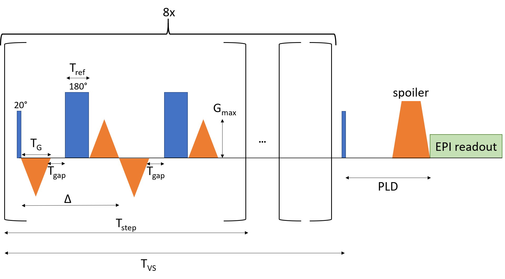
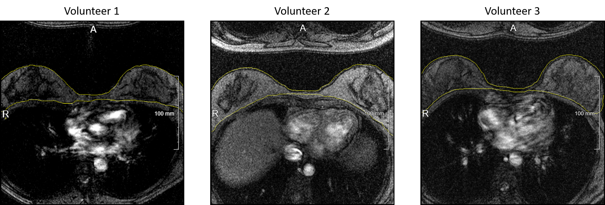
Image acquisition of breast tissue of three female volunteers (30-31y) using the VSI-ASL sequence described by Qin et al. [4] was performed on a 3T MR system (MAGNETOM Skyra, Siemens Healthineers, Erlangen, Germany) implemented in the vendor independent sequence development framework gammaSTAR [5, 6] with following parameters: TVS=44.3 ms, Tstep=5.52 ms, Tref=0.84 ms, TG=0.7 ms, Tramp=0.35 ms, Gmax=27.5 mT/m, Tgap=0.3 ms, Δ=2.5 ms, vc=2.5 cm/s with velocity-encoding in foot-head direction (sequence diagram in Figure 1).The post labeling delay (PLD) was 1000 ms. Additionally, a spoiling gradient was used after the pulse train and PLD to disrupt the remaining transverse magnetization. Only one VSASL module was used to measure just the signal above vc. One transverse slice along the isocenter was acquired with 8 mm thickness and 2.0x2.0 mm² resolution, FoV=256x256 mm², TE=25 ms, TR=2000 ms. An EPI readout [7] was used with anterior-posterior phase encoding and additional fat saturation, but without any further fat or background suppression. For averaging, 20 measurements were taken including three control-label pairs (120 ASL measurements in total). This resulted in a total scan time of 3:59 mins. For better visualization of the perfusion-weighted images, contours of the breast were created with MeVisLab (MeVis Medical Solutions, Bremen, Germany) using the localizer image of every volunteer (Figure 2).Results
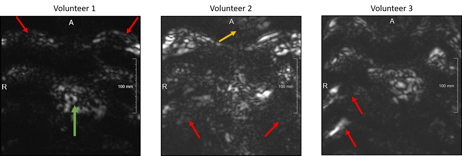
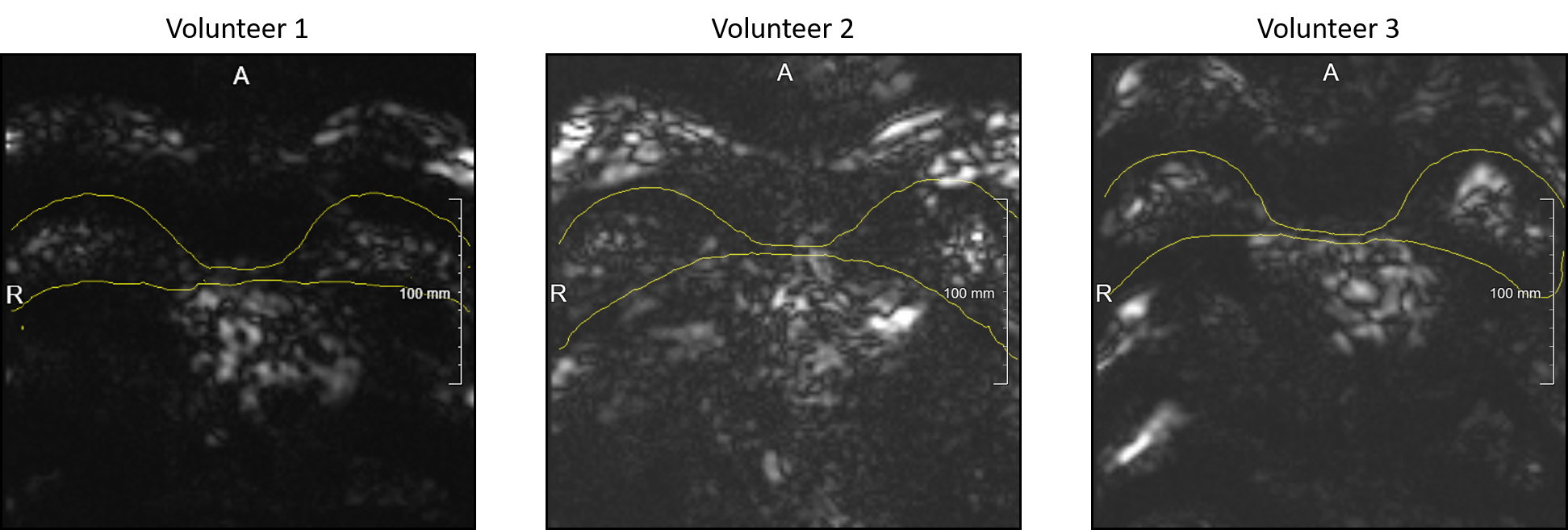
Figure 3 shows the subtracted images of the averaged control and label images for every volunteer. It can be seen that the images are mainly influenced by fat artifacts, resulting in a superposition of the measured signal in the breast in coronal direction. This is indicated illustratively by red arrows. Additionally, the blood signal inside the heart is visible (green arrow). Furthermore, motion artifacts of the blood inside the heart are visible (yellow arrow). In Figure 4, the created contours of the breast are overlaid using the world coordinates in the perfusion-weighted images to get a better focus on the organ of interest. Here, a low signal can be seen within the breast tissue in all cases.Discussion and Conclusion
This work shows the first application of velocity-selective inversion ASL in the breast. The perfusion-weighted images show a low signal in the breast, which could be interpreted as perfusion. However, the strong fat artifacts make a clear assignment difficult. Therefore, for the next step, it is important to include background suppression to reduce the strong influence of fat signal in the images to acquire the pure perfusion signal. In addition, a smaller FoV can be selected to drop the cardiac signal with phase encoding in right-left direction to minimize fold ins.Despite the visible artifacts in these initial proof-of-principle measurements, this work demonstrates that VSI-ASL imaging could be a viable technique for perfusion measurements in the breast. Hence, VSI-ASL imaging could be an important step in bringing non-invasive breast tumor detection to the clinic, with all the advantages that contrast agent free measurement brings to the examination workflow and patients.
Acknowledgements
No acknowledgement found.References
[1] Mann et al. Contrast-enhanced MRI for breast cancer screening. J Magn Reson Imaging 2019; 50: 377-390
[2] Alsop et al. Recommended Implementation of Arterial Spin Labeled Perfusion MRI for Clinical Applications: A consensus of the ISMRM Perfusion Study Group and the European Consortium for ASL in Dementia, Magn Reson Med. 2015; 73(1): 102–116.
[3] Wong et al. Velocity selective arterial spin labeling. Magn Reson Med 2006; 55(6): 1334-1341.
[4] Qin et al. Velocity-Selective-Inversion Prepared Arterial Spin Labeling, Magn Reson Med. 2016; 76(4): 1136–1148.
[5] Cordes et al. Portable and platform-independent MR pulse sequence programs, Magn Reson Med. 2020; 83(4): 1277-1290
[6] https://gamma-star.mevis.fraunhofer.de/
[7] Mansfield. Multi-planar image formation using NMR spin echoes, J Phys C 1977; 10(3): 55-58
Figures