1291
MRI tractography-guided PET image reconstruction regularisation using connectome-based nonlocal means filtering
Zhuopin Sun1, Georgios Angelis1,2, Steve Meikle3,4, and Fernando Calamante1,4,5
1School of Biomedical Engineering, The University of Sydney, Sydney, Australia, 2Australian National Imaging Facility, The University of Sydney, Sydney, Australia, 3Faculty of Medicine and Health, The University of Sydney, Sydney, Australia, 4Brain and Mind Centre, The University of Sydney, Sydney, Australia, 5Sydney Imaging, The University of Sydney, Sydney, Australia
1School of Biomedical Engineering, The University of Sydney, Sydney, Australia, 2Australian National Imaging Facility, The University of Sydney, Sydney, Australia, 3Faculty of Medicine and Health, The University of Sydney, Sydney, Australia, 4Brain and Mind Centre, The University of Sydney, Sydney, Australia, 5Sydney Imaging, The University of Sydney, Sydney, Australia
Synopsis
Diffusion MRI (dMRI) can provide a wealth of information about brain microstructure and connectivity, but its use in PET-MRI image reconstruction has not been exploited. We incorporated dMRI-derived prior information as a regulariser into the iterative PET One-Step Late Maximum A Posteriori (OSLMAP) reconstruction algorithm. The proposed regularisation method has unique advantages of providing more informative and targeted denoising and regularisation based on the complementary structural connectivity information from dMRI.
Introduction
Combining diffusion MRI (dMRI) and PET data has rarely been exploited in image reconstruction, despite the fact that they provide highly complementary information.1,2 CONNectome-based Non-Local Means (CONN-NLM) filtering3 was recently proposed as a post-reconstruction denoising method to exploit synergies between dMRI-derived structural connectivity (i.e. structural connectome) and the molecular information of reconstructed PET images. This method was shown to effectively suppress noise while preserving or improving lesion contrast. In the present study, we incorporated the CONN-NLM as a regulariser into the iterative One-Step Late Maximum A Posteriori (OSLMAP) reconstruction algorithm. This method, which we call CONN-NLM-OSLMAP, retains the advantages of CONN-NLM and, in addition, it allows for more accurate statistical noise modelling as part of the PET reconstruction.4 Here, we describe the new method and evaluate it using a realistic phantom data framework.Methods
We implemented the proposed regularisation method using the IF-OSEM-OSL algorithm in STIR,5 which allows inter-iteration filtering using a generalised one-step-late (OSL) approach6 within the Ordered Subsets Expectation Maximisation (OSEM) algorithm. The CONN-NLM filter was integrated within the reconstruction framework and applied to the updated image after each full tomographic iteration (Fig. 1A). During the last iteration, the CONN-NLM filter was applied as a post-processing step to obtain the final reconstructed PET image.The CONN-NLM filter combines a nonlocal means (NLM) similarity weighting with a hybrid (local+distal) connectivity measure to form a connectome-based weighting (Fig. 1B):3
$$CONN\_NLM(x_i)=\sum_{j{\in}N(i)}w'_{ij}\times{x_j}=\sum_{j{\in}N(i)}\frac{1}{Z'(i)}\times{exp}(-\frac{\parallel\overrightarrow{x_i}-\overrightarrow{x_j}\parallel^2_{2,a}}{h^2})\times{A_{ij}}$$
where xi is the target voxel PET intensity value being smoothed, and xj denotes all the voxel intensities in a large search window. The similarity weight between voxel intensities xi and xj, demoted as w'i,j, is calculated according to the Gaussian-weighted Euclidian distance between patches around xi and xj (denoted as $$$\overrightarrow{x_i}$$$ and $$$\overrightarrow{x_j}$$$ ). Ai,j measures the connectivity strength between xi and xj, which is derived from tractography-based structural connectivity. Z'(i) is a normalisation factor, and h2 controls the filtering strength.3
We evaluated the proposed CONN-NLM-OSLMAP method on a digital phantom created with a realistic PET/dMRI simulation framework.3 Figure 2 shows representative slices of the simulated phantom. To test CONN-NLM as a regularisation step during PET image reconstruction, we simulated in ASIM7 a low-count (107 total counts, 20% noise) noisy PET dataset with one isolated lesion and two lesions connected through white matter fibres.
Results
Both the post-reconstruction CONN-NLM and the proposed CONN-NLM-OSLMAP reconstruction method effectively improved lesion contrast and suppressed overall image noise (Fig. 3). CONN-NLM-OSLMAP demonstrated further improvements in reducing noise in normal grey matter and within lesions. Quantitative evaluations (Fig. 4) showed that the CONN-NLM method is both effective as a post-reconstruction filter and as a regulariser during image reconstruction to reduce noise while maintaining lesion contrast. All three lesions showed improved contrast-to-noise ratios, suggesting that this technique is reliable on both connected and isolated lesions.Discussion
The CONN-NLM filter has previously been shown to outperform Gaussian, nonlocal means, and total variation filters.3 In the present study, we used CONN-NLM as a baseline against which to compare CONN-NLM-OSLMAP to evaluate the potential performance gains that can be achieved by using it as a regulariser during image reconstruction. Brain lesions do not only result in local anatomical changes, but they can also affect structural connections of the brain at a network level. Connectivity based information can be used as a prior to inform PET reconstruction to improve the overall image quality and enhance lesion contrasts. The current implementation provides flexibility in incorporating additional prior information. As with the CONN-NLM filtering method, the accuracy of this method depends on the accuracy of structural connectivity information. Further work is needed to increase the speed of CONN-NLM-OSLMAP to allow comparable computing resources as conventional reconstruction methods.Conclusion
The proposed method is a first attempt to use dMRI-derived prior information in PET image reconstruction. CONN-NLM-OSLMAP has the unique advantages of providing more informative and targeted denoising and regularisation based on the complementary structural connectivity information from dMRI. Various types of connectome-based information can potentially be incorporated into this method to further enhance data synergies between MRI and PET.Acknowledgements
We acknowledge the technical assistance provided by the Sydney Informatics Hub and Sydney Imaging, two Core Research Facilities of the University of Sydney, and the scientific and technical assistance of the National Imaging Facility, a National Collaborative Research Infrastructure Strategy (NCRIS) capability, at the University of SydneyReferences
- Jacobs, H. I. L., Hedden, T., Schultz, A. P., Sepulcre, J., Perea, R. D., Amariglio, R. E., Papp, K. V, Rentz, D. M., Sperling, R. A., & Johnson, K. A. (2018). Structural tract alterations predict downstream tau accumulation in amyloid-positive older individuals. Nature Neuroscience, 21(3), 424–431.
- Wen, Q., Risacher, S. L., Xie, L., Li, J., Harezlak, J., Farlow, M. R., Unverzagt, F. W., Gao, S., Apostolova, L. G., Saykin, A. J., & Wu, Y. C. (2021). Tau-related white-matter alterations along spatially selective pathways. NeuroImage, 226, 117560.
- Sun, Z., Meikle, S., & Calamante, F. (2021). CONN-NLM: a novel CONNectome-based Non-Local Means Filter for PET-MRI denoising. ISMRM 2021. p. 2193.
- Reader, A. J., & Zaidi, H. (2007). Advances in PET Image Reconstruction. PET Clinics, 2(2), 173–190.
- Thielemans, K., Tsoumpas, C., Mustafovic, S., Beisel, T., Aguiar, P., Dikaios, N., & Jacobson, M. W. (2012). STIR: software for tomographic image reconstruction release 2. Physics in Medicine and Biology, 57(4), 867–883.
- Green, P. J. (1990). Tomography Data Using a Modified EM Algorithm. IEEE Transactions on Medical Imaging, 9(893), 84–93.
- ASIM: Analytic PET simulator Home Page (Online). University of Washington.,From: http://depts.washington.edu/asimuw/, (2011).
- Calamante, F. (2019). The seven deadly sins of measuring brain structural connectivity using diffusion MRI streamlines fibre-tracking. Diagnostics, 9(3).
Figures
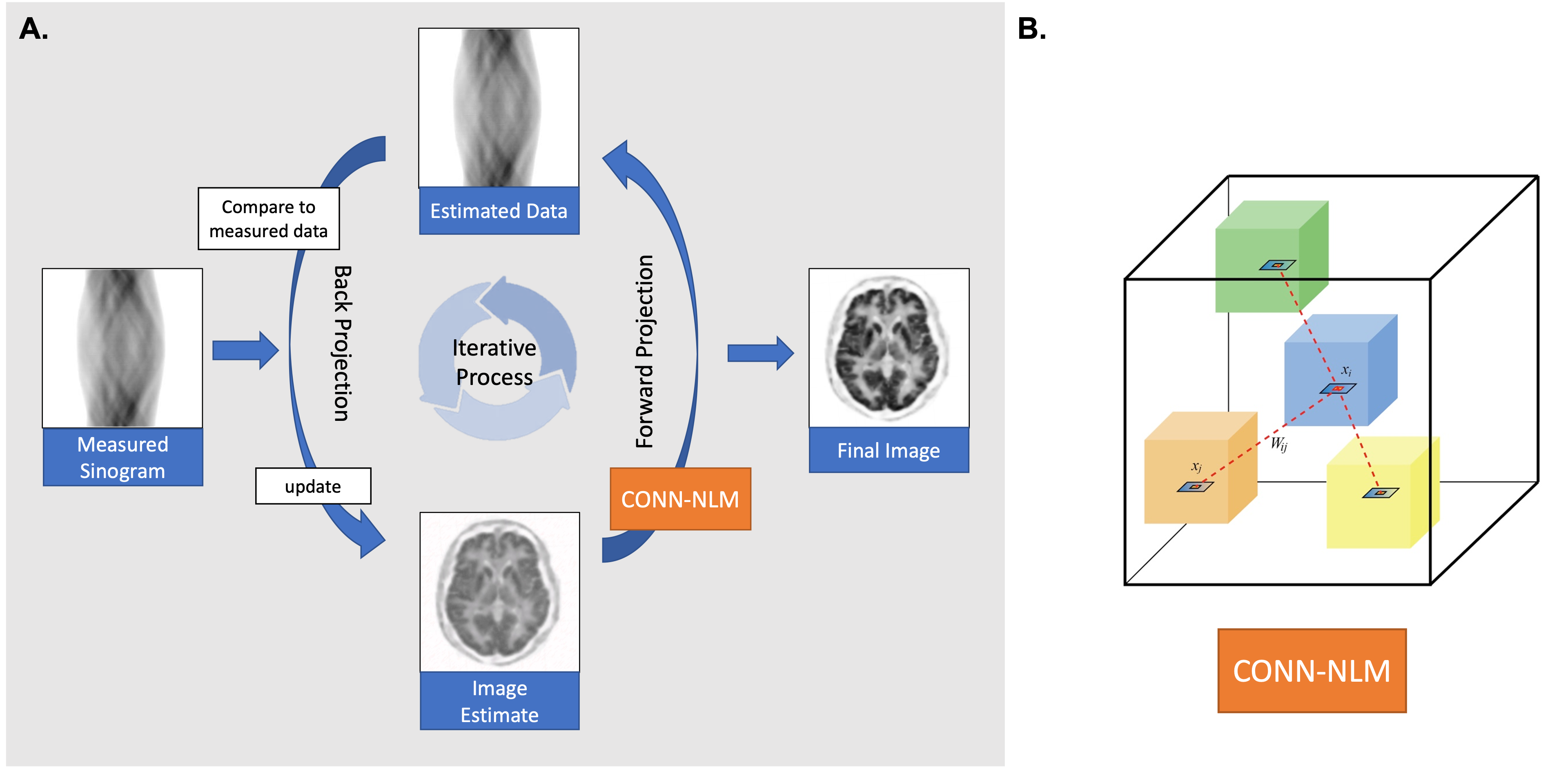
The iterative PET reconstruction framework with CONN-NLM regularisation. A) Schematic overview of the iterative process, which involves back projecting measured data, applying CONN-NLM regularisation, forward projecting the estimated image, and comparing with the measured sinogram data. B) Schematic illustration of the connectome-based NLM filter applied to voxel xi (inside the blue cube), which incorporates connectivity (both local and distant) and intensity similarity information. Each coloured cube schematically represents a node of the brain parcellation.
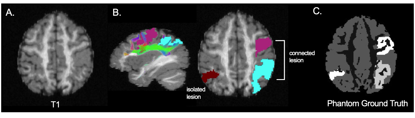
Simulated phantom to generate PET data. A) Tissue segmentation was performed on T1. B) Three lesions were simulated including one isolated lesion and two lesions connected through white matter pathways. T1-w and diffusion weighted images were acquired from the ISMRM 2015 tractography challenge. C) the resulted ground truth phantom image, which combines segmented tissue and lesions, for generating raw PET data.
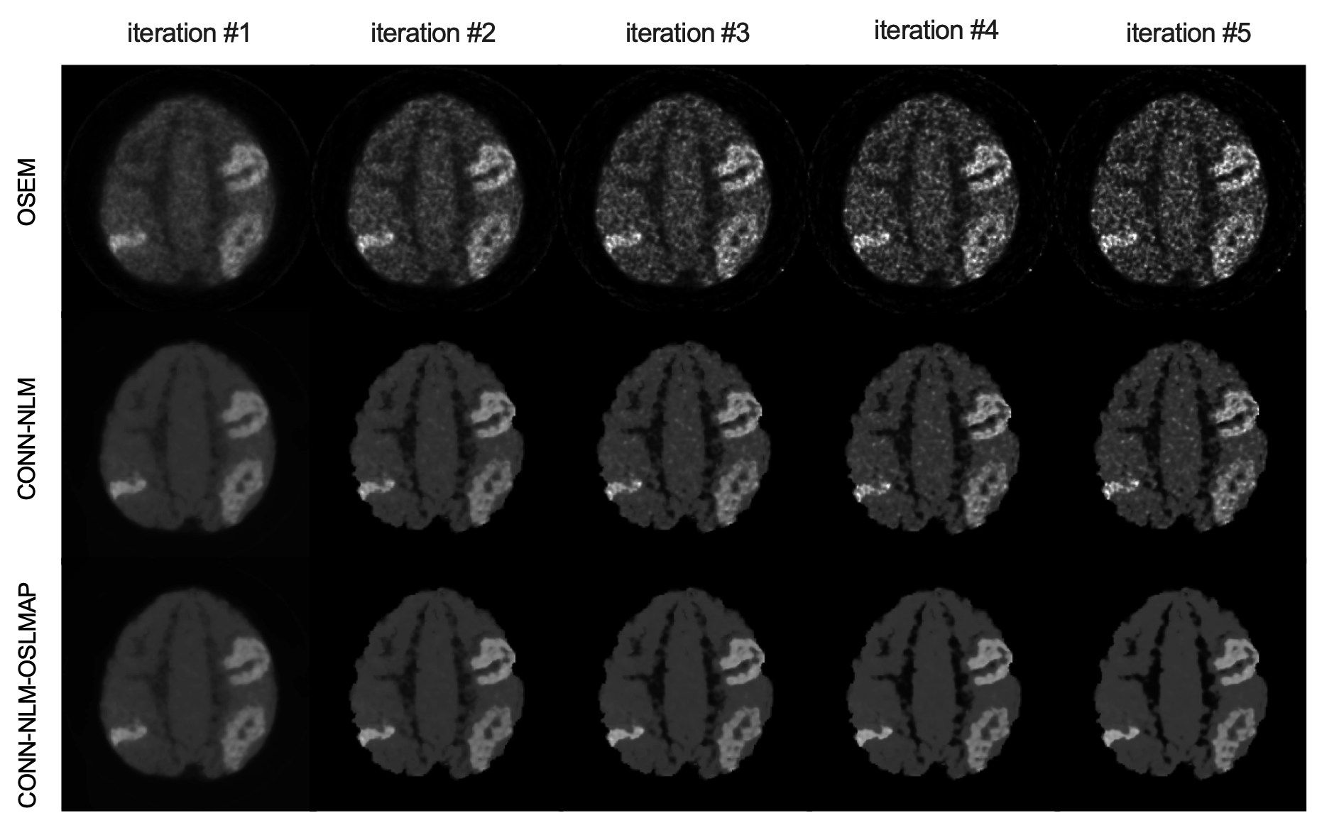
Comparison of the effects of CONN-NLM as a post-reconstruction filter and as a reconstruction regulariser (i.e. CONN-NLM-OSLMAP). The filter is applied to the updated image after each full iteration. Top: OSEM reconstruction without any filtering (‘baseline’ method). Middle: CONN-NLM post-reconstruction filter. Bottom: CONN-NLM-OSLMAP regularised reconstruction.
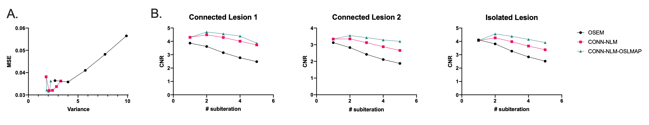
Quantitative evaluations of A) the overall image mean-squared error (MSE) vs. noise variance. B) lesion contrast-to-noise ratios (CNR) plotted for each iteration.
DOI: https://doi.org/10.58530/2022/1291