1225
Shear-rate values in the carotid bifurcation in subjects with carotid webs, subjects with atherosclerotic lesions, and healthy subjects.1Department of Biomedical Engineering, Georgia Institute of Technology & Emory University, Atlanta, GA, United States, 2Department of Radiology & Imaging Sciences, Emory University, Atlanta, GA, United States, 3Department of Neurology, Emory University, Atlanta, GA, United States
Synopsis
Carotid webs (CaWs) have been linked to cryptogenic strokes and therefore quantifying different hemodynamic parameters is essential to understand the mechanism of clot formation in patients. Computational fluid dynamics (CFD) based on patient-specific geometry and PCMR inlet flow conditions was conducted on five subjects with CaWs, five subjects with mild atherosclerosis (with a similar degree of luminal narrowing), and three healthy control subjects. The total area within the carotid bifurcation that contained low shear rate values associated with clot formation is significantly larger in CaW subjects compared to subjects with atherosclerotic lesions or healthy subjects.
Introduction
Carotid webs (CaWs) have previously been associated with stroke and they may account for up to one-third of cryptogenic strokes in younger patients between the ages 30-48 without vascular risk factors1,2. CaWs are intraluminal shelf-like projections in the internal carotid artery bulb that cause focal luminal narrowing of 20-50%. Unlike carotid atherosclerosis, CaWs are not characterized by inflammation, wall thickening, or plaque deposition. The shelf/pyramidal shape of the CaW may result in significant hemodynamic disturbance resulting in flow separation, recirculation, stagnation, and ultimately thrombus formation. Understanding the hemodynamic alterations caused by CaWs is essential for determining the associated stroke risk. The goal of this study is to understand the effect of CaW geometry on local hemodynamic parameters, which may lead to the formation of clots. Shear rate is considered an important hemodynamic parameter in clot formation, and low shear values have been linked to activation of the coagulation cascade3,4. We compared the area of low shear rate in the carotid bifurcation in subjects with CaW, subjects with mild atherosclerosis, and healthy subjects using computational fluid mechanics (CFD) simulations based on 3D time-of-flight (TOF) MRA and 2D PCMR. We hypothesized that CaWs will be associated with a larger area of low shear rate than mild atherosclerosis or healthy carotid bifurcations.Methods
This was an IRB-approved study. The study population included patients with CaWs (n=5, 4 female, age 48.3±10.8) with history of stroke or TIA and focal luminal narrowing near the web (32.8%±13.0), healthy volunteers (n=3, 2 female age 50±1.4), and subjects with mild atherosclerosis (n=5, 2 female, age 71±14.60, luminal stenosis=41%±13%). All CaW subjects have computed tomography angiography of the carotids available in addition to MRA. All subjects were imaged on a 3T system (MAGNETOM Prismafit, Siemens Medical Solutions). Multi-slab, transverse, 3D time of flight (TOF) images were acquired to cover the bifurcation (0.5mm3, TR=23ms, TE=3.1ms). 2D, ECG-gated, cine phase-contrast (PCMR) images were acquired 10mm below and 10mm above the bifurcation (1x1x5mm3, VENC=80cm/sec, TR=43.6, TE=7). Mimics and 3-Matics (Materialise, 2019) were used to segment TOF images for healthy subjects and subjects with atherosclerosis, and either CTA or TOF MRA images for patients with CaW, to create the smoothed 3D geometry. Fluent (ANSYS, 2021 R1) was then used to mesh the geometry and conduct transient flow simulations based on 2D PCMR at the inlet and constant pressure at the outlet with the assumption of rigid walls and no-slip conditions. Outlet 2D PCMR data was used for validation of our CFD method (Figure 1). Based on the literature review, when the shear rate is below a value of 10 s-1, it has been linked to coagulation and clot formation3,4. Therefore, the fractional area of low shear rate was quantified using the surface area below the threshold of 10 s-1 divided by the surface area of the entire carotid bifurcation (starting from 10mm below the bifurcation to 10mm above the bifurcation). An unpaired t-test was performed for statistical analysis comparing the areas of low shear.Results
For the 5 CaWs, the fractional area of low shear rate was 0.33±0.24, and for the 5 atherosclerotic lesions the fractional area of low shear rate was 0.05±0.05 (Figure 2). The difference was statically significant (p=0.05). In the healthy carotid bulb, there was no area of low shear rate. The results are also presented visually in (Figure 3) in an example of each of the cases. The results show that CaWs with similar degree of luminal narrowing to atherosclerosis are associated with a larger area of low shear rate and with more complex flow patterns.Conclusion
CaWs showed a statistically significant larger area of area of low shear rate compared to atherosclerotic lesions and healthy carotid. The results indicate that CaW caused a greater degree of flow disturbance, recirculation and stagnation in comparison to atherosclerosis with similar degree of stenosis.Acknowledgements
This work was funded by NIH R21 (Grant # R21NS114603) and NSF GRFP.References
1. Haussen DC, Grossberg JA, Bouslama M, et al. Carotid Web (Intimal Fibromuscular Dysplasia) Has High Stroke Recurrence Risk and Is Amenable to Stenting. Stroke. 2017;48(11):3134-3137.
2. Park CC, El Sayed R, Haussen DC, Risk BB, Oshinski JN, Allen JW. Assessment of Time-Density Curve based Hemodynamic Parameters in Patients with Carotid Webs versus Atherosclerotic Carotid Artery Stenosis. The 59th Annual Meeting of the American Society of Neuroradiology; 2021; San Francisco.
3. Casa LD, Deaton DH, Ku DN. Role of high shear rate in thrombosis. J Vasc Surg. 2015;61(4):1068-1080.
4. Davie EW, Fujikawa K, Kisiel W. The coagulation cascade: initiation, maintenance, and regulation. Biochemistry. 1991;30(43):10363-10370.
Figures
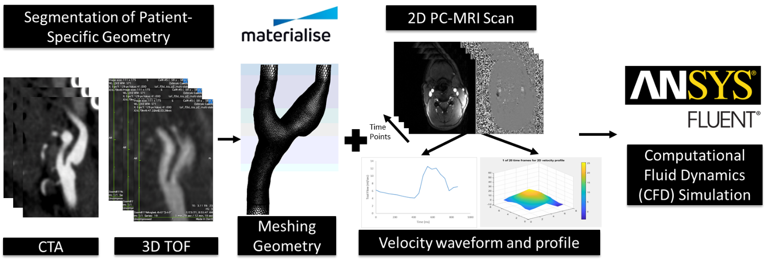
Figure 1. Overview of methods. Patient-specific MRA geometry segmentation and smoothing using Materialize Mimics and 3-Matic. Fluent was used for the meshing and simulation of flow (ANSYS, 2021 R1). Velocity data from Patient-specific PCMR data.
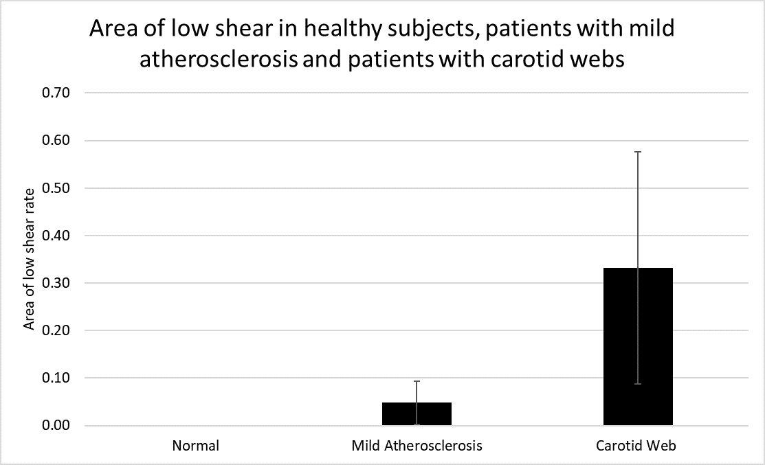
Figure 2. Area of low shear rate based on CFD in normal patients, in patients with mild atherosclerosis and patients with CaW
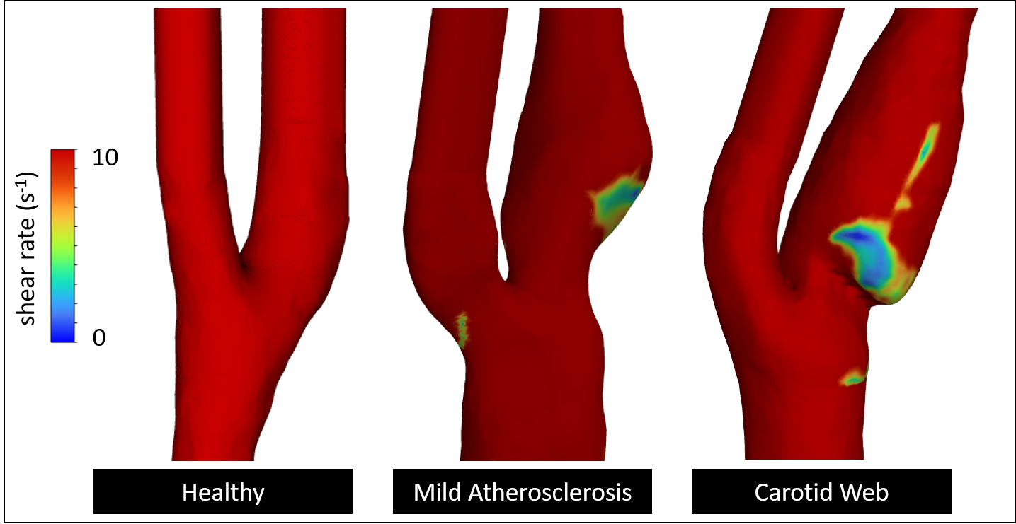
Figure 3. Area of low shear rate (<10 sec-1) are shown in three example geometries: healthy, carotid web and mild atherosclerosis.