1223
Comparing Single and Multiple Delays methods for ASL Perfusion Quantification on Pediatric Neuroimaging1Department of Radiology, Stanford University, Stanford, CA, United States, 2Department of Neuroradiology, Hospital Beatriz Ângelo, Lisbon, Portugal
Synopsis
This study compared the CBF measurements using single and multiple delay ASL on 28 children without neurological or cerebrovascular disorders. Results showed that CBF of multiple delay ASL was significantly higher than the values of single delay ASL by a mean of 18%. CBF of both ASL techniques increased with age.
Introduction
Cerebral perfusion (or cerebral blood flow, CBF) is an important hemodynamic parameter for studying cerebrovascular diseases and brain development. Pseudo-continuous arterial spin labeling with a single post labeling delay (single-PLD PCASL) has been recommended as the clinical application for CBF mapping and widely applied in adults 1. However, the application of ASL on pediatric neuroimaging has been limited because the accuracy of measurement can deteriorate when the expected CBF of children is significantly lower and higher than the normal range in adults 2. Multi-PLD PCASL can account for the variations in arterial transit time (ATT) and has shown superiority over single-PLD PCASL in measuring the CBF of adults with cerebrovascular and neurological diseases 3,4. However, the application of multi-PLD PCASL on pediatric population has been limited. In this study, we compare the CBF measured by single- and multi-PLD PCASL and examine the impact of age on CBF.Methods
Single- and multi-delay PCASL data were acquired from 28 children (average 8.2 years, 12 boys) without cerebrovascular or neurological diseases using a 3T GE Signa Premier MRI. The main scanning parameters included: single-delay PCASL PLD = 1.525s; labeling duration = 1.45s; scanning duration = 4:56min; multi-delay PCASL PLD = 0.7, 2.0, 3.3s; labeling duration = 1.3s (Hadamard encoded); scanning duration = 4:23min. The other parameters were consistent with our previous work 4. A T2-weighted structural image was collected from each child. CBF and ATT were computed using by fitting the ASL difference data to the general kinetic model 5. The mean CBF and ATT of the whole brain was computed for each subject. The relationship between age and CBF was examined by fitting an exponential recovery curve to the whole brain mean CBF data.Single- and multi-delay PCASL data were acquired from 28 children (average 8.2 years, 12 boys) without cerebrovascular or neurological diseases using a 3T GE Signa Premier MRI. The main scanning parameters included: single-delay PCASL PLD = 1.525s; labeling duration = 1.45s; scanning duration = 4:56min; multi-delay PCASL PLD = 0.7, 2.0, 3.3s; labeling duration = 1.3s (Hadamard encoded); scanning duration = 4:23min. The other parameters were consistent with our previous work 4. A T2-weighted structural image was collected from each child. CBF and ATT were computed using by fitting the ASL difference data to the general kinetic model 5. The mean CBF and ATT of the whole brain was computed for each subject. The relationship between age and CBF was examined by fitting an exponential recovery curve to the whole brain mean CBF data.Results
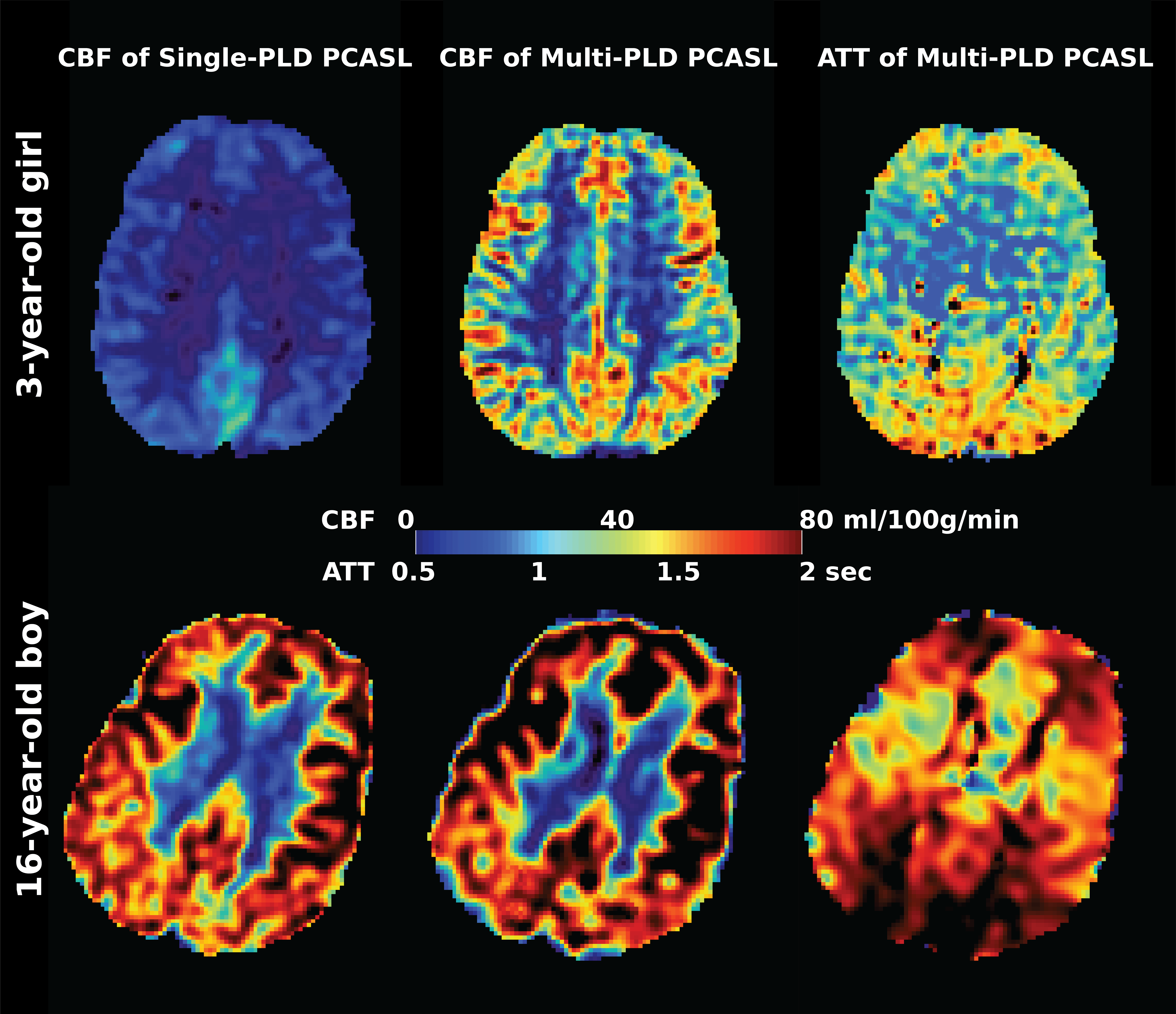
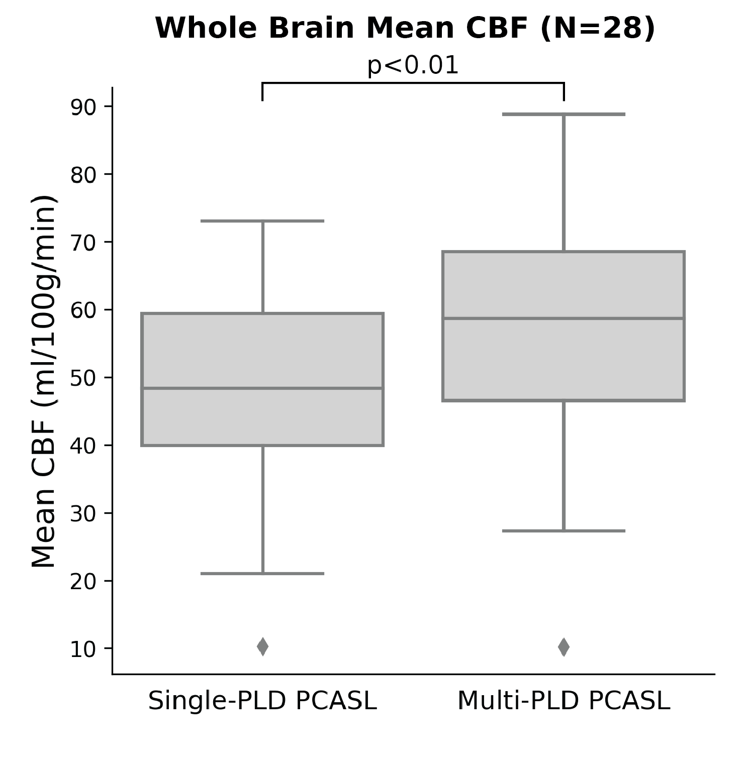
Figure 1 shows the CBF and ATT maps of a younger and older subject. Figures 2 and 3 show the quantitative comparison between CBF measured by single- and multi-PLD PCASL. Figure 4 shows the relationship between age and CBF. Overall, CBF measured by multi-PLD PCASL appeared to be higher than the results obtained by single-PLD PCASL in all brain regions. For this cohort, there is a systematic difference of 18% between single- and multi-PLD PCASL CBF. CBF measured by both techniques increased with age, implying a significant impact on CBF due to brain development.Discussion
In this work, we demonstrated the effectiveness of applying multi-delay ASL onto measuring CBF of children. The primary finding was that CBF measured by multi-PLD PCASL was consistently higher than the results of single-PLD PCASL. A possible explanation could be the impact due to ATT that single-PLD PCASL was unable to account for. Additionally, we demonstrated that the whole brain CBF increased with age, which was consistent with the data measured by single-PLD PCASL with similar scanning parameters and the work using phase contrast MRI 2,6. A cross-validation of these techniques may be desired in future studies to examine the normal range of CBF across age.Conclusions
Multi-delay ASL was effective in measuring CBF and ATT of children using the same scanning time in single-delay ASL. CBF measured by Multi-delay ASL was consistently higher than the results of single-delay ASL and CBF increased with age.Acknowledgements
This work is supported by the American Heart Association (Grant: 826254) and National Institutes of Health (Grant: R01EB025220-02).References
1 Alsop DC, Detre JA, Golay X, Günther M, Hendrikse J, Hernandez-Garcia L et al. Recommended implementation of arterial spin-labeled perfusion MRI for clinical applications: A consensus of the ISMRM perfusion study group and the European consortium for ASL in dementia. Magnetic resonance in medicine 2015; 73: 102–116.
2 Paniukov D, Lebel RM, Giesbrecht G, Lebel C. Cerebral blood flow increases across early childhood. NeuroImage 2020; 204: 116224.
3 Fan AP, Guo J, Khalighi MM, Gulaka PK, Shen B, Park JH et al. Long-Delay Arterial Spin Labeling Provides More Accurate Cerebral Blood Flow Measurements in Moyamoya Patients: A Simultaneous Positron Emission Tomography/MRI Study. Stroke 2017. doi:10.1161/STROKEAHA.117.017773.
4 Zhao MY, Fan AP, Chen DY-T, Sokolska MJ, Guo J, Ishii Y et al. Cerebrovascular reactivity measurements using simultaneous 15O-water PET and ASL MRI: Impacts of arterial transit time, labeling efficiency, and hematocrit. NeuroImage 2021; 233: 117955.
5 Buxton RB, Frank LR, Wong EC, Siewert B, Warach S, Edelman RR. A general kinetic model for quantitative perfusion imaging with arterial spin labeling. Magnetic resonance in medicine : official journal of the Society of Magnetic Resonance in Medicine / Society of Magnetic Resonance in Medicine 1998; 40: 383–396.
6 Liu P, Qi Y, Lin Z, Guo Q, Wang X, Lu H. Assessment of cerebral blood flow in neonates and infants: A phase-contrast MRI study. Neuroimage 2019; 185: 926–933.
Figures