1221
The Value of 3D arterial spin labeling in early diagnosis and prognostic grouping of Full-Term neonatal hypoxic-ischemic encephalopathy
Chang Liu1 and Zhang Yong2
1Department of Radiology, Southern District, The First Affiliated Hospital of USTC, Division of Life, HEFEI, China, 2MR Research,GE Healthcare,Shanghai,China, Shanghai, China
1Department of Radiology, Southern District, The First Affiliated Hospital of USTC, Division of Life, HEFEI, China, 2MR Research,GE Healthcare,Shanghai,China, Shanghai, China
Synopsis
3D-ASL technology can reflect cerebral blood flow perfusion status,which is helpful for early diagnosis of HIE.The predictive value of ASL for the prognosis of full-term HIE patients is higher than that of DWI.
INTRODUCTION
To explore the early diagnosis value of three-dimensional arterial spin labeling(3D-ASL) perfusion imaging in full-term neonates with hypoxic ischemic encephalopathy(HIE) and to compare the predictive value of 3D-ASL with diffusion-weighted imaging(DWI).METHODS
A total of 40 full term newborns who were clinically diagnosed as HIE in our hospital from january 2018 to october 2019 were enrolled .All HIE neonates were divided into favourable outcome group and adverse outcome group according to their ourcome.Both groups of subjects underwent 3D-ASL and DWI.The relative cerebral blood flow (rCBF) and apparent diffusion coefficient(ADC) in each region of in terest were compared .The consistency of rCBF /ADC value and predictive value were analyzed.Results
The rCBF value and the ADC value of the basal ganglia 、thalamic and gray matter in adverse outcome group was higher than that in avourable outcome group,with statistically significant difference (P<0.05).There was no statistically significant difference in the rCBF value and the ADC value of the white matter in adverse outcome group and the avourable outcome group (P>0.05)1.The area under the concentration-time curve of rCBV value was 0.972.The specificity and the sensitivity of rCBV value were 75.36% and 100%.The area under the concentration-time curve of ADC value was 0.842.The mean rCBF value and the ADC value were correlated by pearson correlation analysis(r=-0.61,P<0.05).DISCUSSION
The CBF value and NBNA score of the basal ganglia and thalamus were significantly different between poor prognosis and good prognosis 2(P<0.01). The cerebral perfusion of the poor prognosis group was higher than that of the good prognosis group, indicating that the CBF value can be evaluated and Reperfusion phenomenon related to delayed cell death, the evaluation effect is better than ADC value.Conclusion
3D-ASL technology can reflect cerebral blood flow perfusion status,which is helpful for early diagnosis of HIE.The predictive value of ASL for the prognosis of full-term HIE patients is higher than that of DWI.Acknowledgements
No acknowledgement found.References
1.Lee YK, Penn A, Patel M, et al. Hypothermia-treated neonates withhypoxic-ischemic encephalopathy: Optimal timing of quantitative ADC measurement to predict disease severity[J]. The Neuroradiology Journal,2016, 30(1): 28-35.
2.Wintermark P, Hansen A, Gregas MC, et al. Brain perfusion in asphyxiatednewborns treated with therapeutic hypothermia[J]. AJNR Am JNeuroradiol, 2011, 32(11):2023-2029.
Figures
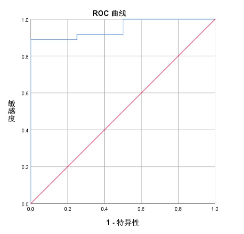
The area under the concentration-time curve of rCBV value was 0.972.The specificity and the sensitivity of rCBV value were 75.36% and 100%.
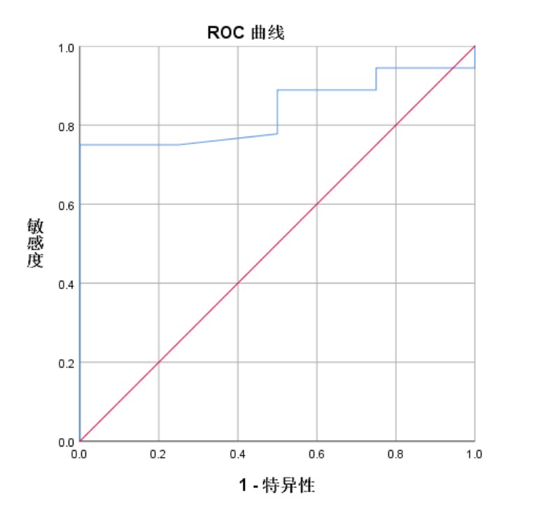
The area under the concentration-time curve of ADC value was 0.842.The mean rCBF value and the ADC value were correlated by pearson correlation analysis(r=-0.61,P<0.05).
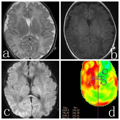
A,T2-weighted image;B,T1-weighted image;C,rCBF color map;D,DWI map.Fig.1 MR images of an infant with HIE in adverse outcome group
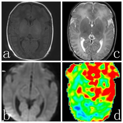
A,T2-weighted image;B,T1-weighted image;C,rCBF color map;D,DWI map.Fig.1 MR images of an infant with HIE in adverse outcome group
DOI: https://doi.org/10.58530/2022/1221