1206
Caffeine acutely reduces aqueductal CSF flow in humans11Department of Experimental Medical Science, Lund University, Lund, Sweden, 2Wallenberg Centre for Molecular Medicine, Lund University, Lund, Sweden, 3Department of Clinical Sciences, Diagnostic Radiology, Lund University, Skåne University Hospital, Lund, Sweden, 4Department of Medical Radiation Physics, Lund University, Lund, Sweden, 5Lund University Bioimaging Center, Lund University, Lund, Sweden
Synopsis
In this ultra-high field 7T MRI study, we find that caffeine acutely reduces CSF net flow in humans. Interestingly, regular caffeine consumption seems to be protective against this reduction. We used non-invasive 2D PC 7T MRI in healthy volunteers to measure CSF flow in the cerebral aqueduct. Ventricular CSF flow is upstream of the glymphatic system and may thus reflect or affect the function of CSF-mediated clearance pathways that have been suggested to be important for waste product removal in the brain. Better understanding of CSF dynamics may help us find new targets for treating proteinopathies such as Alzheimer’s disease.
Introduction
Cerebrospinal fluid (CSF) mediated clearance of waste products from the brain via the glymphatic system has been hypothesised to play an important role in pathophysiology of neurodegenerative diseases. The glymphatic system consists of perivascular CSF flow pathways that allow interchange between CSF and interstitial fluid1. This interchange removes accumulating toxic molecules, such as amyloid beta linked to Alzheimer’s disease2. CSF production has even been suggested to decrease with aging3, which may contribute to the pathology of age-related neurodegenerative diseases.CSF is produced in the choroid plexus of lateral and 3rd and 4th ventricles and circulates to its downstream pathways via the cerebral aqueduct. Therefore, adequate CSF production rates and maintenance of CSF flow homeostasis are needed for driving the perivascular flow and the glymphatic clearance process. Since the majority of glymphatic research has been performed in animal models there is a great need for translational studies examining CSF dynamics in humans.
In this ultra-high field 7 Tesla magnetic resonance imaging (7T MRI) study, we use caffeine to modulate ventricular CSF flow in order to study CSF flow dynamics in humans. Caffeine has been reported to chronically increase and acutely decrease CSF production in rats4. Here, we examine caffeine’s effects in healthy volunteers by studying CSF flow dynamics via the cerebral aqueduct as well as blood flow in internal carotid and vertebral arteries to the brain non-invasively with 2D phase contrast (2D-PC) MRI at 7T. The use of 7T MRI allows high resolution to reliably measure flow even in small structures, such as the aqueduct with a diameter of 1-3mm.
Methods
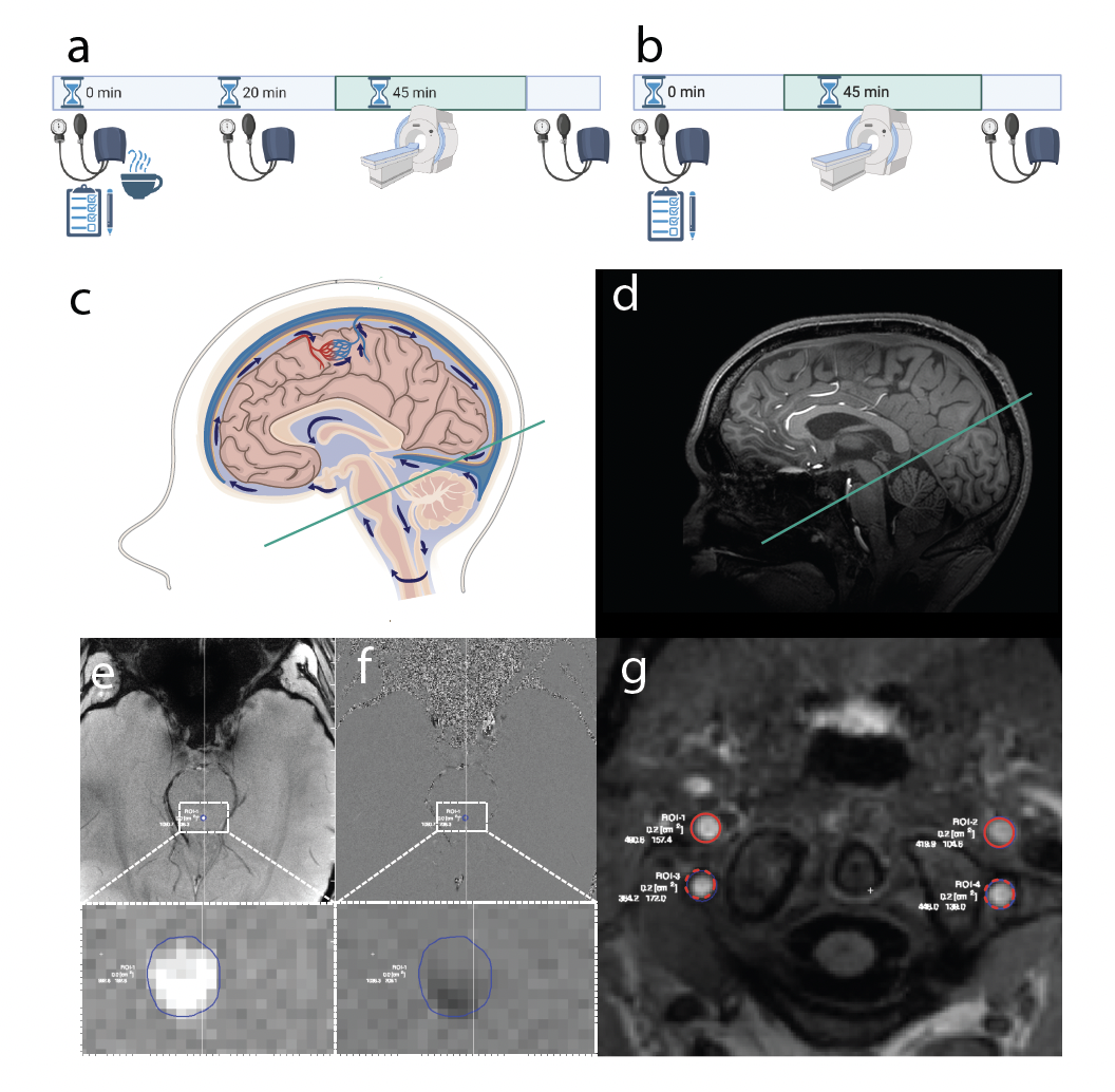
We included 77 healthy volunteers, 58 for baseline (BL) and 19 for the caffeine and the BL scan (Fig 1a-b). 10 individuals performed the BL first, followed by caffeine. 9 performed the caffeine scan first. For the caffeine scan, volunteers ingested 100mg caffeine in tablet form and the scan was performed 45 minutes after caffeine intake (Fig 1a). Blood pressure was measured before and 20min after caffeine intake, as well as after the scan.MRI scans were performed using a 7T MRI scanner (Achieva, Philips Healthcare, Best, the Netherlands) with a 2-channel transmit, 32-channel receive head coil (Nova Medical, Wilmington, MA, USA). Cardiac triggered 2D-PC was used to measure CSF flow in the cerebral aqueduct 5 (Fig 1c-f) and blood flow in the internal carotid and vertebral arteries (Fig 1e). The scan planes were manually placed at 90-degree angle to the aqueduct or artery using a T1-weighted image. The 2D-PC of the aqueduct was scanned at resolution of 0.3x0.3 mm2 and a 5 mm thick imaging slice. VENC was 15cm/s. For arteries, the resolution was 0.5x0.5 mm2 with 5 mm slice thickness. Flow data were analysed using Segment v2.2 R7052 (Medviso, http://segment.heiberg.se). Statistical analysis was performed with Prism v8.2.1 (GraphPad, La Jolla, Ca, USA). The ROUT method (Q=1%) was used for outlier detection. Additionally, scans with poor image quality or incorrectly placed scan planes were excluded (BL 7, Caffeine 4 ). Poor scan quality was related to poor VCG triggering, or background noise in the arterial flow scans placed askew or too caudally, where signal quality is not optimal.
Results
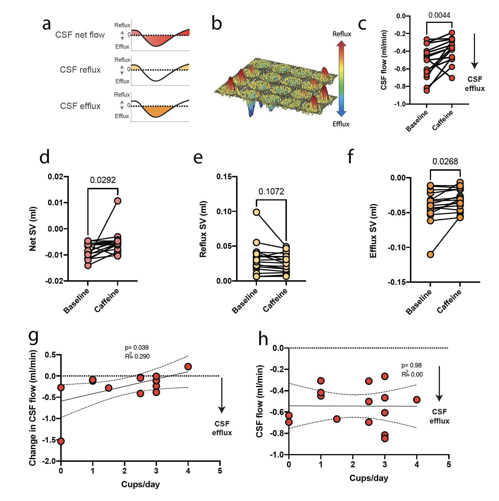
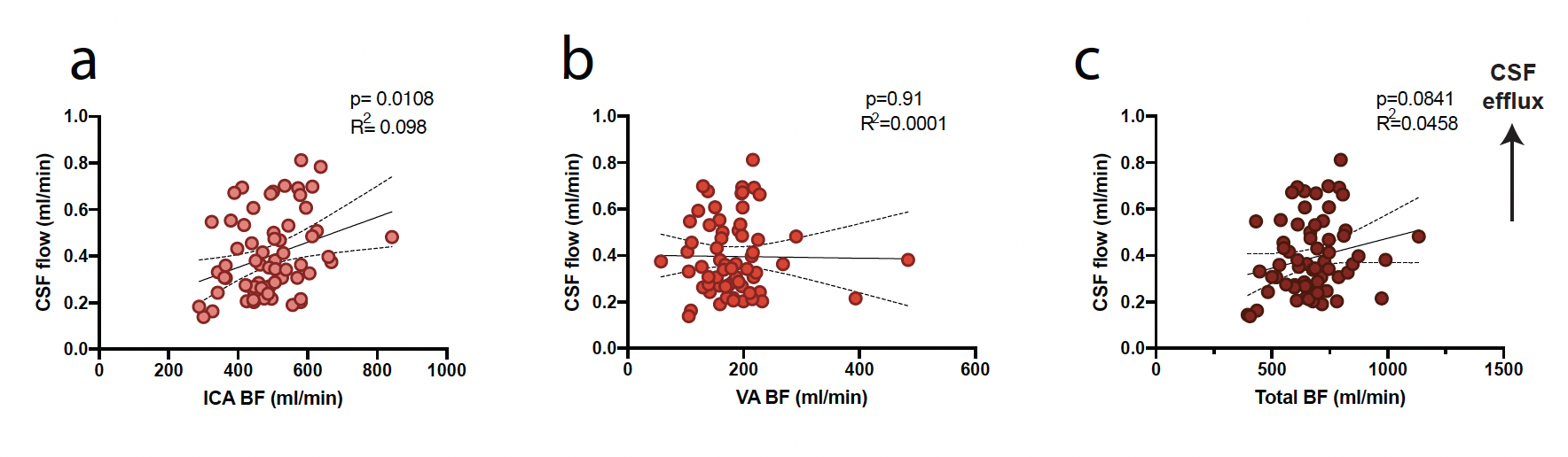
Our data suggest that 100 mg of caffeine acutely reduces CSF net flow in the cerebral aqueduct (p=0.004, paired t-test; Fig 2a-f). Interestingly, CSF flow reduction as a result of acute caffeine consumption was mitigated by higher daily consumption of caffeinated beverages (R2=0.290, p=0.039, LR; Fig 2g-h).Furthermore, we found a reduction in blood flow to the brain via internal carotid (ICA) and vertebral arteries (VA). Linear regression analysis suggests that blood flow in the internal carotid arteries is correlated with the CSF flow (R2=0.098, p=0.010, LR; Fig 3a). Interestingly, there was no correlation between VA flow or combined arterial (ICA+VA) flow (Fig 3b-c)
Discussion
This study suggests caffeine acutely reduces CSF net flow in humans, which in agreement with data from animal studies4. Interestingly, regular caffeine intake during daily life seems to be protective against this decrease: CSF net flow rate drops less in people who consume more coffee per day.Furthermore, we found a correlation between CSF flow and blood flow in the ICA but not the VA, which suggests that ICA has more potential for driving CSF flow. This natural anatomical variation may therefore lead to natural variation in CSF flow rates. The clinical significance of this is unclear, although lower ICA flow has previously been connected to worse cognitive performance in patients with mild cognitive impairment6.
Conclusion
Our study shows that caffeine acutely reduces CSF flow in humans. Caffeine also causes an acute reduction in CSF flow and blood flow. Furthermore, we find a correlation between ICA blood flow and net CSF flow. This link suggests anatomical variation that may play a role in determining CSF flow rates.Acknowledgements
This work was funded by the Knut & Alice Wallenberg Foundation, Torsten Söderberg's Foundation, Tore Nilson's Foundation, Vetenskapsrådet (Dnr: 2018-02340), Wenner-Gren Foundations, Hjärnfonden and The Royal Physiographic society of Lund.
We thank the National 7T facility at the Lund University Bioimaging Centre for the experimental resources needed for the MRI data acquisition.
References
1. Iliff, Jeffrey J et al. “A paravascular pathway facilitates CSF flow through the brain parenchyma and the clearance of interstitial solutes, including amyloid β.” Science translational medicine vol. 4,147 (2012): 147ra111. doi:10.1126/scitranslmed.3003748
2. Jessen, Nadia Aalling et al. “The Glymphatic System: A Beginner's Guide.” Neurochemical research vol. 40,12 (2015): 2583-99. doi:10.1007/s11064-015-1581-6
3. May C, Kaye JA, Atack JR, Schapiro MB, Friedland RP, Rapoport SI. Cerebrospinal fluid production is reduced in healthy aging. Neurology. 1990 Mar;40(3 Pt 1):500-3. doi: 10.1212/wnl.40.3_part_1.500. PMID: 2314595.
4. Han, Myoung-Eun et al. “Regulation of cerebrospinal fluid production by caffeine consumption.” BMC neuroscience vol. 10 110. 3 Sep. 2009, doi:10.1186/1471-2202-10-110
5. Markenroth Bloch K, Töger J, Ståhlberg F. Investigation of cerebrospinal fluid flow in the cerebral aqueduct using high-resolution phase contrast measurements at 7T MRI. Acta Radiol. 2018 Aug;59(8):988-996. doi: 10.1177/0284185117740762. Epub 2017 Nov 15. PMID: 29141450.
6. Berman, Sara E et al. “Intracranial Arterial 4D Flow in Individuals with Mild Cognitive Impairment is Associated with Cognitive Performance and Amyloid Positivity.” Journal of Alzheimer's disease : JAD vol. 60,1 (2017): 243-252. doi:10.3233/JAD-170402
Figures