1193
Feasibility of MRI-guided Interventions at a Large-bore New-generation 0.55T Low-Field MRI System
Iman Khodarahmi1, Inge M Brinkmann2, Mary Bruno1, Jerzy Walczyk1, Ryan Brown1, and Jan Fritz1
1Department of Radiology, New York University School of Medicine, New York, NY, United States, 2Siemens Medical Solutions USA Inc, Malvern, PA, United States
1Department of Radiology, New York University School of Medicine, New York, NY, United States, 2Siemens Medical Solutions USA Inc, Malvern, PA, United States
Synopsis
New generation low-field MRI systems can be ideal for MRI-guided interventions due to wide bore diameters and lower susceptibility artifacts improving patient access and needle visualization, respectively. We evaluated a set of pulse sequences for their suitability for needle visualization using a new-generation 0.55T system and compared the results with those of a clinical 3T system. Interventional needles can be adequately visualized at 0.55T. Using TSE pulse sequences, needle artifacts display more favorable at 0.55T than 3T. SEMAC and HASTE pulse sequences may not facilitate diagnostic needle visualization at 0.55T but are not needed for clinical MR interventions.
Introduction
The new generation of low-field MRI systems is equipped with the state-of-the-art hardware and software developed for high-field systems and is projected to provide broader world-wide access due to its lower cost. Such systems can be an ideal platform for MRI-guided procedures and interventions for two reasons: First, the lower magnetic field, gradient strength and slew rate have enabled the development of wide bore diameters of 80 cm that provide space for interventional apparatus. Second, lower susceptibility artifacts at low field strengths are expected to improve the visualization of the metallic devices used in interventional procedures (1,2). In this study, we aimed to propose a set of pulse sequence parameters suitable for needle visualization at a new generation 0.55T system and compare the results with those of a 3T system used in clinical practice.Methods
A commercially available MR-conditional 20-gauge cobalt-chromium injection needle (Cook Medical, Bloomington, IN, USA) was placed in the central slot of a phantom designed to assess the metal artifacts (Figure 1). The phantom was made of eight rows of six parallel plastic strips with different thicknesses and spacings, and was filled with coconut oil. Similar to human interventions, this setup was placed in the axial plane at the iso-center of a commercial MRI system (1.5T MAGNETOM Aera; Siemens Healthcare GmbH, Erlangen, Germany) modified to operate as a prototype system at 0.55T field strength with a maximum gradient strength of 25 mT/m and a maximum slew rate of 40 T/m/s. An 11 cm loop coil and an 18-channel spine array tuned to operate at 0.55T were used for signal reception. Various pulse sequences, each with different parameters, including turbo spin-echo (TSE), true fast imaging with steady-state free precession (TRUFI), volumetric interpolated breath-hold examination gradient-echo (VIBE), slice encoding for metal artifact correction (SEMAC), and half-Fourier acquisition single-shot TSE (HASTE) were evaluated for optimal needle visualization. The extent of the metal artifacts and overall image quality was qualitatively assessed by two readers by noting acquisitions that provided clear visualization of the strips adjacent to the needle. For comparison with the standard 3T field strength, the same experimental setup was imaged with a clinical system (MAGNETOM Skyra, Siemens Healthcare) using the clinical pulse sequences and signal reception by 3-channel surface and 32-channel spine arrays.Results and Discussion
For the TSE sequence, the effect of various signal averages, parallel imaging acceleration factors (GRAPPA), receiver bandwidths, and turbo factors were assessed (Figure 2) with the optimized parameters being a signal average of 3, no parallel imaging, a receiver bandwidth of 300 Hz/pixel, and no turbo factor dependence. Findings of other pulse sequences are summarized in Table 1. Figure 3 compares the images acquired at 0.55T using the optimized parameters and 3T using the clinical protocols. For the TSE pulse sequence, metal artifacts were substantially smaller at 0.55T than 3.0T, although there is a time-penalty cost to reach diagnostic signal-to-noise ratios. TRUFI images, considered not diagnostic at 3T, can be rapidly acquired at 0.55T and used for fluid visualization during MRI-guided injections. Metal artifacts at 0.55T are smaller with the VIBE pulse sequence with comparable acquisition times. SEMAC is not needed at 0.55T because the through-plane artifacts associated with the needle in the axial plane are minimal and images are degraded by the blurring that is caused by view-angle-tilting. Within the range of tested parameters, HASTE images were considered non-diagnostic for needle visualization at 0.55T.Conclusion
Interventional needles can be adequately visualized at 0.55T, suggesting that the performance of MRI-guided procedures is feasible at the new generation of low-field systems. The TSE pulse sequence, with a longer acquisition time, can result in smaller metal artifacts at 0.55T than 3T. Within the range of tested parameters, SEMAC and HASTE pulse sequences were non-diagnostic, but also not needed for needle visualization at 0.55T, whereas images acquired with TRUFI can be used for rapid tracking of the injected fluid.Acknowledgements
The authors would like to acknowledge the assistance of Siemens Healthcare in the modification of the MRI system for operation at 0.55T under an existing research agreement between NYU and Siemens Healthcare.References
- I. Khodarahmi, I.M. Brinkmann, D. Lin, M. Bruno, P. Johnson, F. Knoll, M.B. Keerthivasan, H. Chandarana , J. Fritz. Advanced Low-Field MRI of Hip Arthroplasty Implants: First Experience at 0.55 T. International Society for Magnetic Resonance in Medicine (ISMRM). May 2021.
- Campbell-Washburn AE, Ramasawmy R, Restivo MC, et al. Opportunities in Interventional and Diagnostic Imaging by Using High-Performance Low-Field-Strength MRI. Radiology. 2019;293(2):384-393.
Figures
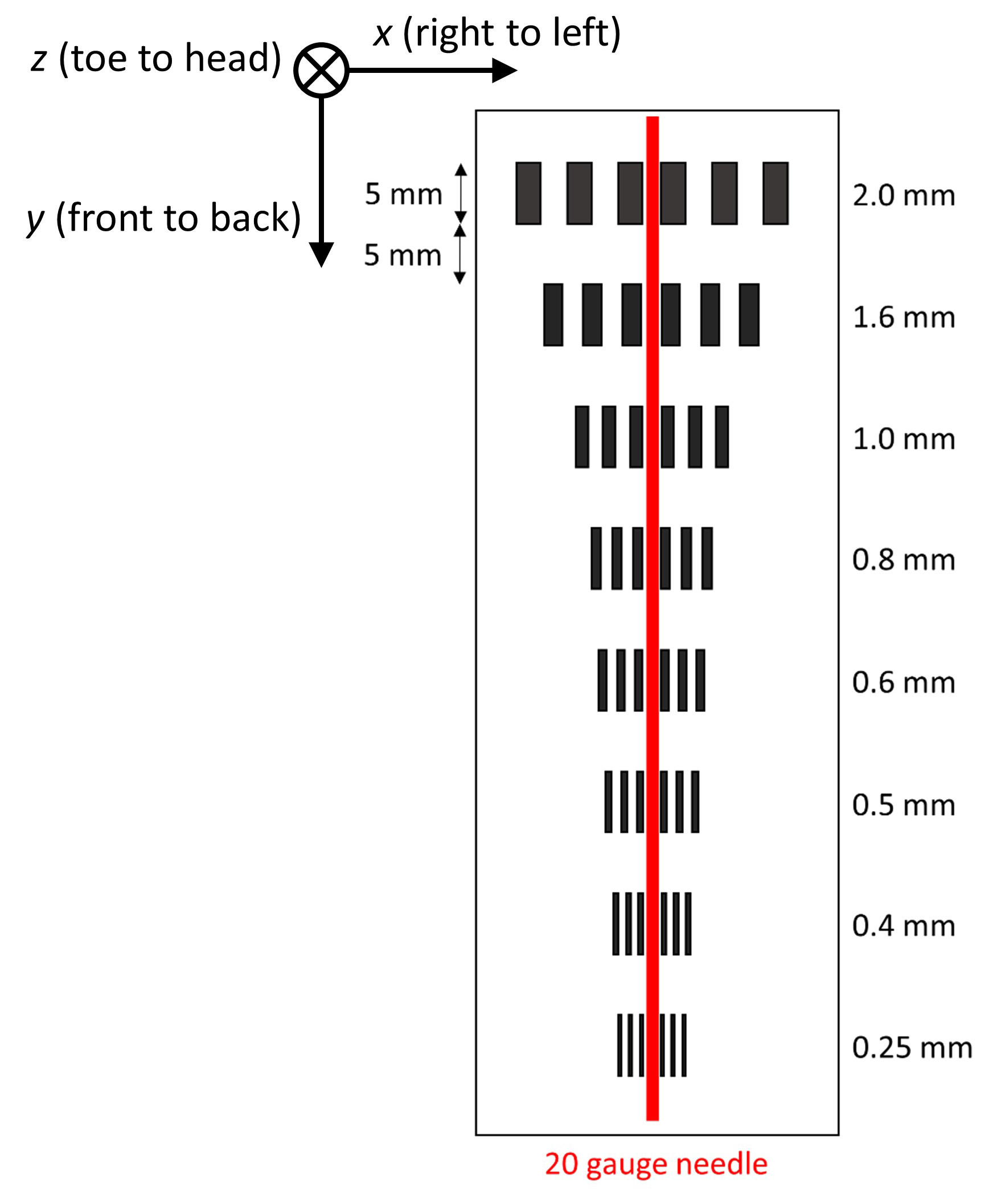
Figure 1: Schematic image of the needle containing phantom
used to assess the metal artifacts and overall image quality.
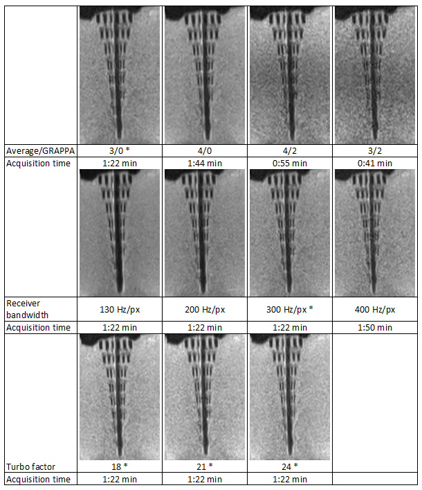
Figure 2: The effect of various acquisition parameters on
metal artifacts using the TSE pulse sequence for image acquisition. Readers’
preference for each parameter set is marked by asterisks.
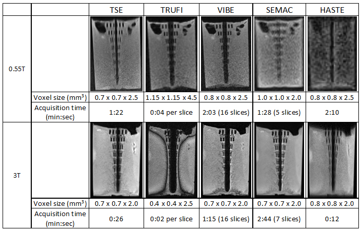
Figure 3: Comparison of optimized pulse sequences at 0.55T
with those of the 3T which are used for MRI-guided procedures in clinical
practice.
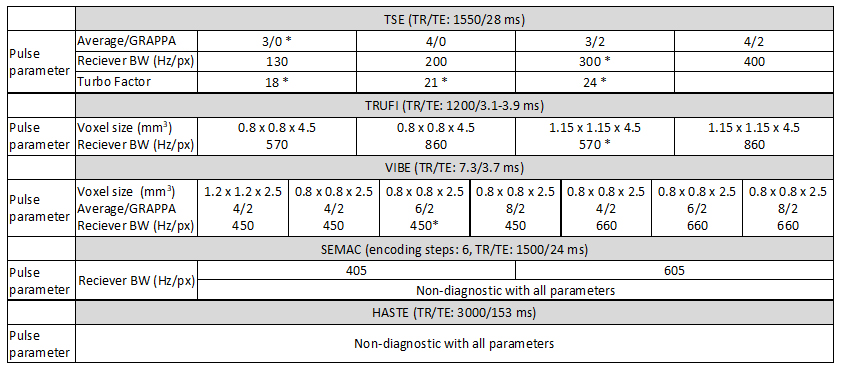
Table 1. Various 0.55T acquisition parameters of different
pulse sequences tested for metal artifacts. Readers’ preference for each parameter
set is marked by asterisks.
DOI: https://doi.org/10.58530/2022/1193