1189
Hyperpolarized [1-13C]Pyruvate MRI of Spleen: Monitoring Immune Activation for Cervical Cancer Patient following Radiotherapy
Gigin Lin1, Ying-Chieh Lai1, Kuan-Ying Lu1, Albert P Chen2, and Ching-Yi Hsieh3
1Medical Imaging and Intervention, Chang Gung Memorial Hospital, Linkou, Taiwan, 2GE Healthcare, Toronto, ON, Canada, 3Medical Imaging Research Center, Institute for Radiological Research, Chang Gung University, Taoyuan, Taiwan
1Medical Imaging and Intervention, Chang Gung Memorial Hospital, Linkou, Taiwan, 2GE Healthcare, Toronto, ON, Canada, 3Medical Imaging Research Center, Institute for Radiological Research, Chang Gung University, Taoyuan, Taiwan
Synopsis
Successful cancer therapy depends on an optimal immune response, but noninvasive tools are lacking to monitor this dynamic process. In this proof-of-concept human study, we demonstrated hyperpolarized [1-13C]pyruvate MRI of the spleen monitoring the immune activation for cancer patients at the early time point following radiotherapy, by measuring the pyruvate-to-lactate conversion rate. The ADC-based cellularity of the spleen, however, did not detect remarkable changes.
Introduction
Successful cancer therapy depends on an optimal immune response, but noninvasive tools are lacking to monitor this dynamic process. In this proof-of-concept human study, we investigated whether hyperpolarized [1-13C]pyruvate MRI of the spleen is capable of monitoring immune activation for cancer patients at the early time point following radiotherapy.Methods
Hyperpolarized [1-13C]pyruvate MRI of spleen was scheduled for a 53-year old female patient with bulky cervical cancer staging T2bN1M0, at the baseline and 2 weeks following radiation therapy.Imaging was performed using a clinical 3T MRI system with imaging sequences from the Multinuclear Spectroscopy (MNS) research pack, using a 13C/1H multinuclear transmit-receive surface coil. Hyperpolarized [1-13C]pyruvate for intravenous injection was prepared less than 24 hours prior to injection and polarized in a 5T SPINlab polarizer, with a quality control (QC) system ensured safety parameters. The time interval between the dissolution of hyperpolarized [1-13C]pyruvate and intravenous injection ranged from 66-72 sec, and an extra 30 sec was waited following injection of 0.43 ml/kg hyperpolarized [1-13C]pyruvate, before the start of 13C MRI acquisition. The 13C MRI sequences included (1) pulse-and-acquire sequence (TR/FOV/FA/ST: 2000msec/200x200 mm2/10/20mm; scan time 28 sec); (2) spectral-Spatial (SPSP) excitation with spiral readout (TR 1000msec for pyruvate, lactate, alanine and bicarbonate, scan time 14 sec); (3) generic chemical shift imaging (TR/FOV/matrix/FA/ST: 80msec/200x200 mm2/8x8/10/20mm, scan time 5.12 sec). Inputless K fitting method was used to estimate the conversion rates1. Additional 1H MRI protocol included a diffusion-weighted imaging (DWI) sequence consisting of a spin echo echo planar imaging (EPI) with b-values of 0 and 1000 s/mm2 (TR/TE/FOV/matrix/slice thickness (ST): 3300msec/79msec/320x320mm2/128x128/5mm), to measure the apparent diffusion coefficient (ADC) value.Results
At the day 14 following treatment, the total blood leukocytes amount ranged within normal limit, but their differential count demonstrated an increased proportion of neutrophils from 71% to 79%, and a decreased proportion of lymphocytesfrom 71% to 79%, indicating an activation of immune activity. Hyperpolarized [1-13C]Pyruvate MRI of the spleenshowed an increased pyruvate-to-lactate conversion rate (KPL) from 3.1x10⁻³ s-1 to 4.3x10⁻³ s-1, and an increased pyruvate-to-alanine conversion rate (KPA) from 5.9x10⁻⁴ s-1 to 8.5x10⁻⁴ s-1, and a 1.5-fold increase of area-under-curve (AUC) of the lactate curve, normalized with the pyruvate curve. The ADC values of the spleen, however, did not detect remarkable changes (990 x 10-6 mm2/s vs 980 x 10-6 mm2/s). A successful anti-cancer response was observed after completion of the entire radiotherapy course.Discussion
We are presenting the first human study focusing on monitoring immune activation using hyperpolarized [1-13C]pyruvate MRI of the spleen. Despite no changes in cellularity of the spleen, demonstrated by the DWI, the rates of pyruvate-to-lactate and pyruvate-to-alanine were both increased. Although not being able to obtaining tissues from the spleen, monitoring early metabolic flux alterations following radiotherapy in cancer and immune cells has been initially observed in a hyperpolarized 13C MRI in vitro study 2. Cancer therapy is accompanied by distinct metabolic patterns in spleen observed by non-invasive in vivo 18F-FDG-PET, as supported by a retrospective analysis of clinical 18F-FDG-PET/CT scans revealed enhanced 18F-FDG uptake in the spleens of some successfully treated patients with metastatic melanoma 3.Conclusion
Hyperpolarized [1-13C]pyruvate MRI of the spleen may be applicable of monitoring immune activity for cancer patients at the early time point following radiotherapy. We are actively recruiting more cases to support this initial finding (ClinicalTrials.gov ID: NCT04951921).Acknowledgements
This research was funded by Chang Gung Foundation (CLRPG3K0022) and the Ministry of Science and Technology Taiwan (MOST-109-2628-B-182A-018). We are grateful for helps from HMTRC, UCSF, supported by NIBIB and NIH (P41EB013598).References
1. Larson, PEZ, Chen, HY, Gordon, JW, et al. Investigation of analysis methods for hyperpolarized 13C-pyruvate metabolic MRI in prostate cancer patients. NMR Biomedicine. 2018;31:e3997. 2. Lai YC, Hsieh CY, Lu KY, et al. Monitoring Early Glycolytic Flux Alterations Following Radiotherapy in Cancer and Immune Cells: Hyperpolarized Carbon-13 Magnetic Resonance Imaging Study. Metabolites. 2021;11(8): 34436459. 3. Schwenck J, Schorg B, Fiz F, et al. Cancer immunotherapy is accompanied by distinct metabolic patterns in primary and secondary lymphoid organs observed by non-invasive in vivo (18)F-FDG-PET. Theranostics. 2020;10(2):925-937.Figures
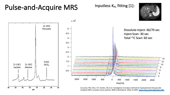
Figure 1. MRS of the pulse-and-acquire sequence clearly demonstrated the signals of pyruvate, lactate, alanine until the end of scan.
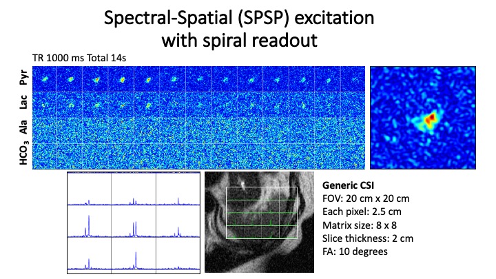
Figure 2. Spectral-spatial (SPSP) sequence and generic chemical shift imaging confirmed the origin of signal from the spleen and captured the dynamic changes of pyruvate, lactate, alanine and bicarbonate.
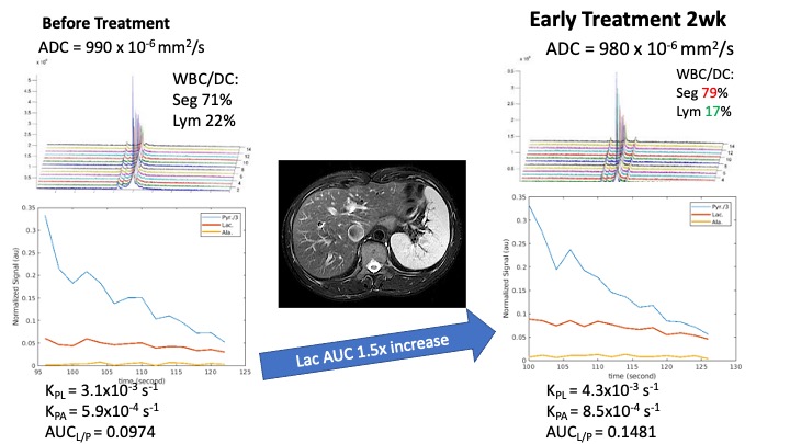
Figure 3. Hyperpolarized [1-13C]Pyruvate MRI of the spleen showed an increased pyruvate-to-lactate conversion rate (KPL) from 3.1x10⁻³ s-1 to 4.3x10⁻³ s-1, and an increased pyruvate-to-alanine conversion rate (KPA) from 5.9x10⁻⁴ s-1 to 8.5x10⁻⁴ s-1, and a 1.5-fold increase of area-under-curve (AUC) of the lactate curve, normalized with the pyruvate curve.
DOI: https://doi.org/10.58530/2022/1189