1166
Sampling Pattern Optimization for Joint Acceleration of Multi-contrast MRI using Deep Learning1Bio and Brain Engineering, Korea Advanced Institute of Science and Technology, Daejeon, Korea, Republic of, 2Radiology, Seoul National University Hospital, Seoul National University College of Medicine, Seoul, Korea, Republic of
Synopsis
Usage of multiple-acquisition MRI is one field of study that proved its effectiveness and practicality since routine MR scan protocol typically acquires multiple information for the same anatomical structures. In this study, we propose simultaneous optimization of sampling pattern and reconstruction for joint acceleration of multi-contrast MRI. The simultaneous optimization of sampling pattern and reconstruction provided superior performance over single contrast imaging and over single sampling pattern for multi-contrast MRI. The proposed technique can be adopted in routine clinical scan without forcing extra scans during acquisition.
INTRODUCTION
Deep learning MR acceleration is an on-going heated research field in MR community. Physics-guided1 approach has become popular, and optimizing the sampling pattern for the reconstruction of images using deep learning has also become an effective way to accelerate MR imaging2,3. One such approach that applied a physics-guided scheme for deep learning, is a method named joint model-based deep learning (J-MoDL)3. In J-MoDL, the sampling scheme and reconstruction were optimized together in single-contrast imaging. In routine scan protocol, however, it is common to acquire multiple MR data such as multi-contrast imaging. Previous studies showed that MRI reconstruction can be improved through joint optimization of multi-contrast MR acquisitions. In this study, we extended the idea of jointly optimizing the sampling pattern and image reconstruction for multi-contrast MRI for further application in routine multiple-acquisition MRI environment.METHODS
As shown in Figure 1, the overall scheme is an unrolled process, where each iteration comprises of a denoiser neural network and data consistency blocks following the baseline of J-MoDL. For the denoiser network, multi-contrast images were optimized together. Each of the contrast images was then processed in the data consistency block separately, and the sampling location parameters, Θ, were separately optimized for each of the contrasts, where sampling parameters for N contrast MR images (e.g. T2, FLAIR, T1) can be denoted as [Θ1, Θ2,…, ΘN]. The scheme enabled simultaneous sampling pattern optimization and reconstruction of multiple contrast images at once.T2, FLAIR, and T1 weighted brain images from 31 healthy subjects were acquired on a 3T MRI scanner (SIEMENS Verio, Siemens Healthcare, Erlangen, Germany). Datasets from 20 subjects were used for training, 1 subject for validation, and 10 subjects for testing. Turbo spin echo was applied to obtain T2 and FLAIR images and spin echo to obtain T1 images with following parameters: TR= 3700ms(T2), 9000ms(FLAIR), and 500ms(T1) ; Inversion Time= 2500ms(FLAIR); TE= 91ms(T2/ FLAIR) and 9.8ms(T1); FOV= 230´184mm2; number of slices= 15; slice thickness= 5mm; flip angle= 150°(T2), 140°(FLAIR), and 70°(T1); matrix size=320´256; pixel bandwidth=260Hz/Px; and number of channels= 12.
Undersampled T2, FLAIR, and T1 weighted images were used together to reconstruct full sampled images of each contrast, and sampling patterns were jointly optimized for different contrasts simultaneously, resulting in three separate sampling pattern masks, [Θ1, Θ2, Θ3]. U-net4 was used for the denoiser network. Conjugate gradient algorithm with 10 iterations was used for data consistency blocks5. The initial sampling patterns were uniformly distributed, where sampling lines were equidistant with approximately 4% of phase encoding lines consecutively sampled at the center. The equidistantly sampled lines were shifted across contrast to reduce redundancy. The unrolled scheme was applied with iteration K=5. Coil sensitivity maps were acquired using ESPIRiT6
As a main comparison study, our proposed multicontrast scheme was compared against the performance of single contrast reconstruction, which is equivalent to the baseline J-MoDL. Then, a sampling pattern test that served as an ablation study was performed to investigate the effectiveness of simultaneously optimizing the sampling pattern for each of the contrasts. Two models were compared: 1) the proposed model that optimized [Θ1, Θ2, Θ3] for three contrasts and 2) the model in which all three contrasts shared one Θ set. This comparison could demonstrate the effect of learning different Θ sets for each contrast simultaneously. The same initial uniform sampling pattern across contrasts was used for both of the models.
The acceleration factors of R= 8 and 10.67 (32 and 24 phase encoding lines, respectively) were investigated. The quantitative analysis was done using 3 metrics: normalized mean squared error (NMSE), peak signal-to-noise ratio (PSNR), and structural similarity index (SSIM). The significance of the comparison was tested using Wilcoxon signed rank test, where p values <0.05 were considered statistically significant.
RESULTS
As shown in Table 1, our proposed scheme demonstrated overall superior performance over the single contrast reconstruction method across all tested contrast types and acceleration factors except for SSIM for T1 contrast in R=8. All of the improvements were statistically significant. Figure 2 also shows visual improvement by using our proposed multi-contrast method over single contrast method. The difference map shows the overall performance improvement through the proposed method, and the red arrows demonstrate that details were better reconstructed by our scheme. The qualitative analysis aligns well with the quantitative results.Table 2 shows the sampling pattern test results. The performance was improved by optimizing sampling patterns for each of the contrasts [Θ1, Θ2, Θ3] rather than using single Θ set for all the contrasts. All of the quantitative improvements were statistically significant except for the SSIM of FLAIR in R=10.67 and T1 in R=8 and 10.67.
DISCUSSION and CONCLUSION
The proposed study is a seamless incorporation of two widely researched fields of MR acceleration: multiple-acquisition MRI and sampling optimization. We demonstrated that sampling patterns can be optimized for different contrasts, which improved the performance over single contrast imaging (J-MoDL) and over single sampling pattern set for multi-contrast MRI. Usage of multi-contrast information for the acceleration takes into account the routine MR protocol, since acquisition of multi-contrast information for the same anatomical structure is common. Therefore, the proposed technique can be adopted without forcing extra scans during acquisition.Acknowledgements
No acknowledgement found.References
1. Liang D, Cheng J, Ke Z, Ying L. Deep Magnetic Resonance Image Reconstruction: Inverse Problems Meet Neural Networks. IEEE signal processing magazine 2020;37(1):141-151.
2. Bahadir CD, Dalca AV, Sabuncu MR. Learning-based optimization of the under-sampling pattern in MRI. 2019. Springer. p 780-792.
3. Aggarwal HK, Jacob M. J-MoDL: Joint model-based deep learning for optimized sampling and reconstruction. IEEE Journal of Selected Topics in Signal Processing 2020;14(6):1151-1162.
4. Ronneberger O, Fischer P, Brox T. U-Net: Convolutional Networks for Biomedical Image Segmentation. Medical Image Computing and Computer-Assisted Intervention – MICCAI 2015; 2015; Cham. Springer International Publishing. p 234-241. (Medical Image Computing and Computer-Assisted Intervention – MICCAI 2015).
5. Aggarwal HK, Mani MP, Jacob M. MoDL: Model-based deep learning architecture for inverse problems. IEEE transactions on medical imaging 2018;38(2):394-405.
6. Uecker M, Lai P, Murphy MJ, Virtue P, Elad M, Pauly JM, Vasanawala SS, Lustig M. ESPIRiT--an eigenvalue approach to autocalibrating parallel MRI: where SENSE meets GRAPPA. Magn Reson Med 2014;71(3):990-1001.
Figures
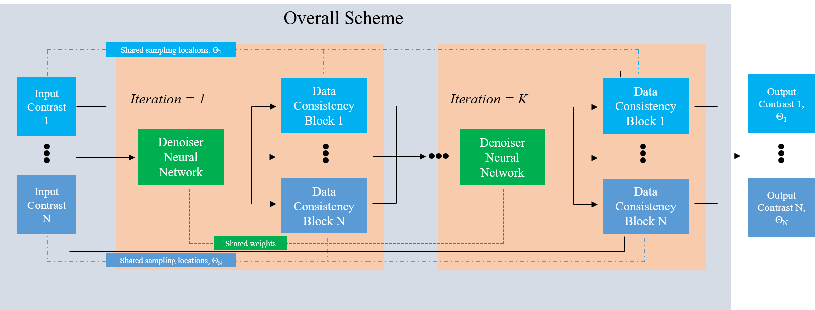
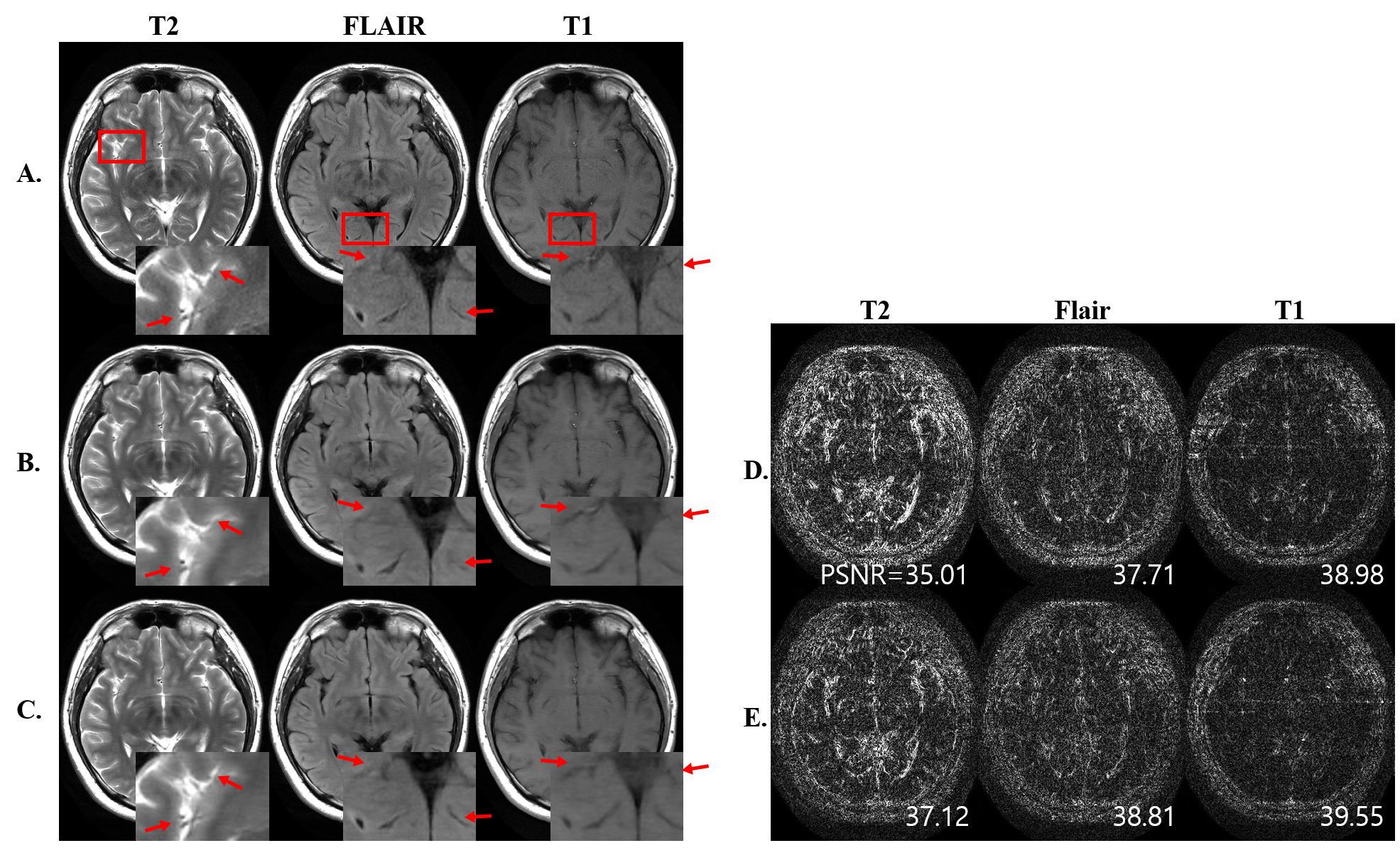

Table 1. The comparison results of proposed multi-contrast scheme
Data is given as mean ± standard deviation across 10 test sets.
R: acceleration factor.
Single: single contrast optimization (J-MoDL).
Proposed: multi-contrast optimization.
*: The signed rank test showed significant improvement compared with single contrast method (p<0.05).

Data is given as mean ± standard deviation across 10 test sets.
R: acceleration factor.
Single Θ set: multi-contrast optimization through single shared Θ set.
[Θ1, Θ2, Θ3]: Proposed multi-contrast optimization where different Θ set was learned for contrast 1,2, and 3 (T2, FLAIR, and T1).
*: The signed rank test showed significant improvement compared with single contrast method (p<0.05).