1147
Intra-Subject Variability of Skeletal Muscle Glycogen Using 13C/1H MRS at 3T Using a Novel Standardization Method1Human Magnetic Resonance Center, Institute for Applied Life Sciences, University of Massachusetts Amherst, Amherst, MA, United States, 2Kinesiology, University of Massachusetts Amherst, Amherst, MA, United States
Synopsis
Given its central role in substrate metabolism, the ability to quantify muscle glycogen (Gly) continuously and noninvasively using natural abundance 13C MR spectroscopy presents an important opportunity for understanding human metabolism. This study evaluated the reproducibility of Gly measured by 13C MRS in the quadriceps muscle using three methods for signal standardization at 3T: Gly/total creatine (TCr) from 13C, Gly/TCr using 13C and 1H, and Gly/water. The novel results from this study suggest that Gly/TCr measurements using the creatine peak from 1H MRS is highly reproducible and may be an advantageous approach to measuring muscle glycogen in the future.
Introduction
Skeletal muscle is a primary site of glucose disposal and plays a key role in whole-body glucose homeostasis1. Following uptake by the muscle cells, glucose is either broken down as a source of energy for muscle contraction or stored in the form of glycogen (Gly) such that basal concentration of glycogen can provide information about metabolic dysregulation under some specific experimental conditions2,3. Carbon Magnetic Resonance Spectroscopy (13C MRS) offers a non-invasive and accurate quantification of skeletal muscle glycogen levels. Specifically, despite a relatively low sensitivity due to the low natural abundance and gyromagnetic ratio of 13C, the glycogen‐C1 resonance can be detected at 100.5 ppm. A reliable method of evaluating muscle glycogen noninvasively could help with the diagnosis and management of metabolic disease4,5. The goals of this study were to 1) determine the intra-subject reproducibility of glycogen measurement using 13C MRS, and 2) compare 13C and 1H MRS normalization methods for the estimation of glycogen at 3T.Materials and Methods
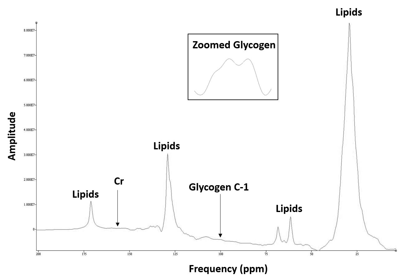
Carbon MRS data from 6 scans performed on different days were collected using a Siemens 3T Skyra MRI scanner from the vastus lateralis muscle of a 45-year-old healthy, vegetarian male 1-hour post-meal. A double-tuned 1H/13C circular surface coil was used for the MRS acquisition. 13C spectra were acquired with the following parameters, using a pulse and acquire sequence: TR=300ms, TE=0.15ms, 4096 averages, FA 90 degrees, 2048 points, 20K bandwidth, hard pulse, 50% decoupling with Waltz4 pulse, NOE count 1, 5.0ms duration, carrier frequency close to glycogen C-1 peak, 21 minutes scan time. For the 1H MRS, a PRESS sequence was used with the following parameters: 2000Hz spectral width, 2048 points, 2000ms repetition time, 30ms echo time, and 64 averages with voxel coverage of 40x40x40mm3 (2:15 min scan time). Non-water suppressed spectrum was acquired with two averages. Proton MRS spectra were utilized to quantify water and total creatine, which are not as readily observed in 13C MRS as in 1H MRS. This measurement allowed us to calculate the total ratios of Gly/TCr and Gly/Water. Spectra were processed using jMRUI 6.0 and glycogen, creatine and water peaks quantified using the AMARES non-linear least squares algorithm. To evaluate reproducibility, the mean, standard deviation (SD) and coefficient of variation (CV, %) of the 6 measurements were determined for each variable.Results
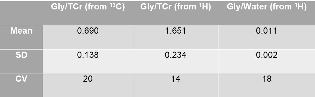
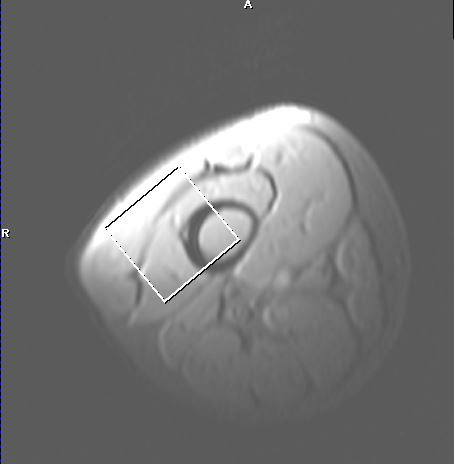
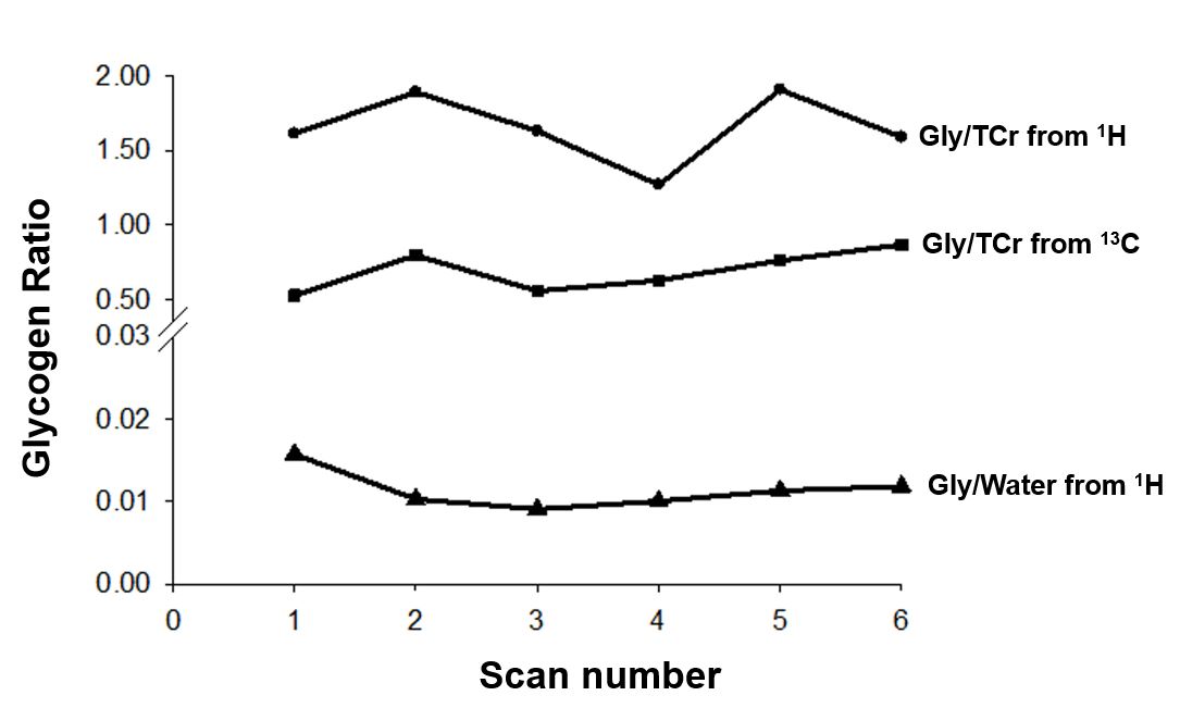
Figure 1 shows the MRS location of 1H and 13C in thigh muscle. Figure 2 illustrates the natural abundance 13C MRS spectrum from the vastus lateralis muscle. Our experiment shows that the glycogen doublet consistently appeared at 100.5 ppm in all 6 measurements in this subject. The variability of the Gly/TCr from 13C, and Gly/TCr and Gly/Water ratios from the 13C and 1H MRS measurements are shown in Figure 3. The mean, SD and CV for each ratio across the 6 experimental days are given in Table 1.Discussion and Conclusions
The detection and reproducibility of glycogen content in skeletal muscle have been demonstrated previously in high-field MR studies6,7. Our results confirm that glycogen content can be reproducibly detected at a lower magnetic field (3T). To improve glycogen quantification reproducibility, we introduced a new method that combines 13C MRS with localized 1H MRS, as Cr and water are easily detectable using 1H. The use of a dual-tuned coil provided the capability to collect proton and carbon data in one setup, ensuring identical shim coverage area between the two acquisitions. The use of two different techniques (localized and non-localized) and nuclei to normalize the glycogen signal were novel design aspects of this study. Using this multi-nuclear spectroscopic approach, we provide preliminary evidence for improved standardization of muscle glycogen concentration by normalizing the 13C Gly signal to TCr using the creatine peak from 1H MRS. This new method may allow more reproducible measures of muscle glycogen with shorter acquisition times in the future.Acknowledgements
We thank Elena Bliss for assistance with MRS data collection.References
1. Shulman GI, Rothman DL, Jue T, Stein P, DeFronzo RA, Shulman RG: Quantitation of muscle glycogen synthesis in normal subjects and subjects with non-insulin-dependent diabetes by 13C nuclear magnetic resonance spectroscopy N Engl J Med 1990; 322: 223– 228.
2. Petersen KF, Hendler R, Price T, Perseghin G, RothmanDL, Held N et al. 13C/31P NMR studies on the mechanism of insulin resistance in obesity. Diabetes, 1998;47:381–6.
3. Price TB, Rothman DL, Avison MJ, Buonamico P, Shulman RG. 13C NMR measurements of muscle glycogen during low-intensity exercise. J. Appl. Physiol. 1991; 70: 1836–1840.
4. Bally L, Buehler T, Dokumaci AS, Boesch C, Stettler C. Hepatic and intramyocellular glycogen stores in adults with type 1 diabetes and healthy controls. Diabetes Res. Clin. Pract. 2015; 109: e1–e3.
5. van den Bergh AJ, Tack CJ, van den Boogert HJ, Vervoort G, Smits P, Heerschap A. Assessment of human muscle glycogen synthesis and total glucose content by in vivo 13C MRS. Eur. J. Clin. Invest. 2000; 30: 122–128.
6. Wary C, Laforêt P, Eymard B, Fardeau M, Leroy-Willig A, Bassez G, Leroy JP, Caillaud C, Poenaru L, Carlier PG. Evaluation of muscle glycogen content by 13C NMR spectroscopy in adult-onset acid maltase deficiency. Neuromuscular disorders. 2003 Sep 1;13(7-8):545-53.
7. Avison MJ, Rothman DL, Nadel E, Shulman RG (1988) Detection of human muscle glycogen by natural abundance 13C NMR. Proc Natl Acad Sci USA 85: 1634–1636.
Figures