1115
The importance of optimized post-processing for accurate T1-mapping using shMOLLI: A phantom and in vivo evaluation at 3 T1Department of Medical Physics and Biomedical Engineering, Sahlgrenska University Hospital, Gothenburg, Sweden, 2Department of Paediatric Radiology, Queen Silvia Children's hospital, Sahlgrenska University Hospital, Gothenburg, Sweden, 3Institute of Clinical Sciences, Sahlgrenska Academy, University of Gothenburg, Gothenburg, Sweden
Synopsis
We demonstrate the effect of not having relevant post-processing algorithm implemented when using the shMOLLI scan sequence for T1-mapping of myocardium and propose an optimization strategy where we use only the five first acquired images in post-processing. Phantom and in vivo measurements of T1-values in myocardium and blood pool in a pediatric population show the importance of using relevant post-processing: a large heart rate dependence is introduced in estimated T1-values without it. This heart rate dependency can, however, be effectively reduced by using only the five first acquired images in post-processing, resulting in measurements with high accuracy.
Introduction
The use of T1-mapping techniques in cardiovascular imaging (CMR) has proved to be useful in the diagnosis of several myocardial diseases such as myocarditis, amyloidosis, Fabry’s disease and congenital heart disease. The quantitative nature of T1-mapping makes it valuable for long-term follow-up of patients with myocardial disease. However, good reliability of the T1-mapping technique, including both scanning and post-processing, is essential. Various T1-mapping techniques have been developed for application in CMR. Compared to saturation recovery (SR)-based scan sequences (e.g. SASHA, SAPPHIRE), inversion recovery (IR)-based scan sequences (e.g. MOLLI, shortened MOLLI: shMOLLI) reveal poorer accuracy but better precision and reproducibility1. Both MOLLI and shMOLLI have an inherent heart rate (HR) dependence in accuracy and in shMOLLI, this dependence is minimized using a conditional data analysis2. In pediatric patients, large differences in HR between individuals are expected, ranging between 40-140 bpm and expected to change with age. Hence, it is important and desirable to use T1-mapping techniques with low HR dependence for evaluation of myocardial tissue in the pediatric population.The aim of this study was to confirm with phantom measurements if ShMOLLI can offer reliable T1-measurement with low HR dependence and in phantoms and patients investigate how the T1 measurements improves after optimized post-processing.
Methods
PhantomThe QalibreMD System Standard Model 130 phantom was used with measurements made in the T1-spheres with T1-values in the clinically relevant range: 509, 726, 998, 1398 and 1838 ms3.
Patients
Seventeen patients with various heart diseases were included in the study and divided into three groups based on HR: 41-60 bpm (n=6), 71-90 bpm (n=7) and 91-110 bpm (n=4).
Scanning
The shMOLLI 5(1)1(1)1 scan sequence was used. Scans of the phantom were performed with varying HR in the 40-140 bpm interval with 10 bpm increment. In all patients, native scans of myocardium were performed in a mid short-axis plane. In one patient with presence of late gadolinium enhancement (LGE) in the myocardium, a post contrast scan was performed.
Post-processing
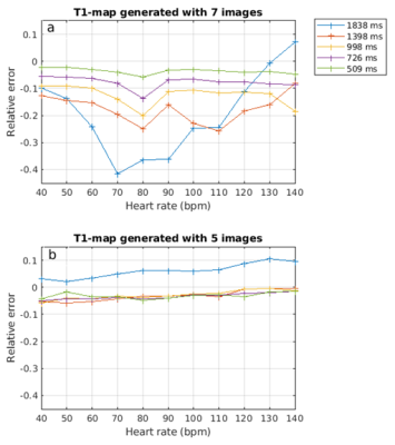
T1-maps were generated using all seven images and the five first acquired images (i.e. the images in the first Look-Locker block), without conditional data analysis. Regions in septal myocardium and blood pool and in the phantom were manually segmented. In the phantom, relative errors in T1-values were measured to investigate the differences in accuracy and HR dependence. In patients, the effect of using five compared to seven images was demonstrated by calculating differences in both myocardial and blood T1 values and tested using Wilcoxon signed rank test with a significance level of 0.05. Also, the variance in myocardial and blood T1 was compared. For the one patient with LGE, we also compared the effect in contrast between the region with and without LGE in both native and post contrast T1-maps.
Results
PhantomThe HR dependence was substantial in phantom T1-maps generated using all seven images with an underestimation of T1 of as much as 40 % (Figure 2). In these T1-maps, the error was shown to increase with increasing T1-value. T1-maps generated with the first five images displayed low HR dependence and accurate estimates.
Patients
In patients, the estimated T1-value increased significantly in both myocardium and blood (p<0.001) when five images were used in generation of the T1-maps (Figure 3). By using this optimized postprocessing strategy, the variance in the blood T1-value decreased significantly. The difference was larger in the blood pool compared to the septal myocardium and largest for the HRs in the range 71-90 bpm (table 1). Table 2 shows the relative difference of T1-values in the region of LGE compared to region without in the patient with both native and post-contrast scans. The relative difference was larger when five instead of seven images was used in the generation of the T1-maps.
Discussion
Present study demonstrated clinically relevant errors in the T1-value with a substantial HR dependence if conditional data optimization is not implemented in the post-processing pipeline. We showed that the HR dependency can be effectively reduced and the accuracy can be highly improved by including only the five first acquired images in the generation of the T1-maps. As shown by our findings, such optimized postprocessing strategy is of outmost importance for the evaluation of patients with myocardial disease, not only for accurate T1-estimations of the myocardial tissue but also for improved differentiation between affected and unaffected myocardium. Moreover, the concomitant low HR dependence is beneficial in the long-term follow-up of these patients. With lower variance in the blood pool, more accurate synthetic extracellular volume fraction measurements might be achieved. Future studies are warranted to confirm this.The small change in the variance of the myocardial T1-value after optimized post-processing could be due to the fact that myocardium has a lower T1-value and thereby is less sensitive to HR variations and that the difference in myocardial T1-value between the included individuals thereby was larger than the HR effect.
Conclusion
Our findings suggest that ShMOLLI can offer reliable T1-measurement with low HR dependence after optimized post-processing by including only the first five images in the generation of the T1-maps and as such is suitable for the pediatric population.Acknowledgements
No acknowledgement found.References
1. Kellman P, Hansen MS. T1-mapping in the heart: accuracy and precision. J Cardiovasc Magn Reson. 2014;16(1):2.
2. Piechnik SK, Ferreira VM, Dall'Armellina E, et al. Shortened Modified Look-Locker Inversion recovery (ShMOLLI) for clinical myocardial T1-mapping at 1.5 and 3 T within a 9 heartbeat breathhold. J Cardiovasc Magn Reson. 2010;12(1):69.
3. Keenan, K. , Stupic, K. , Boss, M. , et al. Comparison of T1 measurement using ISMRM/NIST system phantom. Proceedings of the 24th annual meeting of ISMRM, Singapore, Singapore 2016.
Figures

