1104
VASO fMRI USING 2D-SMS SPIRAL READOUTS1Maastricht University, Maastricht, Netherlands, 2Athinoula A. Martinos Center for Biomedical Imaging, MGH, Boston, MA, United States
Synopsis
High-resolution blood volume-sensitive fMRI VASO can capture functional signal changes with high localization specivity compared to conventional BOLD sequences. However, it suffers from reduced sampling efficiencies and lower detection sensitivity. Here, we overcome these limitations by combining VASO contrasts with the high efficiency of multi-echo enabled spiral k-space sampling and CAIPI-optimized SMS acceleration. We find that spiral sampling enables faster VASO acquisitions than possible with Cartesian EPI sampling. For high spatial resolutions (1.25mm), we confirm that the proposed method is insensitive to large draining veins while having more stable fMRI signals (higher tSNR).
INTRODUCTION
Blood-volume-sensitive (VASO) fMRI[1] can be advantageous compared to conventional GE-EPI-based BOLD methods, especially at ultra-high field or when high spatial resolution is required. VASO provides improved spatial specificity down to the level of cortical layers and columns, and allows the quantification of meaningful physiological parameters. Its neuroscientific application, however, is limited by BOLD-contrast contamination and temporal sampling inefficiency. VASO fMRI acquisitions are conventionally performed with Cartesian EPI readouts, which put limits on the shortest echo time (TE) and readout durations achievable, especially for high resolution studies.To overcome these VASO fMRI limitations, specifically to achieve faster acquisitions at a shorter echo time, free of BOLD contamination, we combined the efficiency of spiral k-space sampling and SMS[2] acceleration with blood-volume-sensitive VASO contrast. We used two different spiral readout schemes to perform interleaved VASO-BOLD acquisitions to explore the feasibility of VASO fMRI with 2D-SMS spiral readout at ultra-high field. The flexibility and efficiency of spiral readouts have been previously demonstrated at 7T, including BOLD fMRI acquisitions[3,4,5]. Spiral sampling enables faster VASO acquisitions than previously possible for given resolution and coverage requirements. Furthermore, for high spatial resolution we show that the proposed method is less sensitive to large draining veins; which allows for better separation of fMRI signals from ‘kissing’ gyri, e.g. as in sensory and motor cortex.METHODS
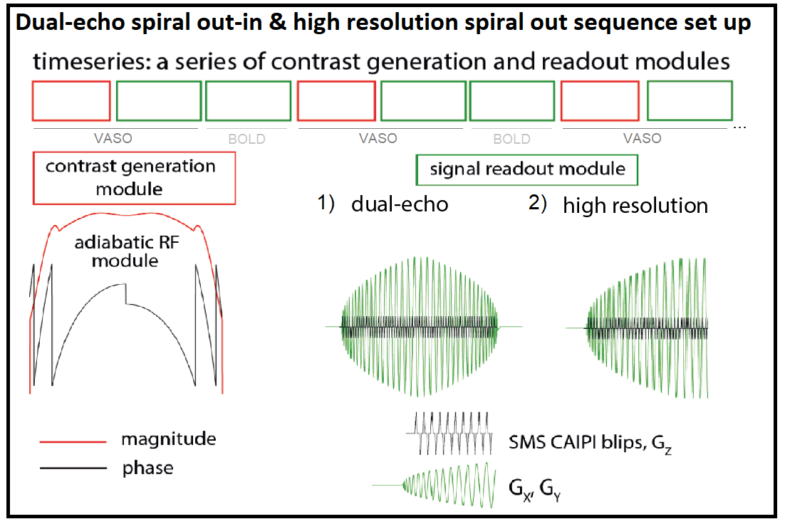
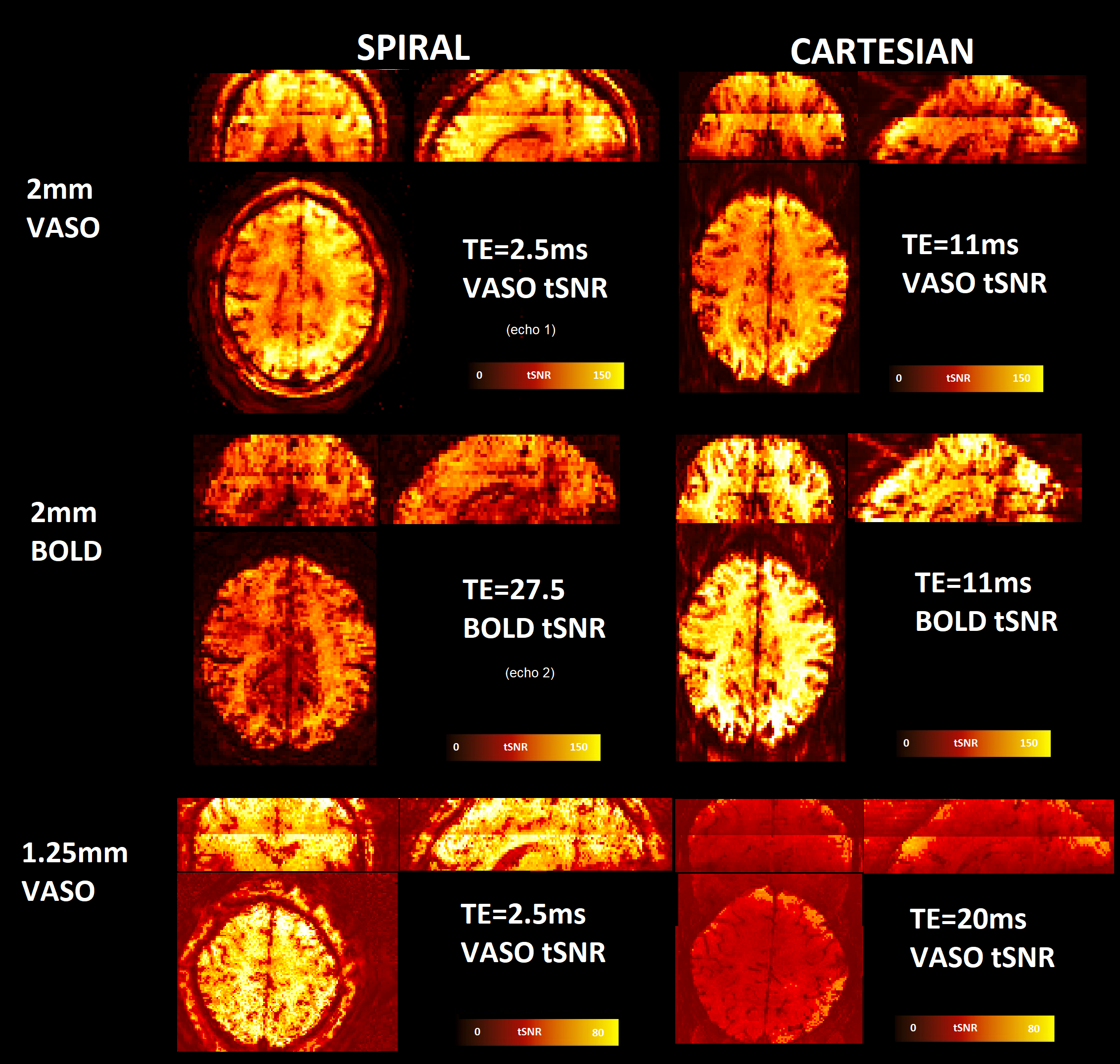
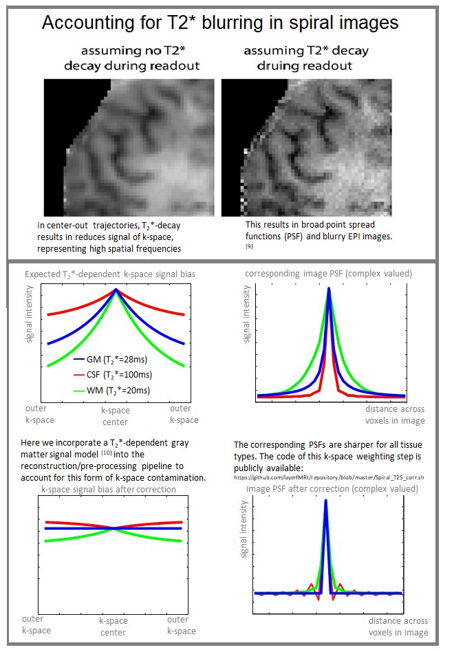
Two spiral readout schemes were implemented in a slice-selective slab inversion (SS-SI) VASO[6] sequence in the vendor-provided IDEA environment (VB17A-UHF) (Figure 1): 1) dual-echo spiral out-in readout for simultaneous acquisition of VASO and BOLD contrasts at 2mm3 iso, nominal TE1/TE2=2.5ms/27.5ms, TRVASO=1350ms, TRBOLD=350ms 24 slices, SMS-factor=2. 2) High resolution spiral-out readout at 1.25x1.25x1.2mm3 resolution, TE=2.5ms, TRVASO=1500ms, TRBOLD=500ms, 30 slices, SMS-factor=2. Data was acquired on a 7T Siemens scanner (SIEMENS Healthineers). Cartesian acquisitions with matched parameters were also performed (TE2mm/TEhighres=11/19ms). The VASO specific inversion was implemented by means of a 10 ms TR-FOCI pulse 950 ms before the first excitation pulse of the readout module. 9-minute runs were acquired with a visuo-motor functional block-design task (30/30 TRs ON/OFF). The image reconstruction of the spiral data was performed with Minimal Linear Network reconstruction that is trained on session-specific B0 maps [7]. No smoothing was applied during any stage of the analysis in SPM (MOCO), AFNI, FSL (for GLM), and LayNii (for profile plot). We applied a T2* deblurring in the pre-processing of the spiral data (Figure 4).RESULTS
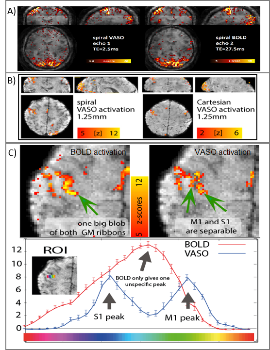
Figure 2 shows the mean image and tSNR maps of the VASO time series, corrected for BOLD contamination. Compared to Cartesian acquisitions, spiral acquisitions at very short TE show superior temporal stability for the VASO contrast. The difference between the tSNR of spiral and Cartesian acquisitions is especially highlighted in the high resolution acquisitions as the Cartesian EPI TE becomes longer. For the dual-echo VASO-BOLD acquisitions we show that the visuomotor task leads to strong signal changes in both CBV and BOLD. In the high resolution VASO acquisitions, spiral images resulted in stronger activation maps compared to Cartesian images (figure 3B). Additionally, VASO shows higher spatial specificity compared to BOLD (figure 3C): this is visible in the profiles as two separate activations for M1 and S1 in the VASO signal, whereas the BOLD signal generates an unspecific peak over sensory and motor cortices.DISCUSSION AND CONCLUSIONS
We combined non-Cartesian spiral readouts with blood volume sensitive VASO contrast preparations and showed the feasibility of such technique in a task-based fMRI experiment at 7T. The proposed approach provides superior signal stability, unprecedented sampling efficiency (sub-second TRs), and avoids unwanted sensitivity to large veins. The technique might become a valuable tool for neuroscientific applications that require high spatio-temporal resolutions. Extension of the proposed 2D approach to 3D stack-of-spirals is desirable as benefits of 3D VASO acquisitions have previously been shown[8].Acknowledgements
Funding support: LH NWO VENI 016.198.032, BAP, DK VIDI 016.178.052,We thank Christopher J Wiggins from Scannexus (Maastricht, NL) for his enthusiastic support of this project.References
1- Lu, Hanzhang, Xavier Golay, James J Pekar, and Peter C M van Zijl. 2003. “Functional Magnetic Resonance Imaging Based on Changes in Vascular Space Occupancy.” Magnetic Resonance in Medicine 50: 263–74.
2- Setsompop, K., Gagoski, B. A., Polimeni, J. R., Witzel, T., Wedeen, V. J., & Wald, L. L. (2011). “Blipped-controlled aliasing in parallel imaging for simultaneous multislice echo planar imaging with reduced g-factor penalty. Magnetic Resonance in Medicine.” 67(5), 1210–1224.
3- Engel, M., Kasper, L., Barmet, C., Schmid, T., Vionnet, L., Wilm, B., & Pruessmann, K. P. (2018). “Single-shot spiral imaging at 7 T.” Magnetic Resonance in Medicine 80(5), 1836–1846.
4- Kasper, Lars; Engel, Maria; Heinzle, Jakob; Mueller-Schrader, Matthias; Reber, Jonas; Schmid, Thomas et al. (2019): “Advances in Spiral fMRI. A High-resolution Study with Single-shot Acquisition.” In bioRxiv.
5- Kurban, D., Liberman, G., Kashyap, S., Ivanov, D., Poser, AB. (2019) “Dual-echo simultaneous multi-slice spiral acquisition for concurrent CBF and BOLD fMRI at 7T.” Proceedings of ISMRM, Montreal
6- Huber, Laurentius et al. 2014. “Slab-Selective, BOLD-Corrected VASO at 7 Tesla Provides Measures of Cerebral Blood Volume Reactivity with High Signal-to-Noise Ratio.” Magnetic Resonance in Medicine 72(1): 137–48.
7- Liberman G, Poser BA. (2019) “Minimal Linear Networks for Magnetic Resonance Image Reconstruction.” Sci Rep 9, 19527
8- Huber, L., Ivanov, D., Handwerker, D. A., Marrett, S., Guidi, M., Uludağ, K., … Poser, B. A. (2018). “Techniques for blood volume fMRI with VASO: From low-resolution mapping towards sub-millimeter layer-dependent applications.” NeuroImage, 164, 131–143.
9- Huber L, Guidi M, Goense JBM, et al. (2015). The magnitude point spread function is an inadequate measure of T2*-blurring in EPI. Proc Int Soc Magn Reson Med.;23:2056.
10- Kasper L, Engel M, Barmet C, et al. (2018). Rapid anatomical brain imaging using spiral acquisition and an expanded signal model. Neuroimage.;168:88-100.
11. Shao X, Guo F, Shou Q, et al. Laminar perfusion imaging with zoomed arterial spin labeling at 7 Tesla. bioRxiv. 2021:1-20.
Figures