1074
Investigating liver fibrosis using water T1 estimated by flip angle corrected multi-parameter MRS
Ashley L Louie1, Gavin Hamilton1, Alexandra N Schlein1, Walter C Henderson1, Danielle N Batakis1, Lael K Ceriani1, Yesenia Covarrubias1, Tanya Wolfson1, Nikolaos Panagiotopolous2, David T Harris2, McMillan Alan2, Daiki Tamada2, Kathryn J Fowler1, Scott B Reeder2, and Claude B Sirlin1
1Radiology, University of California, San Diego, La Jolla, CA, United States, 2Radiology, University of Wisconsin, Madison, WI, United States
1Radiology, University of California, San Diego, La Jolla, CA, United States, 2Radiology, University of Wisconsin, Madison, WI, United States
Synopsis
The purpose of this study was to examine the relationship between water T1 and T2 estimated by Flip Angle Corrected Multi-Parameter (CMP) MRS and biopsy-determined fibrosis. The sample of twenty-eight subjects with biopsy and MRS data is part of a dual-center, weight loss surgery study. Subjects without fibrosis were found to have significantly higher CMP MRS T1 water values than patients with fibrosis.
Introduction
Liver Water T1 (T1w), has been proposed as a possible non-invasive MR based biomarker for quantifying liver fibrosis (1). MRI based measures of T1 cannot directly estimate T1w but rather estimate liver T1, a value confounded by the presence of fat. MRS techniques have been developed that can directly estimate T1w, as well as water T2 (T2w), fat T1 and fat T2, and proton density fat fraction (PDFF), all within a single breath-hold (2). The main limitation of these techniques is bias related to B1 related flip angle calibration errors. To address these issues, we developed a single breath-hold flip-angle Corrected Multi-Parameter MRS sequence (CMP MRS) that estimates these parameters independent of flip angle calibration errors. In this pilot dual-center study, we examined whether T1w estimated by CMP MRS was significantly different in patients with liver fibrosis compared to those without.Methods
Study designThis dual-site study was IRB approved and HIPAA compliant, and all subjects provided written informed consent. Adult subjects (n = 28 mean age 45.7 years, range 29-65 yrs, 3 male, 25 female) who were undergoing Laparascopic Sleeve Gastrectomy surgery for treatment of obesity at two different sites had CMP MRS acquired at 3.0T (Discovery 750, Signa Excite HD, GE Healthcare, Waukesha, WI). Liver biopsies were acquired during weight loss surgery performed 1-3 days after MRI, and were scored based on NASH CRN criteria (3) to assess fibrosis.
MRS protocol
The CMP MRS sequence acquires 39 STEAM spectra in a single 21 s acquisition (timings shown in Table 1). A 20x20x20 mm voxel was selected within the liver avoiding major blood vessels, bile ducts, the gallbladder, and liver edges. No spatial or chemical saturation was used. Signals from different array elements were combined using an SVD technique (4) and a single experienced observer analyzed the spectra using custom prior knowledge (1) in the AMARES algorithm (5) included in the JMRUI software package (6). To estimate T1 and T2, longitudinal magnetization at each acquisition (Mn) was calculated by,
Mn = Mn-1e(-τn⁄T1)cos α + M0(1-e(-τn⁄T1)),
with signal given by
Sn = Mne-TEn/T2) sin (α)
where α is the flip angle, M0 the equilibrium longitudinal magnetization, and τn and TEn as listed in Table 1.
Statistical analysis
Twenty-eight subjects with both MRS and biopsy were included in this analysis. The relationship between T1 and T2 of water with fibrosis, was assessed. Due to sample size, fibrosis was binarized into two categories “none” versus “any.” T1 and T2 data were compared between fibrosis using the Wilcoxon-Mann-Whitney test.
Results
T1w and T2w were estimated for twenty-eight subjects and was included in this analysis. Five subjects showed evidence of fibrosis on histological analysis compared to 23 showed who with no evidence of fibrosis. Subjects without fibrosis had significantly (p=0.026) higher T1w (982 ms) than patients with fibrosis (887 ms) (Figure 1). There were no significant relationship between fibrosis and T2w.Discussion
In this dual-center study we found a significant difference between the T1w for patients with and without biopsy-proven fibrosis. Due to the small sample size, we could only investigate fibrosis vs non-fibrosis. This analysis is exploratory and not adjusted for multiple comparisons. Results will need to be validated with independent data.Conclusion
We found that liver T1w estimated by CMP MRS was significantly different in fibrotic subjects compared to non-fibrotic subjects. It is surprising the T1 change seen in this study is opposite to that previously observed (1).Acknowledgements
The parent study for this analysis was funded by the NIH and Pfizer, Inc.References
- Banerjee R, Pavlides M, Tunnicliffe EM, et al. Multiparametric magnetic resonance for the non-invasive diagnosis of liver disease. Journal of Hepatology 2014; 60; 69-772)
- Hamilton G, Middleton MS, Hooker JC, et al. In vivo breath-hold 1H MRS simultaneous estimation of liver proton density fat fraction, and T1 and T2 of water and fat, with a multi-TR, multi-TE sequence. J Magn Reson Imaging 2015; 42: 1538-15433)
- Kleiner D.E., Brunt E.M., Van Natta M. et al. Design and validation of a histological scoring system for nonalcoholic fatty liver disease. Hepatology 2005; 41:1313-13214)
- Bydder M, Hamilton G, Yokoo T, Sirlin CB. Optimal phased-array combination for spectroscopy. Magn Reson Imaging 2008;26:847-850.5)
- Vanhamme L, van den Boogaart A, Van Huffel S. Improved Method for Accurate and Efficient Quantification of MRS Data with Use of Prior Knowledge. J Magn Reson 1997;129:35-43.6)
- Naressi A, Couturier C, Devos JM, et al. Java-based graphical user interface for the MRUI quantitation package. Magn Reson Mater Physics, Biol Med 2001;12:141-152.
Figures
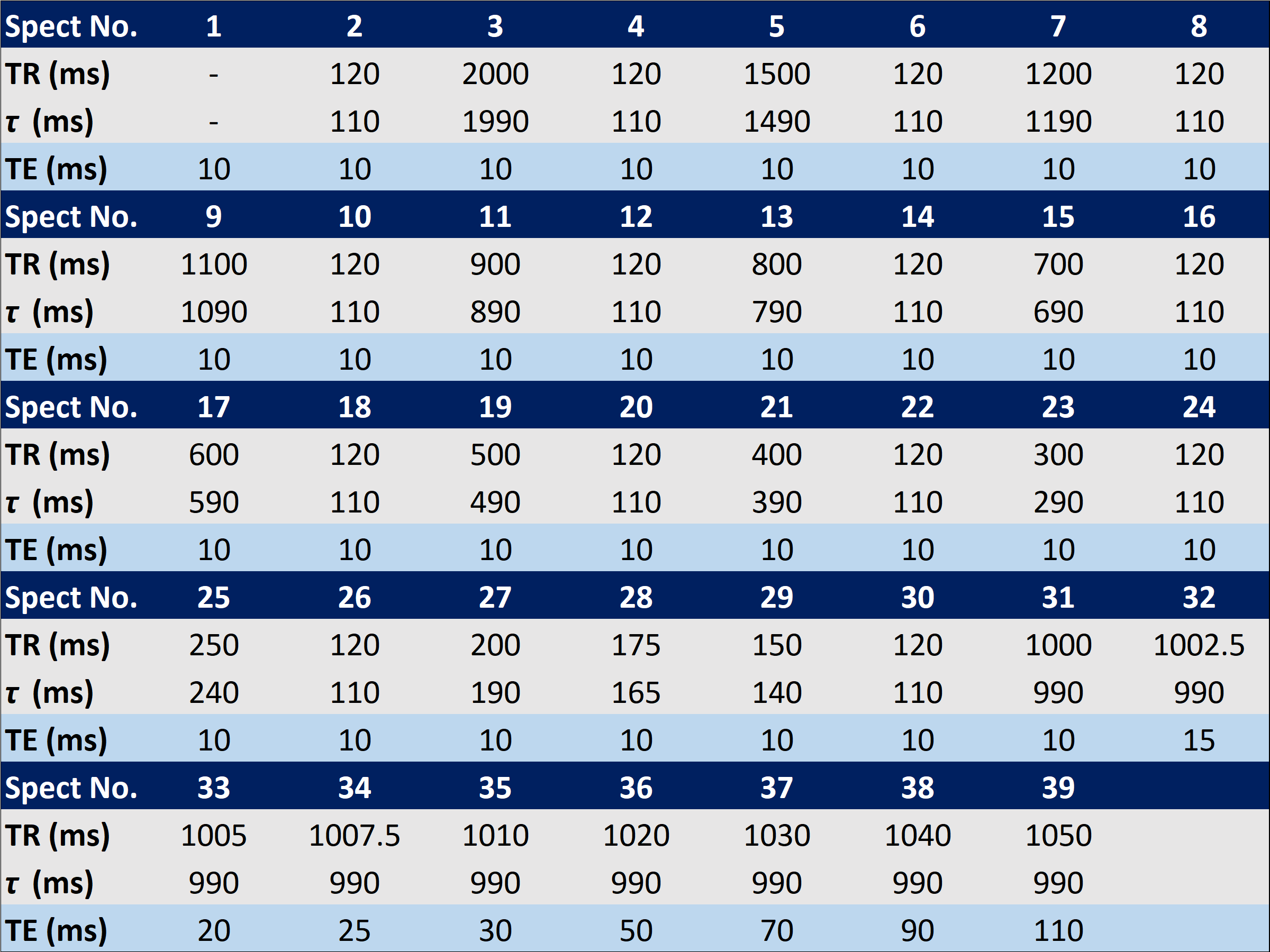
Table 1: Timings of the Flip Angle Corrected Multi-Parameter (CMP) MRS Sequence
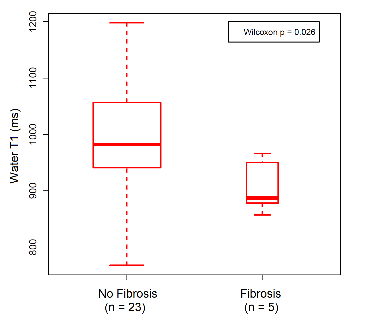
Figure 1: Comparison of Liver T1 water estimated by Flip Angle Corrected Multi-Parameter MRS in fibrotic and non- fibrotic subjects.
DOI: https://doi.org/10.58530/2022/1074