1036
Highly Asymmetric Acquisitions for Diffusion MRI Tractography
Manoj Shrestha1, Pavel Hok1,2, Ulrike Nöth1, and Ralf Deichmann1
1Brain Imaging Center (BIC), Goethe University Frankfurt, Frankfurt am Main, Germany, 2Department of Neurology, Palacky University Olomouc and University Hospital Olomouc, Olomouc, Czech Republic
1Brain Imaging Center (BIC), Goethe University Frankfurt, Frankfurt am Main, Germany, 2Department of Neurology, Palacky University Olomouc and University Hospital Olomouc, Olomouc, Czech Republic
Synopsis
Frequently, diffusion-weighting modules such as schemes based on the Stejskal-Tanner concept or the STEAM are combined with EPI readout for fast k-space sampling. In contrast to standard sequences, highly asymmetric spin-echo and highly asymmetric STEAM sequences (HASE, HASTEAM) offer shorter TE, maintaining constant diffusion preparation duration, independent of the matrix-size. In this study, HASE and HASTEAM sequences were implemented for neuronal fibre tracking, and results were compared with standard sequences. Both asymmetric sequences HASE and HASTEAM allow for more accurate estimations of the subsidiary fibre orientations, which may considerably improve the quality and quantity of the detected fibre tracts.
Introduction
Frequently, diffusion-weighting (DW) modules such as schemes based on the Stejskal-Tanner concept [1] or the stimulated echo acquisition mode (STEAM) [2] are combined with an echo-planar imaging (EPI) readout for fast k-space sampling. Recently, highly asymmetric spin-echo (HASE) and highly asymmetric STEAM (HASTEAM) sequences [3] were developed for improving the signal-to-noise ratio (SNR) and reducing eddy-current related artifacts in DW imaging (DWI) [4]. In this study, these sequences were implemented for neuronal fibre tracking, and results were compared with standard DWI sequences.Methods
Pulse sequences: Figures 1A and 1B show the HASE-EPI and the HASTEAM sequences [3], respectively, where echo formation occurs prior to the EPI-readout, rather than at the k-space centre as in standard SE-EPI. This allows for DW preparation with constant duration, independent of the spatial resolution. In both sequences, there is a T2-decay during TE1, with an additional T1-decay during TM in HASTEAM. Additionally, there is a T2*-decay during TE2 which depends on the matrix-size of the acquisition. HASTEAM offers longer diffusion times and higher b-values at short TE, thus allowing to assess restricted diffusion of brain regions with short T2. Both HASE-EPI and HASTEAM-EPI sequences should yield a higher SNR for T2*>0.5T2 when choosing the shortest available TE [3].Experiments: All experiments were performed on a 3T whole-body MRI scanner (MAGNETOM Prisma, Siemens Healthineers, Germany), using a body transmit and a 64-channel phased-array head/neck receive coil. Sequences were first tested on an Agarose gel phantom, using different matrix-sizes. The optimal protocol was then used for in-vivo experiments on five healthy volunteers (3 females, 2 males, mean age ± SD: 25.5±5.4 years). The in-vivo study was approved by the local ethics committee and written informed consent was obtained from each subject before scanning.
The following parameters were the same for all experiments: FoV=256×256mm2, 72 interleaved axial slices (2mm without gap), b-value=500s·mm-2/1000s·mm-2 (in-vitro/in-vivo), TM=60ms (STEAM/HASTEAM), 2-fold acceleration, partial-Fourier=6/8, five pairs of reference images at b=0 with positive and negative phase blips. Experiment-specific parameters for HASE, HASTEAM and standard DW-sequences based on STEAM, single-refocused spin-echo (srSE) and twice-refocused spin-echo (trSE) were:
In-vitro experiments:
(a) matrix-size=128×128: TR=10.5s, TE=60/72/41ms (srSE/trSE/STEAM), TE1/TE2=47/11ms (HASE), TE1/TE2=31/11ms (HASTEAM), echo spacing Tes=0.6ms, bandwidth=1953 Hz/pixel.
(b) matrix-size=160×160: TR=11.5s, TE=65/77/50ms (srSE/trSE/STEAM), TE1/TE2=48/15ms (HASE), TE1/TE2=32/15ms (HASTEAM), Tes=0.67ms, bandwidth=1736 Hz/pixel.
(c) matrix-size=192×192: TR=13s, TE=75/83/60ms (srSE/trSE/STEAM), TE1/TE2=49/20ms (HASE), TE1/TE2=33/20ms (HASTEAM), Tes=0.78ms, bandwidth=1447Hz/pixel.
In-vivo experiments: The optimal protocol (i.e. in-vitro protocol with matrix-size=128×128) with a shortened EPI readout was used. For anatomical reference, a T1-weighted dataset (3D MPRAGE [5]) with an isotropic resolution of 1 mm was acquired. The preprocessing and the subsequent data analysis were performed as previously described [4, 6].
Results
Figure 2 shows that the signal gain of HASE versus srSE (left) and of HASTEAM versus STEAM (right) increases with increasing matrix-size. Figure 3 shows that both for mean diffusivity (top) and diffusion anisotropy (bottom) maps, HASE and HASTEAM yield similar results as their standard counterparts srSE/trSE and STEAM, respectively. Figures 4a, 4b and 5 show connectivity distribution maps resulting from probabilistic fibre tracking, overlaid on the anatomical T1-weighted dataset and displayed in three orthogonal planes. Colour intensities represent the number of streamlines binned in each voxel with a lower threshold of 20 fibres per voxel. Each figure compares a sequence S1 with a sequence S2, showing fibre tracks resulting only from S1 (red-yellow), only from S2 (blue) or from both (green). Comparisons refer to S1=HASE, S2=trSE (Fig. 4a), S1=srSE, S2=HASE (Fig. 4b), S1=STEAM, S2=HASTEAM (Fig. 5). HASE-based data show additional connections to the lateral frontal, parietal and temporal cortices as well as to the upper brainstem, compared to data based on standard sequences (Fig 4). In contrast, both STEAM and HASTEAM yielded almost similar fibre tracking results (Fig. 5).Discussion/Conclusion
In contrast to standard sequences, both HASE and HASTEAM offer shorter TE, maintaining constant diffusion preparation duration, independent of the matrix-size. In-vitro results clearly show that the signal gain of HASE/HASTEAM increases with increasing matrix-size. In-vivo results suggest that HASE/HASTEAM improve fibre tracking results in high resolution imaging. This allows for more accurate estimations of the subsidiary fibre orientations, which may considerably improve the quality and quantity of the detected fibre tracts.Acknowledgements
No acknowledgement found.References
- Stejskal EO, Tanner JE (1965). Spin diffusion measurements: spin echoes in the presence of a time‐dependent field gradient. J Chem Phys, 42(1), 288-292.
- Tanner JE (1970). Use of the stimulated echo in NMR diffusion studies. J Chem Phys 52(5), 2523-2526.
- Shrestha M, Nöth U, Deichmann R (2019). Improved signal-to-noise ratio in EPI sequences with highly asymmetric spin echo and highly asymmetric STEAM preparations (HASE-EPI and HASTEAM-EPI). Magn Reson Mater Phy 32(5), 549-558.
- Shrestha M, Hok P, Nöth U, Lienerth B, Deichmann R (2018). Optimization of diffusion-weighted single-refocused spin-echo EPI by reducing eddy-current artifacts and shortening the echo time. Magn Reson Mater Phy 31(5): 585–597.
- Mugler JP, Brookeman JR (1990). Three‐dimensional magnetization‐prepared rapid gradient‐echo imaging (3D MP RAGE). Magn Reson Med, 15(1), 152-157.
- Shrestha M, Hok P, Nöth U, Lienerth B, Deichmann R (2018). Improved fibre tracking with optimized diffusion-weighted single-refocused spin echo EPI. In: Proceedings of the 26th Annual Meeting of ISMRM, Paris, #5209.
Figures
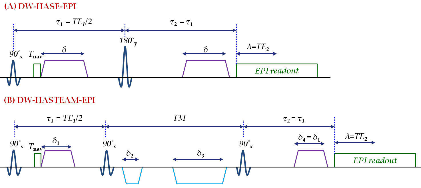
Fig. 1: Schematic representation of highly asymmetric sequences for diffusion-weighted imaging. In DW-HASE-EPI (A) and DW-HASTEAM-EPI (B), the diffusion preparation is independent of the spatial resolution, thus achieving shorter TE. Navigator echoes (duration Tnav) for phase correction are acquired initially.
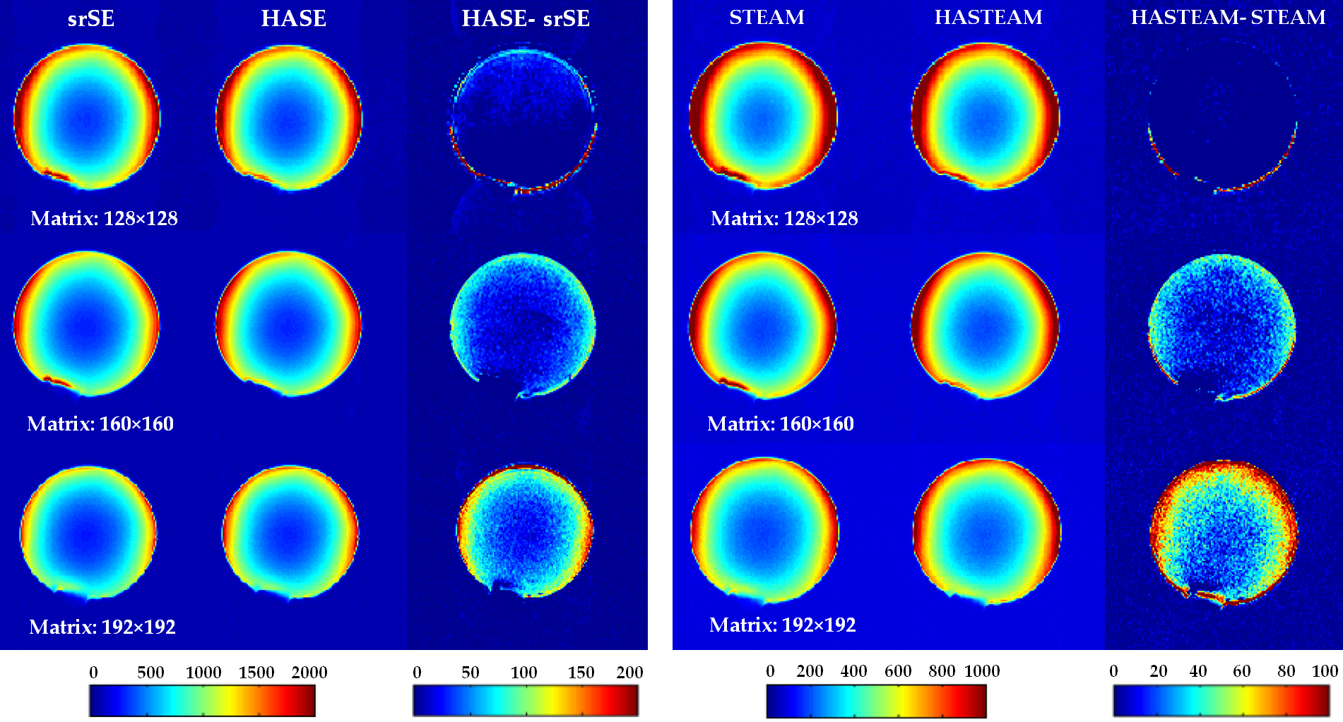
Fig. 2: Left panel: Images acquired with srSE (left column), HASE (central column) and their differences (right column) for different matrix-sizes. Right panel: respective data for STEAM and HASTEAM. Data show a signal gain of HASE and HASTEAM versus their conventional counterparts for large matrix-sizes.
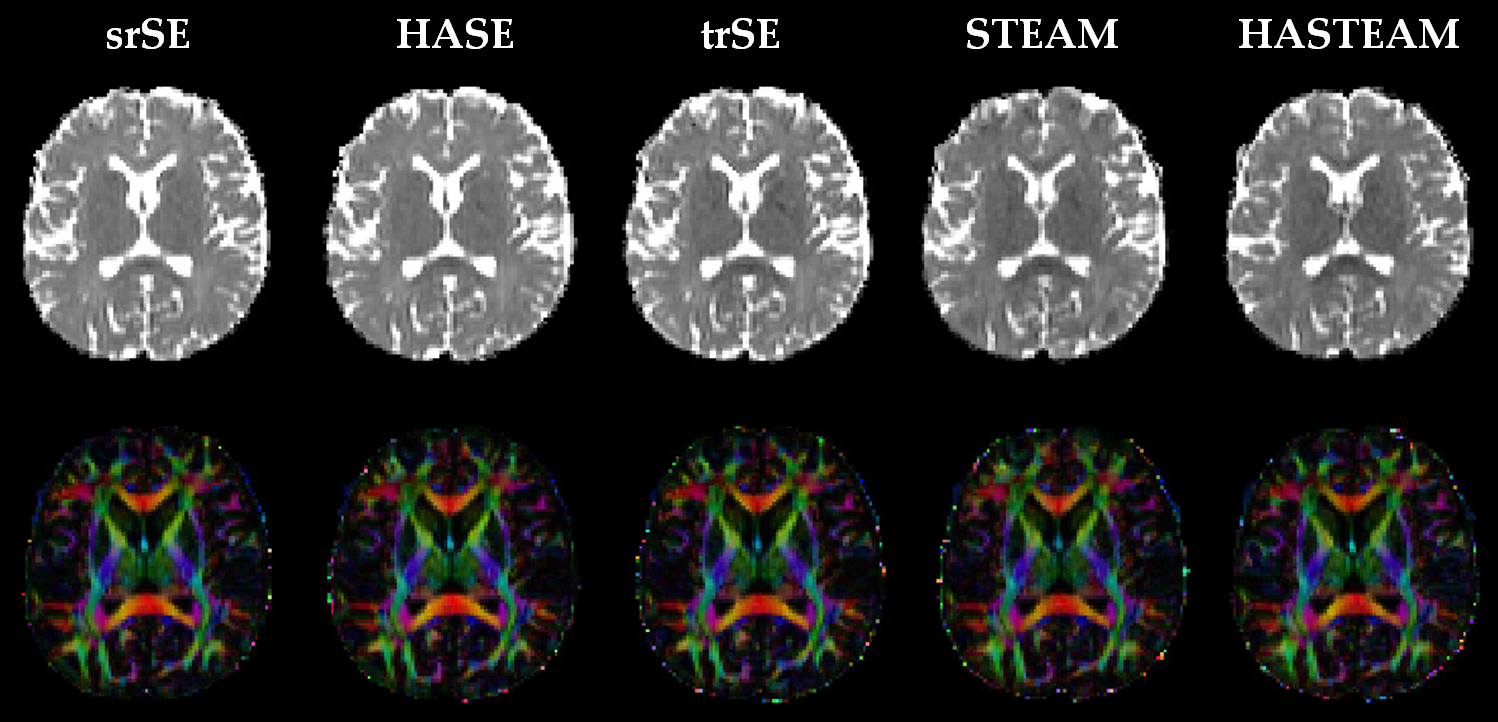
Fig. 3: Mean diffusitivity (top) and diffusion anisotropy (bottom) maps acquired with different sequences. Colors indicate the direction of the first eigenvector of the diffusion tensor. Results of the proposed sequences HASE and HASTEAM match the results of the standard sequences.
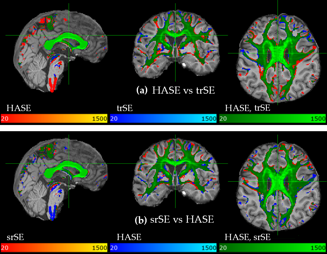
Fig. 4: Connectivity distribution maps resulting from probabilistic fibre tracking, overlaid on a high resolution T1-weighted anatomical image. Colour-overlays represent the number of streamlines binned in each voxel for the corresponding datasets, thresholded at 20 fibres/voxel. HASE traces more fibres than the twice-refocused spin-echo (trSE) sequence (a) but yields almost similar results as the single-refocused spin-echo (srSE) sequence (b); however, additional fibres in the upper brainstem are obtained with HASE only (a, b).
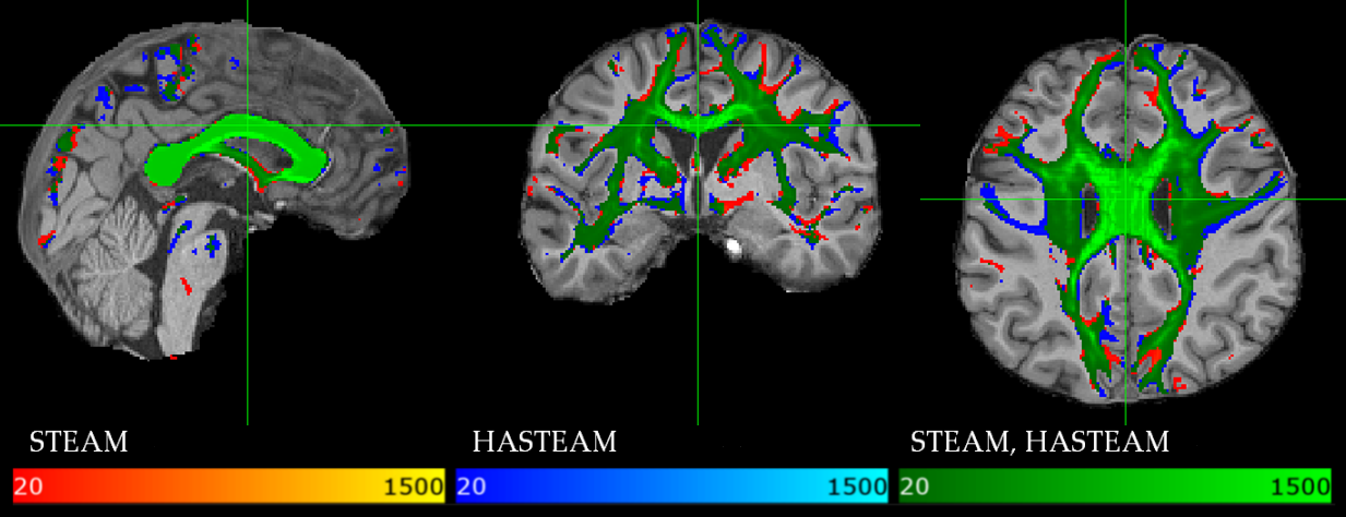
Fig. 5: Similar to Fig. 4, tractography results are compared between STEAM and HASTEAM. Both sequences yield almost similar results.
DOI: https://doi.org/10.58530/2022/1036