1034
Cerebral Blood Flow Quantification using Pseudo-continuous Arterial Spin Labeling at 7T MRI1University of Pittsburgh, PITTSBURGH, PA, United States, 2University of Pittsburgh, Pittsburgh, PA, United States, 3University of Pittsburgh, pittsburgh, PA, United States
Synopsis
The feasibility of implementing pCASL technique at 7T MRI as a routine protocol uncovers the difficulties and challenges that need to be overcome and taking into consideration the many variables that need to be addressed before running the sequence. We were able to successfully implement pCASL sequence with satisfactory results. CBF quantification for the 4 volunteers was within clinical normal ranges and CBF mapping agrees with published data. Future work will include the inclusion of neck coil module for improving labeling efficiency.[GU1] Where is figure 4 referenced [GU1] Please add the references
Introduction
Pseudo-continuous Arterial Spin Labeling (pCASL) is the recommended non-invasive technique to quantify and map cerebral blood flow (CBF) in most clinical MRI settings (1). The advantages of implementing pCASL are: 1) adaptation to the existing system hardware, 2) higher signal to noise ratio that improves outcomes of ASL pair subtraction between label and control images, 3) and higher labeling efficiency. 7T MRI systems can furtherly enhance pCASL SNR as the longitudinal relaxation time of blood (T1) is longer allowing longer post-labeling delay (PLD) and higher temporal resolution(2). However, the inhomogeneity of B0 field, the deterioration of B1 field in the distal region of the brain and higher specific absorption rate (SAR) at 7T MRI are some challenges that require advanced development in radiofrequency (RF) head coil and sequence optimization (3). This work aims to implement pCASL technique at 7T MRI to ultimately quantify CBF using an existing Tic Tac Toe (TTT) RF head coil system that is currently being vastly utilized in more than 30 research studies and has been already used to scan more than 2000 human subjects.Methods
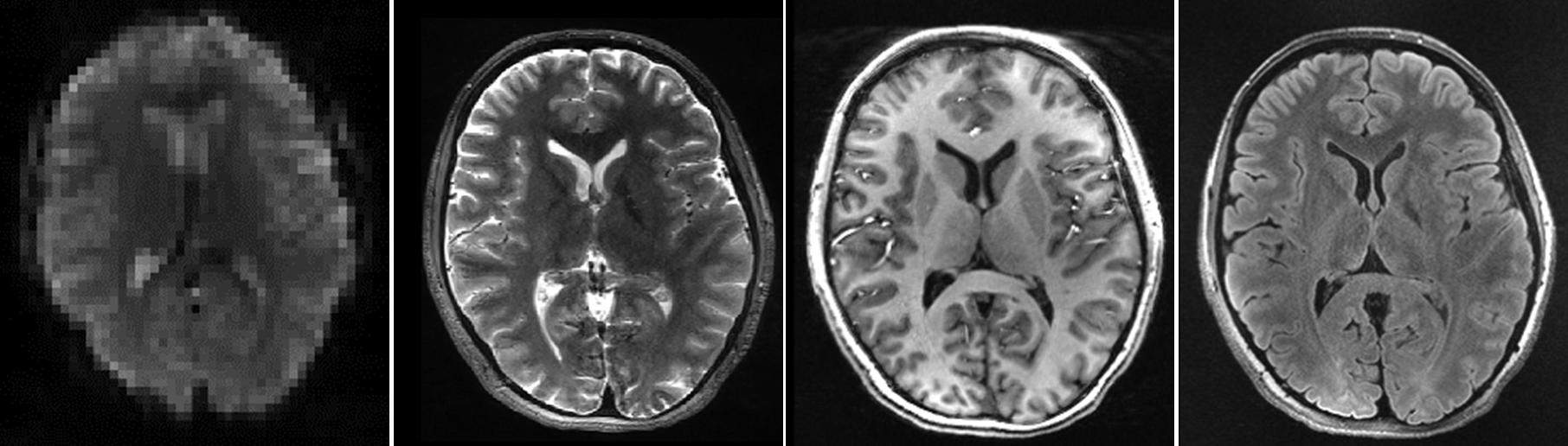
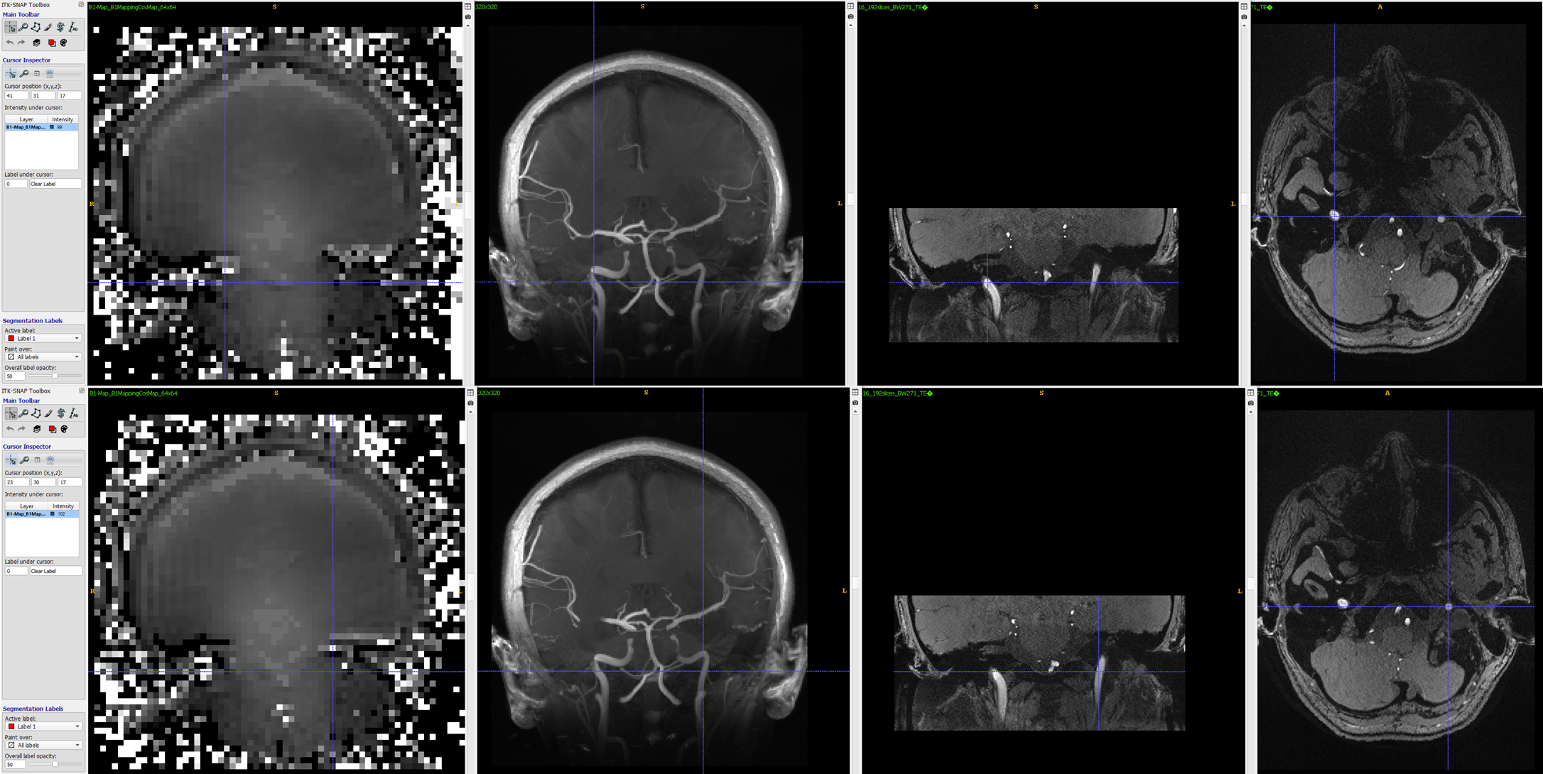
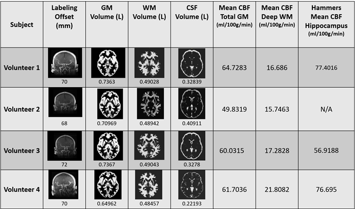
Imaging was performed on a whole-body 7T SIEMENS MRI scanner (Siemens Magnetom, Germany) with a TTT 16 channels RF transmit coil and a 32-channel receive-only head coil that achieves a homogenous B1 field in the tagging area on 4 healthy subjects (Age 21 years old), under a protocol approved by the institutional review board. As shown in figure (1), four sequences were acquired: T1-weighted (T1w) Magnetization Prepared RApid Gradient Echo (MPRAGE), T2-weighted (T2w) SPACE sequence, T2w Fluid-Attenuated Inversion Recovery (FLAIR), pCASL sequence. The pCASL labeling was optimized to the following parameters: labeling time = 1.8 s, Flip angle = 30, post labeling delay (PLD) = 1.7s, and labeling plane offset = 65-70 mm which we manipulate depending on the tagging area of each subject as shown in figure (3).Two‐dimensional EPI readout parameters for 2D PCASL were TR/TE = 4.5 s/18 ms, matrix size = 64 × 64, in‐plane resolution = 3.75 × 3.75 mm2, Echo spacing =0.47. Twenty-two slices with 4 mm thickness and a 20% distance factor were acquired. Sixteen label–control pairs were acquired, and the mean of the control images was used as M0 image.Results
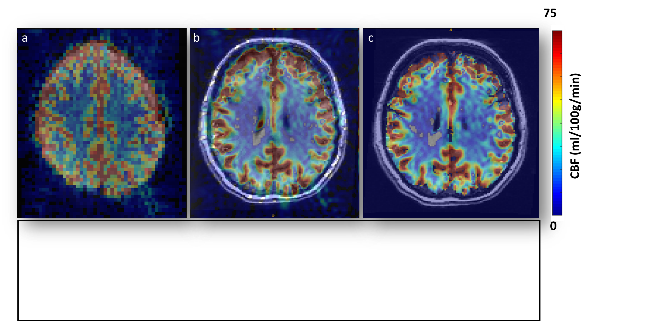
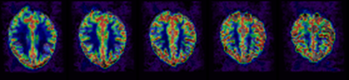
The process of optimizing pCASL sequence parameters to ensure optimal outcomes in young adults included: 1) adjusting the labeling offset between (55 mm - 85 mm); as shown in figure (2), 2) accounting for B1 intensity difference between left and right carotid arteries which were minimal (B1 left =102 degrees, B1 Right = 99 degrees) and at a proximally accepted distance from circle of Willis, 3) variating post labeling delay based on literature review between (1.4 s – 2s); we concluded to use 1.7 s. 3), 4) optimizing tagging flip angle between (23 degree to 45 degree) to compensate for B1 deterioration in the distal brain region, FA= 30 degrees achieved sufficient flipping and the magnetic transfer was tolerable, 5) testing FOV and Image resolution. We decided to adapt the following resolution (3.75mmx3.75mmx4mm), and 6) testing RF pulse duration so it can be tailored to our coil RF capacity and elements. After applying the final parameters, other external factors need to be regulated such as shimming and motion were considered. User determines the pipeline output by modifying a .json file that generates images as shown in figure (3,4) and measurements as shown in table 1. In figure (3) perfusion weighted images and CBF distribution are viewed in both standard and MNI space.Discussion and Conclusion
The feasibility of implementing pCASL technique at 7T MRI as a routine protocol uncovers the difficulties and challenges that need to be overcome and taking into consideration the many variables that need to be addressed before running the sequence. We were able to successfully implement pCASL sequence with satisfactory results. CBF quantification for the 4 volunteers was within clinical normal ranges and CBF mapping agrees with published data. Future work will include the inclusion of neck coil module for improving labeling efficiency.Acknowledgements
No acknowledgement found.References
1. Grade M, Hernandez Tamames JA, Pizzini FB, Achten E, Golay X, Smits M. A neuroradiologist’s guide to arterial spin labeling MRI in clinical practice. Neuroradiology. 2015 Dec;57(12):1181–202.2. Ivanov D, Gardumi A, Haast RAM, Pfeuffer J, Poser BA, Uludağ K. Comparison of 3T and 7T ASL techniques for concurrent functional perfusion and BOLD studies. NeuroImage.
Bandettini PA, Bowtell R, Jezzard P, Turner R. Ultrahigh field systems and applications at 7 T and beyond: Progress, pitfalls, and potential: UHF Systems and Applications at 7 T and Beyond. Magn Reson Med. 2012 Feb;67(2):317–21.4. Pohmann R, Speck O, Scheffler K. Signal-to-noise ratio and MR tissue parameters in human brain imaging at 3, 7, and 9.4 tesla using current receive coil arrays: SNR at 9.4T. Magn Reson Med. 2016 Feb;75(2):801–9.5. Santini T, Kim J, Wood S, Krishnamurthy N, Farhat N, Maciel C, et al. A new RF transmit coil for foot and ankle imaging at 7T MRI. Magn Reson Imaging. 2018 Jan;45:1–6.6. Kim J, Santini T, Bae KT, Krishnamurthy N, Zhao Y, Zhao T, et al. Development of a 7 T RF coil system for breast imaging. NMR Biomed [Internet]. 2017 Jan [cited 2019 Nov 17];30(1). Available
from: https://www.ncbi.nlm.nih.gov/pmc/articles/PMC5943082/7. Santini T, Zhao Y, Wood S, Krishnamurthy N, Kim J, Farhat N, et al. In-vivo and numerical analysis of the eigenmodes produced by a multi-level Tic-Tac-Toe head transmit array for 7 Tesla MRI. PLoS ONE [Internet]. 2018 Nov 27 [cited 2019 Nov 17];13(11). Available from: https://www.ncbi.nlm.nih.gov/pmc/articles/PMC6258503
Mutsaerts et al. - 2020 - ExploreASL An Image Processing Pipeline for Multi.Pdf,” n.d. Mutsaerts, Henk J.M.M., Jan Petr, Paul Groot, Pieter Vandemaele, Silvia Ingala, Andrew D. Robertson, Lena Václavů, et al. “ExploreASL: An Image Processing Pipeline for Multi-Center ASL Perfusion MRI Studies.” NeuroImage 219 (October 2020): 117031. https://doi.org/10.1016/j.neuroimage.2020.117031.
Figures