1023
Comparative Study on MR Imaging Quality of Anterior Talofibular Ligament based on Carotid Plaque Special Surface Coil1Department of Radiology, the Third Xiangya Hospital, Central South University, Changsha, China, 2Philips Healthcare, Guangzhou, China
Synopsis
Ankle sprain is a common athletic injury. Most patients with acute ankle sprain could not be treated properly, without precise diagnosis. Conventional Magnetic Resonance Imaging (MRI) sequences, like T1WI and T2WI, could not provide diagnostic proof as accurately as possible. Proton density weighted imaging (PDWI) collaborating with fat saturation (FS) could be a better alternative to detect the lesions. A comparative study was established, to investigate the performance of 4 sequences, with 8-channel carotid special surface coil. The results showed that PDWI-FS may be the best choice on diagnosis of ankle injuries among 4 sequences
INTRODUCTION
Ankle sprain is a common athletic injury [1,2], of which 85% is lateral ankle sprain (LAS), and up to 80% of LAS are of the inversion type, with the anterior talofibular ligament (ATFL) being the most vulnerable [3]. Due to the lack of diagnostic precision, up to 50% of patients with acute ankle sprain could not receive medical treatment properly, and 34~70% of patients continued to suffer from symptoms such as ankle pain, swelling and instability. Magnetic Resonance Imaging has been widely used in clinical detection of athletic injuries, without radiation exposure about which CT has always been concerned. Conventionally in such scenario, T1WI and T2WI are involved the most, while PDWI [4] is rarely reported. Besides, sequence with FS has been proved to be valuable and more sensitive to diagnose ATFL injuries [5]. In this study, a comparison was established, to investigate the performance of 4 sequences on diagnosis of ankle ATFL, including T1WI, T2WI, T2WI-FS and PDWI-FS, with 8-channel carotid special surface coil. Results from this study would provide diagnosis of ATFL injuries more precisely, and hence offer better treatment plans in case of corresponding symptoms patients would experienceMETHODS
32 healthy subjects were enrolled and examined by magnetic resonance imaging of ATFL with Philips Ingenia 3.0T scanner and 8-channel carotid surface coil. The MRI sequences included T1WI, T2WI, T2WI-FS and PDWI-FS, which were scanned with oblique axial thin layer of 1.2 mm. Protocols: Plain scan. Axial T2WI-FS or PDWI-FS, with the baseline parallel to the ankle space on the sagittal image, and parallel to the line between medial and lateral malleolus on the coronal image. The scanning range covered from the tibiofibular joint to the calcaneus. Coronal T1WI and PDWI-FS or T2WI-FS set the baseline parallel to the connecting line of medial, lateral malleolus, and the long axis of tibia. The scanning range covered the anterior and posterior edges of ankle joint. Sagittal PDWI or T2WI-FS, with the baseline perpendicular to the line between medial and lateral malleolus of tibia, and parallel to the long axis of tibia. The scanning range included medial and lateral malleolus of ankle joint. Enhanced scanning of axial, coronal and sagittal T1WI-FS should also be performed. Parameters: the scanning orientations were mainly coronal plane and sagittal plane, supplemented by axial plane. Small FOV, thin layer and high-resolution scanning were performed. The layer thickness of two-dimensional sequence was 3.0~4.0 mm, and the layer interval ≤ layer thickness × 10%,FOV as 160~200 mm × 160~200 mm, matrix ≥ 256 × 224. The three-dimensional sequence layer thickness was 0.5~2.0 mm, without interval scanning, FOV as 160~200 mm × 160~ 200 mm, matrix ≥ 288×256. The image quality was evaluated subjectively and objectively. Subjective evaluation: double-blind evaluation was performed by 2 senior radiologists, with rich experience in image diagnosis, to check upon image quality according to *-point Likert scale scoring criteria. Moreover, the consistency of scoring results between the radiologists? was also investigated. Objective evaluation: parameters of the anterior peroneal ligament MR images were measured, including the signal intensity, signal-to-noise ratio (SNR) and contrast-to-noise ratio (CNR)RESULTS
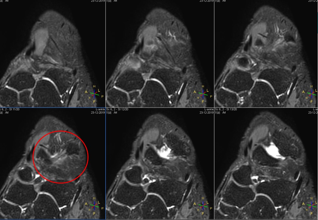
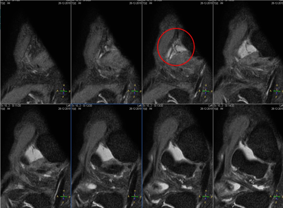
3D PDWI-FS images of Example patient A and B with ATFL injury are shown in Figure 1 and Figure 2.Subjective evaluation: after reaching an agreement between 2 radiologists, T1WI and T2WI-FS scores showed more than 3 points, and high-resolution PDWI-FS scores reached more than 5. The integrity of the start-end points of T1WI, T2WI, T2WI-FS and PDWI-FS images were scored as 0.92, 0.91, 0.89 and 0.98, respectively, indicating that high-resolution PDWI-FS performed the best.
Objective evaluation: the signal intensities of T1WI, T2WI, T2WI-FS and PDWI-FS were recorded as 1246.39, 1345.03, 860.88 and 1560.06, respectively. The SNR of each acquisition from ATFL were measured as 32.56, 33.18, 18.82 and 32.53, respectively. The tissue CNR were 5.89, 5.36, 3.07 and 7.91, respectively. High-resolution PDWI-FS was proved superior to others
DISCUSSION & CONCLUSION
In this study, a comparative experiment was conducted, to investigate the performance of 4 sequences on diagnosis of ankle ATFL, including T1WI, T2WI, T2WI-FS and PDWI-FS. By the utility of the special surface coil for carotid plaque scanning, MR images demonstrated high signal intensity and uniformity, high SNR and CNR, and hence high scores from subjective evaluation. Among all 4 sequences, T2WI-FS performed poorly, while high-resolution PDWI-FS served the best, indicating PDWI-FS might be preferred in ATFL diagnosis scenarioAcknowledgements
No acknowledgement found.References
1. Cavazos GJ Jr., Harkless LB. The epidemiology, evaluation, and assessment of lateral ankle sprains in athletes. J Sports Med Ther. 2021; 6: 008-017
2. Mollon B, Wasserstein D, Murphy GM, White LM, Theodoropoulos J. High Ankle Sprains in Professional Ice Hockey Players: Prognosis and Correlation Between Magnetic Resonance Imaging Patterns of Injury and Return to Play. Orthopaedic Journal of Sports Medicine. September 2019
3. Khor YP, Tan KJ. The Anatomic Pattern of Injuries in Acute Inversion Ankle Sprains: A Magnetic Resonance Imaging Study. Orthopaedic Journal of Sports Medicine. December 2013
4. Park HJ, Cha SD, Kim SS, Rho MH, Kwag HJ, Park NH, Lee SY. Accuracy of MRI findings in chronic lateral ankle ligament injury: comparison with surgical findings. Clin Radiol. 2012 Apr;67(4):313-8.
5. De-an Qin, Zhi-zhen Jin and Jie-fu Song. Combined anterior and posterior ankle impingement syndrome with nonunion of Cedell fracture in a 58-year-old female: a case report. BMC Musculoskeletal Disorders (2020) 21:556
Figures