1022
A Diffusion Tensor Imaging approach to investigate the effects of exercise on quadricep muscle fiber lengths1Department of Mechanical Engineering, Mississippi State University, Mississippi State, MS, United States, 2Department of Radiology, Stanford University, Stanford, CA, United States
Synopsis
There is currently no established method to monitor muscle architecture throughout exercise intervention. This work investigates the value of DTI and fiber tractography to detect changes in fiber length in the quadriceps caused by resistance training. Five human subjects participated in a three-month training regimen, and DTI scans of their quadriceps were acquired. Through DTI tractography, fiber lengths were measured and compared between quadricep muscle components. There were no statistically significant changes in fiber length after three months of exercise, showing further optimization is needed to utilize DTI tractography for monitoring fiber length changes due to exercise.
Introduction
Muscles are a highly plastic tissue that can adapt due to exercise intervention and resistance training. Athletes, particularly those engaging in high-caliber training, are prone to muscle injuries that could have physical, mental, and financial consequences3. To minimize the risk of muscular injuries, preventative strategies (e.g., targeted exercises) are used with the primary focus of increasing muscle fiber length, as longer muscle fibers are less prone to tearing or long-term damage2,5,13. In order to improve muscle adaptation in training regimens and monitor the effects of preventative strategies and rehabilitation over time, it is crucial to not only measure global changes in volume, but also changes in muscle architecture. While ultrasound imaging has investigated changes in superficial muscles throughout preventative strategies, the modality is limited to two-dimensional measurements and a narrow field-of-view that prevents analysis of deeper muscles6-7. A three-dimensional method is needed to easily track the effectiveness of preventative strategies. Diffusion Tensor Imaging (DTI), combined with deterministic fiber tractography, was shown to detect changes in muscle fiber length in the lower leg due to passive stretching8. This study aims to investigate the value of DTI and fiber tractography to detect changes in quadricep fiber length caused by resistance training.Methods
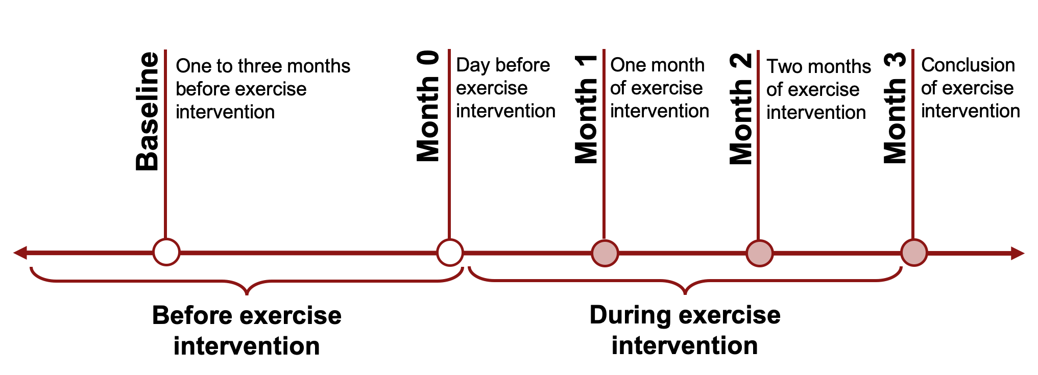
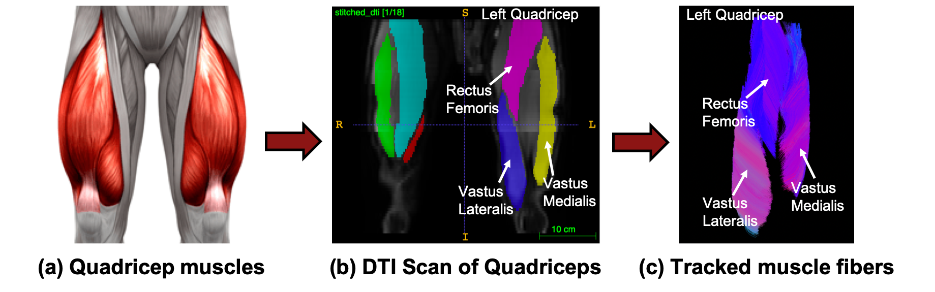
Five healthy participants performed 3x15 repetitions of single-leg decline squats twice daily, four to five days a week for three months. The DTI acquisition was performed in two stacks with the following parameters: single-shot EPI readout, b=0 s/mm2 (3 volumes) and b=400 s/mm2 (15 volumes), matrix size=256x256x26, voxel size=1.56x1.56x6 mm3, TR=3000ms, TE=43ms, 8min15s. Post-processing of DTI data included denoising, registration to anatomical scan, and tensor fitting. Dixon water datasets were also acquired from each subject for anatomical reference, manual muscle segmentation at baseline, and subsequent transfer to the other time points using imaging registration. This process was repeated for five time points, as shown in Fig 1. Then, a deterministic fiber tractography algorithm used these DTI scans to reconstruct the muscle fibers in the quadricep muscles of all five subjects, as seen in Fig 2.The Vastus Lateralis, Vastus Medialis, and Rectus Femoris muscles in the left and right leg were manually segmented for each subject and used to place seed points for initiating fiber tracking. Fiber tract density was derived from the number of tracts crossing each voxel in a whole leg tractography. Tracked fibers were terminated upon reaching a certain fiber density threshold or a dilated version of the segmented volume. The fiber density method allows for semiautomatic segmentation and exclusion of tendons and fascia that have higher tract density values compared to muscle tissue8. The segmentation volumes were slightly dilated to account for small errors in the registration between time points. Tracking parameters included a step length of five and a curvature threshold of 20°. Fiber lengths in the left and right muscle groups were averaged together with a minimum threshold of 30 mm. Differences in average muscle fiber lengths were compared for all subjects between the five time points using the Single Factor ANOVA statistical analysis with a significance level of 0.05.
Results and Discussion
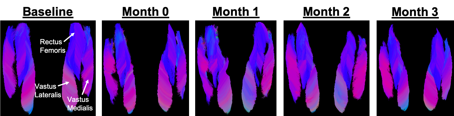
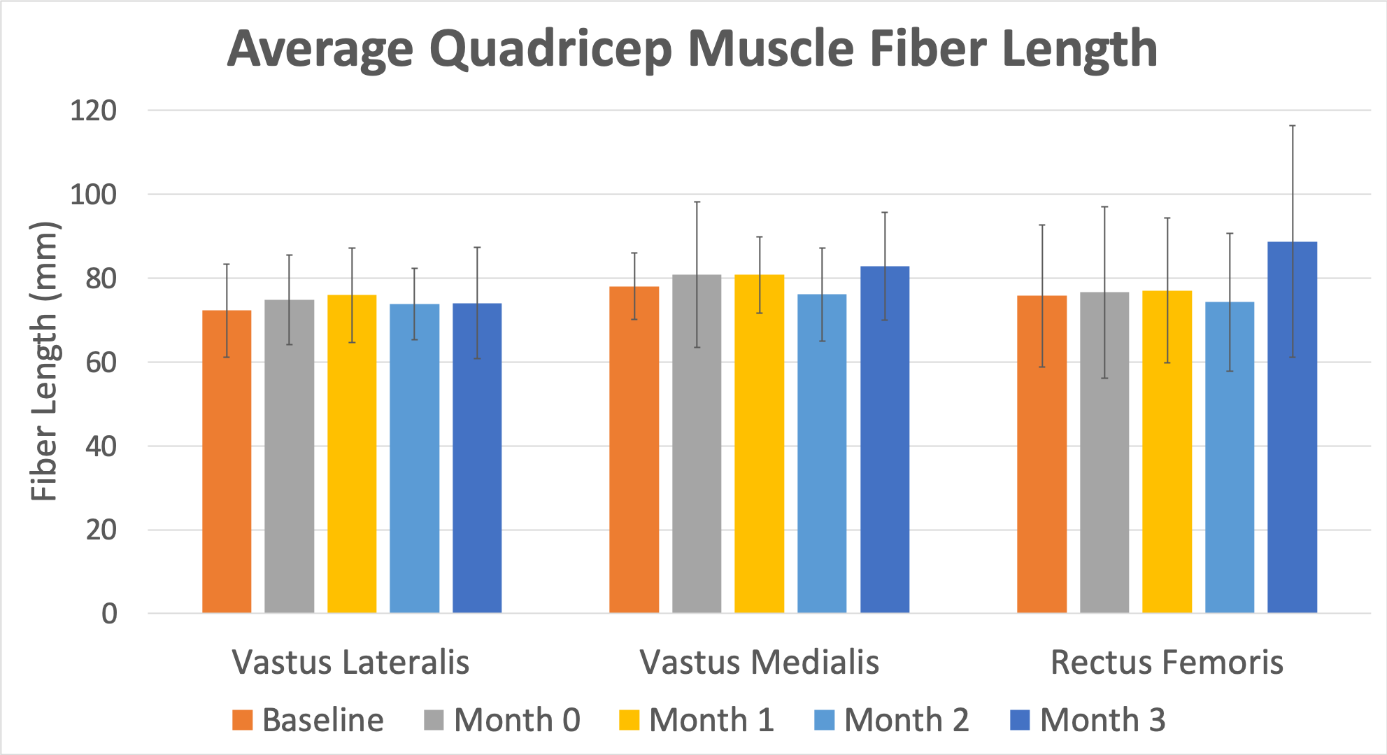
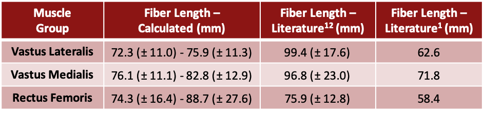
Figure 3 shows three-dimensional renderings of a representative subject’s quadricep muscle fibers for all five time points. Figure 4 quantifies the change in average fiber length between each muscle group throughout the experiment. There were slight changes in average fiber length between the beginning of exercise intervention (Month 0) and the conclusion (Month 3), including a 0.8 mm decrease, 2.0 mm increase, and 12.1 mm increase in the Vastus Lateralis, Vastus Medialis, and Rectus Femoris groups, respectively. However, these differences in average muscle fiber length are not statistically significant (p > 0.05).The lack of significant differences in fiber length could be associated with the specific exercises performed by the subjects. While the single-leg decline squats primarily targeted the human quadricep muscles, implementing exercises that are specifically aimed at increasing muscle fiber length (e.g., Nordic Training) may yield more significant results10. Nevertheless, the DTI muscle tractography approach successfully calculated quadricep muscle fiber lengths that agree with values found in literature, as represented in Table 11,12. Similar experiments used ultrasound and magnetic resonance imaging and observed an approximate 10-11% increase in quadricep muscle fiber length over time due to resistance training9,11. While no statistically significant changes were determined using the DTI tractography method, the Rectus Femoris muscle increased by about 12% from Month 0 to Month 3, which corresponds well with literature. DTI tractography provides short and easy muscle fiber acquisition and could potentially be used to conduct three-dimensional assessments of fiber length changes due to exercise. However, optimized studies with more specific fiber-lengthening exercises and larger sample sizes should be explored in the future.
Conclusion
This work used DTI and deterministic fiber tractography to investigate the effects of resistance training on fiber length in the quadricep muscles. After the three-month resistance training regimen, no statistically significant differences in fiber length were observed. However, DTI and muscle tractography calculated quadricep fiber lengths that agree with values found in literature. Taken together, these results suggest that, with further optimization, DTI fiber tractography could be a useful tool to monitor changes in quadricep muscle fiber length due to exercise.Acknowledgements
No acknowledgement found.References
1. Chow, John W., Warren G. Darling, and James C. Ehrhardt. "Determining the force-length-velocity relations of the quadriceps muscles: II. Maximum muscle stress." Journal of Applied Biomechanics 15.2 (1999): 191-199.
2. Davis, J. F., et al. "The mechanisms of adaptation for muscle fascicle length changes with exercise: Implications for spastic muscle." Medical Hypotheses 144 (2020): 110199.
3. Edouard, Pascal, and Kevin R. Ford. "Great challenges toward sports injury prevention and rehabilitation." Frontiers in sports and active living 2 (2020): 80.
4. Evans, Tracey. “The Quadriceps.” The Islander magazine (n.d.).
5. Kawakami, Yasuo. "The effects of strength training on muscle architecture in humans." International Journal of Sport and Health Science 3.Special_Issue_2005 (2005): 208-217.
6. Klimstra, Marc, et al. "The effect of ultrasound probe orientation on muscle architecture measurement." Journal of Electromyography and Kinesiology 17.4 (2007): 504-514.
7. Kwah, Li Khim, et al. "Reliability and validity of ultrasound measurements of muscle fascicle length and pennation in humans: a systematic review." Journal of applied physiology 114.6 (2013): 761-769.
8. Oudeman, Jos, et al. "A novel diffusion‐tensor MRI approach for skeletal muscle fascicle length measurements." Physiological reports 4.24 (2016): e13012.
9. Reeves, Neil D., Marco V. Narici, and Constantinos N. Maganaris. "Effect of resistance training on skeletal muscle-specific force in elderly humans." Journal of applied physiology 96.3 (2004): 885-892.
10. Richards, Jim, et al. "The effect of different decline angles on the biomechanics of double limb squats and the implications to clinical and training practice." Journal of human kinetics 52 (2016): 125.
11. Seynnes, Olivier Roger, Maarten de Boer, and Marco Vincenzo Narici. "Early skeletal muscle hypertrophy and architectural changes in response to high-intensity resistance training." Journal of applied physiology 102.1 (2007): 368-373.
12. Ward, Samuel R., et al. "Are current measurements of lower extremity muscle architecture accurate?." Clinical orthopaedics and related research 467.4 (2009): 1074-1082.
13. Zhao, Heng, et al. "Changes of calf muscle-tendon biomechanical properties induced by passive-stretching and active-movement training in children with cerebral palsy." Journal of applied physiology 111.2 (2011): 435-442.
Figures