1020
Ultrashort Echo Time Magnetization Transfer Ratio (UTE-MTR) Measurements of Paraspinal Muscles Correlate Well with Lumbar Disc Herniation1Department of Radiology, The Fifth Affiliated Hospital of Sun Yat-Sen University, Zhuhai, China, 2MR Research, GE Healthcare, Beijing, China, 3Department of Radiology, University of California San Diego, San Diego, CA, United States
Synopsis
Ultrashort echo time magnetization transfer (UTE-MT) is suggested to a non-invasive technique to assess collagenous matrix indirectly. In this study, we utilized UTE-MT sequence to investigate the relationship between UTE-MT ratio (UTE-MTR) measurements of paraspinal muscles and disc herniation in patients with disc degeneration. We found that the UTE-MTR showed a good performance in differentiation of patients with and without disc herniation, thus potentially valuable in predicting disc termination.
Introduction
Tissues in musculoskeletal system degenerate with aging, which leads to reduced tissue strength and function at an old age [1]. Degenerative paravertebral muscles may be highly related to lumbar disc herniation. Ultrashort echo time magnetization transfer (UTE-MT) is suggested to a non-invasive technique to assess collagenous matrix indirectly [2]. UTE sequence is able to detect both bound and free water signals in tissues, such as muscle, thus providing more accurate MT measurements. In this study, we utilized UTE-MT sequence to investigate the relationship between UTE-MT ratio (UTE-MTR) measurements of paraspinal muscles and disc herniation in patients with disc degeneration.Methods
A total of 57 patients with disc herniation (age 41 ± 15 years, age range 35-66) were recruited and scanned with UTE-MT sequence in lumbar L4/5 and L5/S1 disc on a 3T MRI scanner (Signa, Pioneer, GE Healthcare). Informed consent was obtained from all participants in accordance with the Institutional Review Board. A Fermi pulse was employed to generate the MT contrast in UTE-MT sequence with duration of 8 ms and bandwidth of 160 Hz. The frequency offset of this MT pulse was 1500 Hz. The UTE-MT sequence was scanned twice with flip angle of 750° for MT-On and 0° for MT-Off. Other UTE-MT sequence parameters were as follows: TR = 100 ms, TE = 0.032 ms, excitation flip angle = 5°, number of spokes per-TR = 5, FOV = 28cm × 28cm, matrix = 140 × 140, slice thickness = 3.6mm, and slice number = 16, oversampling factor = 1.2, and scan time = 3min. The UTE-MTR is calculated by the signal ratio of the difference between UTE-MT-OFF and UTE-MT-ON to the UTE-MT-OFF.Disc herniation was identified in consensus by two musculoskeletal radiologists with 6 and 8 years of experience respectively. 35 patients with herniated discs were found from the total of 57 participants. The region of interest (ROI) of the paraspinal muscles was manually drawn by a radiologist with 8 years of experience. The student’s t-test was used to compare UTE-MTR measurements of paraspinal muscles between patients with and without disc herniation. Receiver operating characteristic (ROC) analysis and computed the area under the curve (AUC) with 95% CI was performed to evaluate the performance of the UTE-MTR of paraspinal muscles in discriminating between patients with and without disc herniation. A value of P < 0.05 was considered as a statistically significant.
Results
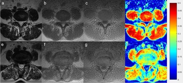
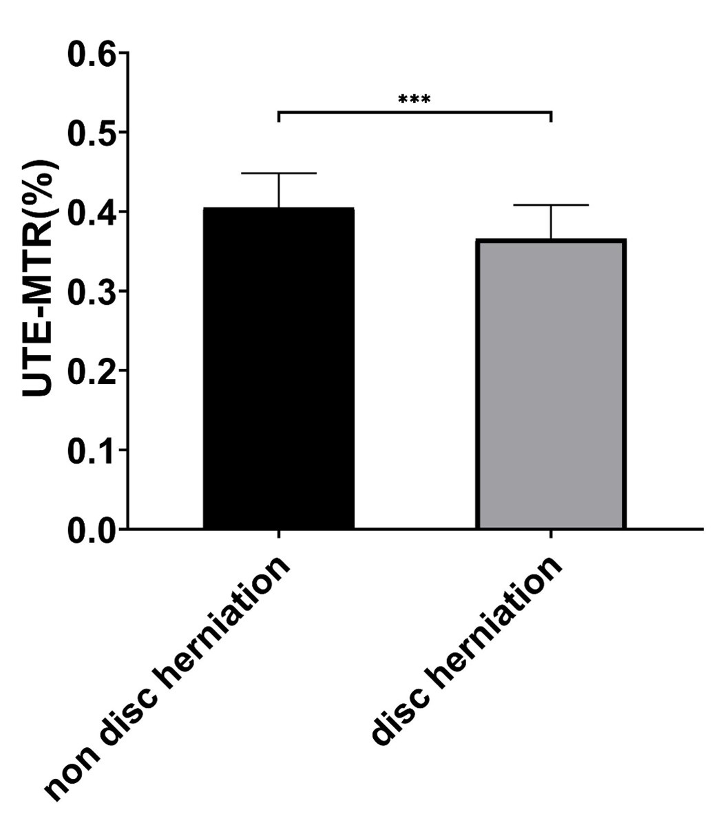
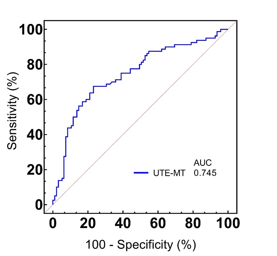
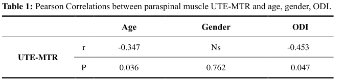
Figure 1 shows the representative UTE-MT images acquired from a 41‐year‐old male subject without disc herniation and a 48‐year‐old male subject with disc herniation. The paraspinal muscle UTE-MTR decreased significantly in patients with lumbar disc herniation compared to the those without disc herniation (Figure 2). The ROC curves of paraspinal muscle UTE-MTR showed good performance in differentiation of the patients with and without disc herniation (AUC = 0.745) (Figure 3). Moreover, our results showed that the paraspinal muscle UTE-MTR was negatively correlated with age (r = -0.347, P = 0.036) and ODI (r = -0.453, P = 0.047), while had no association with gender. The results were summarized in Table 1.Discussion and Conclusion
To our best knowledge, this is the first prospective study of applying UTE-MT measurement in paraspinal muscles to assess disc herniation in lumbar. Our results showed that the patients with disc herniation have significantly lower UTE-MTR values in lumbar paraspinal muscles compared to those without disc herniation. This means the muscle collagen matrix may partially lose its integrity and strength. More mechanical loading was transferred to spinal disc to support the body weight and movement [3], which may lead to a higher possibility of disc herniation. Our study suggests that the UTE-MTR measurements of paraspinal muscles may be a useful biomarker to predict disc herniation in lumbar.Acknowledgements
No acknowledgement found.References
[1] Beasley LE, Koster A, Newman AB, Harris TB, The Health ABC study (2009) Inflammation and race and gender differences in computerized tomography-measured adipose depots. Obesity (Silver Spring) 17:1062–1069.
[2] Ma Y, Chang EY, Carl M, Du J. Quantitative magnetization transfer ultrashort echo time imaging using a time-efficient3Dmultispokeconessequence. Magn Reson Med. (2017) 79:692–700.
[3] Zhu K, Hunter M, James A, Lim EM, Cooke BR, Walsh JP (2017) Discordance between fat mass index and body mass index is associated with reduced bone mineral density in women but not in men: the Busselton Healthy Ageing Study. Osteoporos Int 28:259–268.
Figures