1019
Contraction-Induced Changes in Acetylcarnitine in the Human Vastus Lateralis Muscle Detected by 1H MRS at 3T1Human Magnetic Resonance Center, Institute for Applied Life Sciences, University of Massachusetts Amherst, Amherst, MA, United States, 2Kinesiology, University of Massachusetts Amherst, Amherst, MA, United States
Synopsis
Using 1H-MRS at 3T, vastus lateralis muscle acetylcarnitine was measured in 8 young male subjects at rest and in response to incremental, dynamic knee extension contractions. Intramyocellular [acetylcarnitine] increased >3-fold in response to the contraction protocol, thus buffering mitochondrial acetyl coenzyme A and thereby supporting oxidative metabolism of glucose and fat in the citric acid cycle. Measurement of acetylcarnitine has the potential to be a useful tool for the investigation of muscle substrate use and metabolic flexibility. This work is the first demonstration of dynamic changes in muscle [acetylcarnitine] at 3T.
Introduction
Proton magnetic resonance spectroscopy (1H-MRS) is a non-invasive method for detecting and quantifying chemical compounds involved in metabolism and bioenergetics in vivo. Acetylcarnitine plays an essential role in energy metabolism and can be observed using 1H MRS in skeletal muscle1,2. The measurement of acetylcarnitine permits new insights into mitochondrial buffering of acetyl-CoA during muscular work and, potentially, the relative use of fat and carbohydrate as substrate in working muscle in vivo3. Detection of acetylcarnitine using long echo times has been demonstrated in skeletal muscle4, 5. Resting acetylcarnitine levels have been reported in type 2 diabetes, lean and obese sedentary adults, and endurance-trained athletes at 3T. However, to date changes in intramyocellular acetylcarnitine with high-intensity muscular work have been shown only at 7T. Our primary goal was to investigate potential contraction-related changes of acetylcarnitine in healthy volunteers using localized, single-voxel 1H-MRS at 3T.Materials and Methods
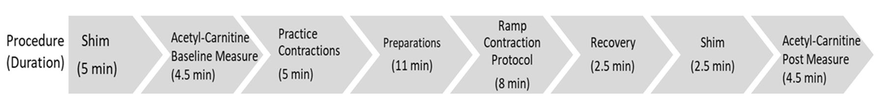
Proton MRS data were collected from the vastus lateralis muscles of 8 healthy male volunteers (median 27.5 y, range 25 – 35) in a Siemens 3T Skyra MRI scanner (Siemens Medical Solutions, Erlangen, Germany) operating on a VE11C platform. A double-tuned 1H/31P circular surface coil was used for the MRS acquisition. Gradient-echo scout images were used to confirm optimal leg positioning in the isocenter and correct coil positioning over the muscle. All volunteers were examined in the supine position. For each contraction protocol, the knee and ankle of the dominant leg were strapped to a custom MR-compatible ergometer6. The protocol consisted of 4 two-min stages of submaximal isotonic knee extensor contractions, with torque incrementing by ~3% of peak isometric torque at each stage, beginning at 6%. Acetylcarnitine was measured in the resting muscle before and after the contraction protocol in all subjects, using a PRESS sequence for volume localization. The voxel was placed in the vastus lateralis muscle, and the following parameters were applied: TR/TE=6000/350 ms (i.e., long echo), BW=2000 Hz, vector size=2048 points, averages=32, voxel size=36 ml (3:12 min scan time). The change in acetylcarnitine following contractions was evaluated by paired t-test. A non-water suppressed spectrum was acquired using a TE of 38 ms with 2 averages. All other parameters were identical. These spectra were acquired to compare the use of water and total creatine (TCr) as internal references. Acetylcarnitine was reported without correcting for T1 and T2 relaxation times, and a [TCr] of 30 mmol/kg wet wt was used as an internal reference for absolute quantification4. Spectra were processed using jMRUI 6.0 and quantified using the AMARES non-linear least squares algorithm. A schematic of the order of measurements is shown in Figure 1.Results
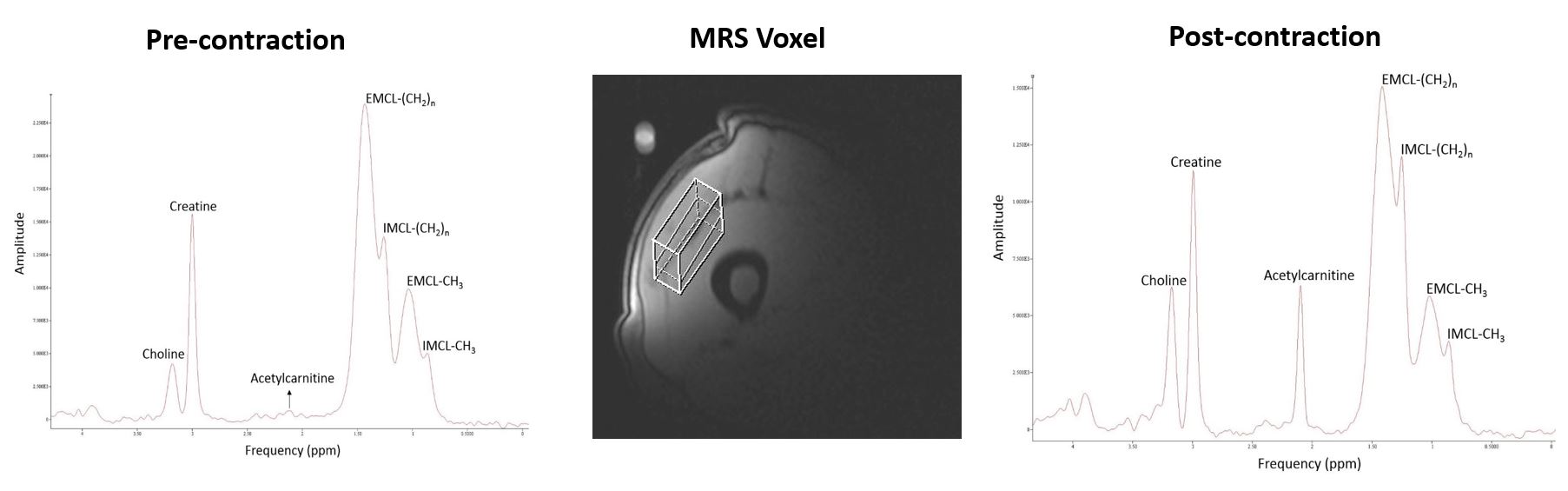
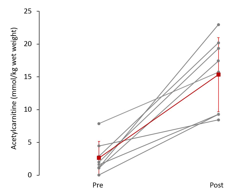
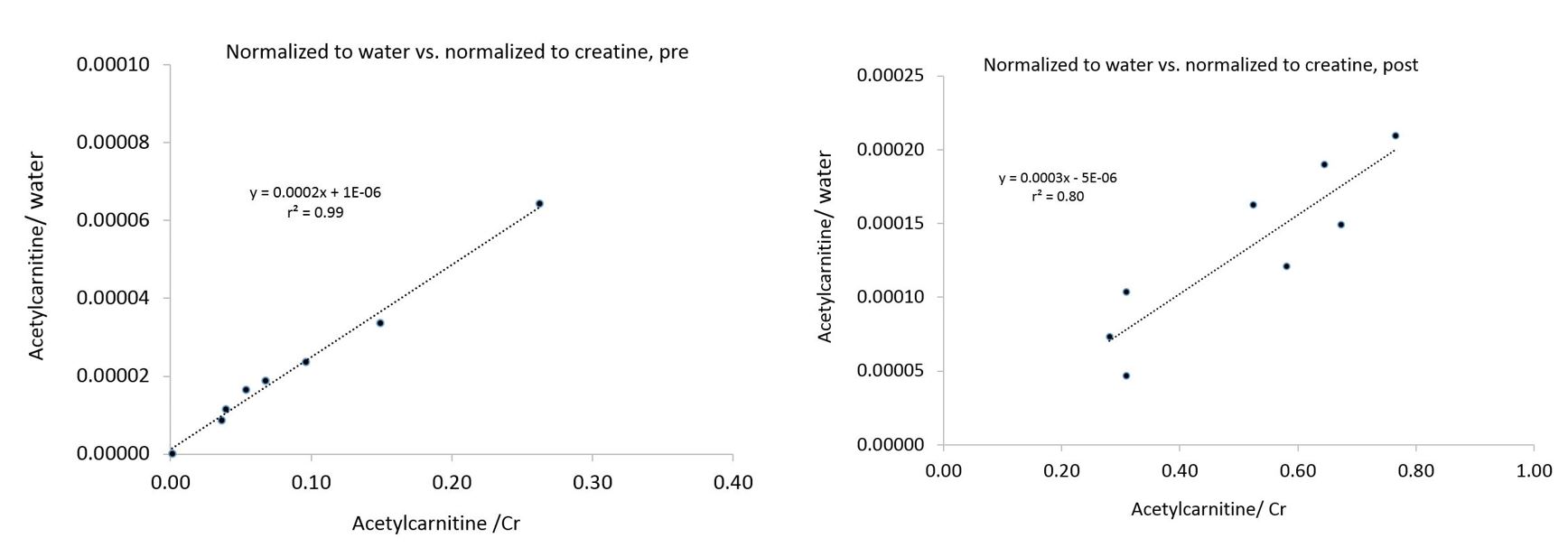
The acetylcarnitine peak was detected at 2.13 ppm both before and after the contraction protocol; in one case acetylcarnitine was not detected at rest prior to exercise. Figure 2 shows representative MRS voxel locations and corresponding spectra for a 29-year old participant before and after the contraction protocol. Overall, vastus lateralis muscle [acetylcarnitine] increased significantly in these healthy male volunteers in response to the contraction protocol (Figure 3), from 2.6 ±2.5 mmol/kg wet wt (mean ±SD) to 15.3 ±5.6 mmol/kg wet wt (p<0.0007). The same result was observed when acetylcarnitine was normalized to either TCr or water (data not shown). Indeed, as shown in Figure 4, there was a strong linear correlation between acetylcarnitine:TCr and acetylcarnitine:water both before (top, r2 =0.99, p<0.001) and after (bottom, r2 =0.80, p=0.003) the contraction protocol.Discussion and Conclusions
Using a long echo time sequence manages to suppress the overlapping lipid resonances and thus improve the sharpness of the acetylcarnitine singlet at 2.13 ppm. Short echo times are commonly used in 1H-MRS, as it helps reduce the T2 relaxation effect by allowing a higher SNR. To compensate for this SNR loss due to our use of a long echo, we used a relatively large voxel size to optimize our SNR. High-intensity contraction protocols are suggested to increase muscle acetylcarnitine. Studies quantifying acetylcarnitine using biopsies show a rapid increase with exercise7, which agrees with our results. Use of this technique in concert with well-established 31P-MRS measurements of muscle bioenergetics will enhance researchers’ ability to interrogate muscle metabolism and will be a powerful tool for the evaluation of muscle substrate utilization and metabolic flexibility.Acknowledgements
We thank Elena Bliss for assistance with all MRS data collections.References
1. Kreis R, Jung B, Rotman S, Slotboom J, Boesch C. Non‐invasive observation of acetyl‐group buffering by 1H‐MR spectroscopy in exercising human muscle. NMR in Biomedicine: An International Journal Devoted to the Development and Application of Magnetic Resonance In Vivo. 1999 Nov;12(7):471-6.
2. Boss A, Kreis R, Jenni S, Ith M, Nuoffer JM, Christ E, Boesch C, Stettler C. Noninvasive assessment of exercise-related intramyocellular acetylcarnitine in euglycemia and hyperglycemia in patients with type 1 diabetes using 1H magnetic resonance spectroscopy: a randomized single-blind crossover study. Diabetes care. 2011 Jan 1;34(1):220-2.
3. Muoio DM, Noland RC, Kovalik JP, Seiler SE, Davies MN, DeBalsi KL, Ilkayeva OR, Stevens RD, Kheterpal I, Zhang J, Covington JD. Muscle-specific deletion of carnitine acetyltransferase compromises glucose tolerance and metabolic flexibility. Cell metabolism. 2012 May 2;15(5):764-77.
4. Lindeboom L, Nabuurs CI, Hoeks J, Brouwers B, Phielix E, Kooi ME, Hesselink MK, Wildberger JE, Stevens RD, Koves T, Muoio DM. Long–echo time MR spectroscopy for skeletal muscle acetylcarnitine detection. The Journal of clinical investigation. 2014 Nov 3;124(11):4915-25.
5. Klepochová R, Valkovič L, Gajdošík M, Hochwartner T, Tschan H, Krebs M, Trattnig S, Krššák M. Detection and alterations of acetylcarnitine in human skeletal muscles by 1H MRS at 7 T. Investigative radiology. 2017 Jul;52(7):412.
6. Jaber Y, Jiminez Francisco E, Bartlett MF, Fitzgerald, Kent JA, Sup FC. Magnetic resonance compatible knee extension ergometer. J Biomech Engineering doi.org/10.1115/1.4046585 March 6, 2020.
7. Stephens FB, Constantin‐Teodosiu D, Greenhaff PL. New insights concerning the role of carnitine in the regulation of fuel metabolism in skeletal muscle. The Journal of physiology. 2007 Jun 1;581(2):431-44.
Figures