0974
Refined feature selection method for the depiction of pathology by texture analysis – Applicated on MR images of intervertebral discs fissures1University of Gothenburg, Gothenburg, Sweden, 2Institute of Clinical Sciences, Sahlgrenska Academy, University of Gothenburg, Gothenburg, Sweden
Synopsis
Texture analysis combined with attention mapping has the potential to identify the position of pathology normally not visible in MR images by exploiting inter-pixel relationships in magnetic resonance (MR) images. However, as pathology can influence adjacent tissue, the features used to identify the position of the pathology have to be selected with care. Here we present an easily implemented method that efficiently selects texture features that are only sensitive to the pathology and can improve the localization of pathology even when not visible in MR images.
Introduction
Within medicine, artificial intelligence is mainly used to automate cognitive tasks and improve consistency by automatically segmenting and classifying tissue and bone structures. However, the full potential of the technique is not yet adopted as it is possible to add new diagnostic information to detect pathology that is not directly visible in MR images. Texture analysis is an emerging technique used to characterize pathology in images by exploiting inter-pixel relationships. Combined with artificial neural networks (ANNs) and attention mapping [1] it has the potential to identify the position of pathology “invisible” to the human eye [2]. However, as pathology can cause tissue changes not only in close proximity to the pathology but also in adjacent tissue [3], the texture features used to identify the position of the pathology have to be selected with care to achieve satisfactory results.This study aims to develop a method that selects texture features which is sensitive to depicting pathology in MR images. To demonstrate the relevance of the method, it will be applied to identify the position of annular fissures in the intervertebral discs (IVDs) that, in MR images, are normally invisible to the naked eye.
Methods
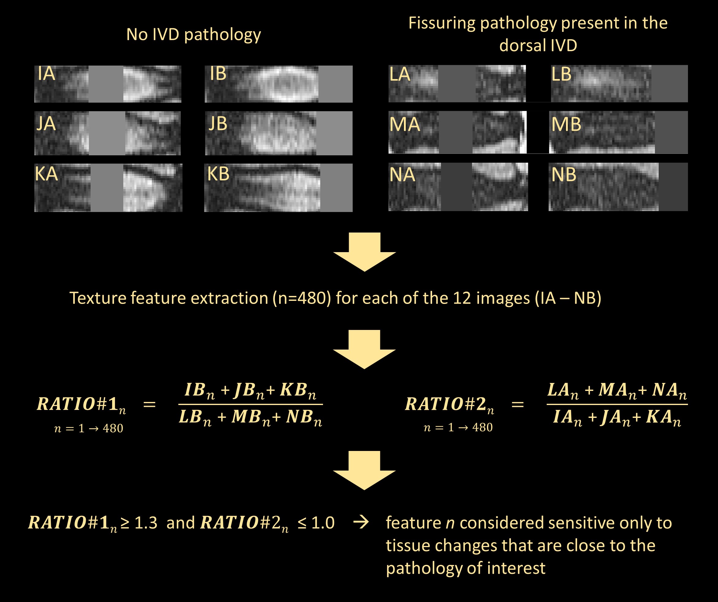
Texture features of five midsagittal T2-weighted (T2W) images of 123 IVDs in 43 patients (age 25-63 years, mean 45 years, 19 male) were calculated using RaCaT v1.18, an open-source Radiomics calculator tool [4]. A total of 480 texture features were calculated for each IVD describing the tissue in the whole IVD. The full list of features and a detailed description of each feature are described elsewhere [4, 5]. As the pathological fissures are not visible in the MR images, the position of the pathology was identified by studying CT discograms of the corresponding IVDs. Texture features sensitive only to the tissue close to the pathological fissures and insensitive to other tissues in the IVD were selected using the following method:First, six L2-L3 IVDs, three with and three without fissuring pathology were randomly selected. A 3D grey patch, sufficiently large to cover the pathology in the image volume, was created. The patch was overlaid onto the dorsal part of all IVDs, occluding possible pathology (Figure 1). The image volumes, overlaid with the occluding patch, were then analyzed with the RaCaT software producing 480 feature values for each image volume. The occluding patch was then moved to the central part of the IVD covering tissue with no pathology and a similar analysis was conducted producing another 6 sets of feature values. Next, for each texture feature, a ratio was calculated between the feature values calculated from the IVDs with pathology and a dorsal occlusion and the feature values calculated from the IVDs with no pathology and dorsal occlusion (RATIO#1). Similarly, ratios were also calculated for the IVDs with the central occlusion (RATIO#2). Finally, unique features that fulfilled RATIO#1 ≥ 1.3 and RATIO#2 ≤ 1.0 were considered to be sensitive mainly to the tissue close to the pathology.
The selected features, calculated from MR images, were analyzed with ANNs and attention mapping to determine the location of annular fissures in intervertebral discs. The method has partly been presented in an earlier ISMRM abstract [2].
Results
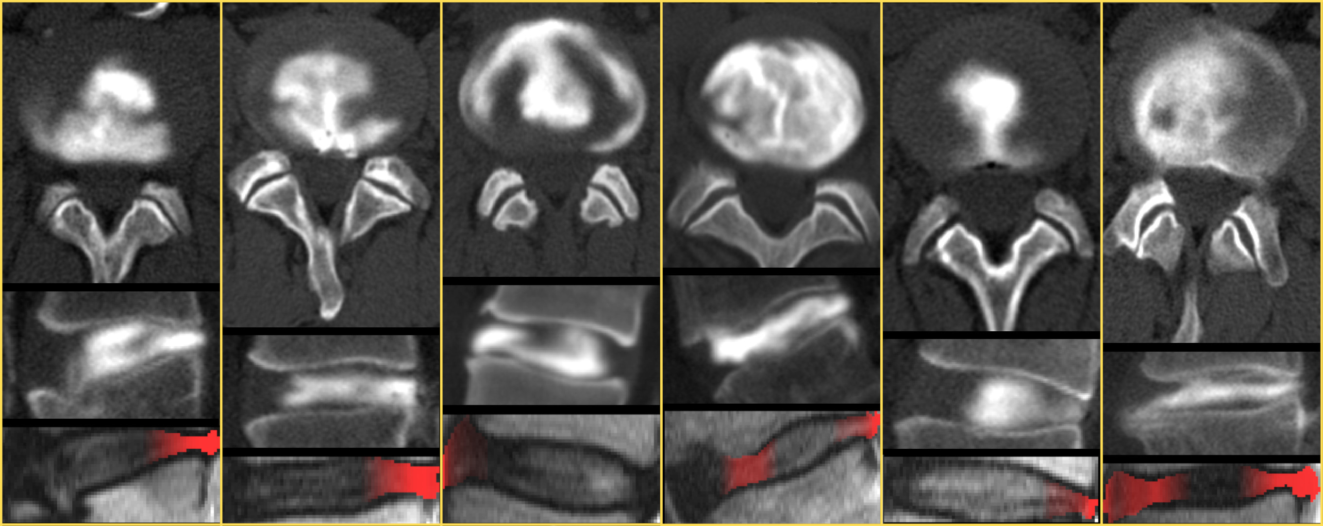
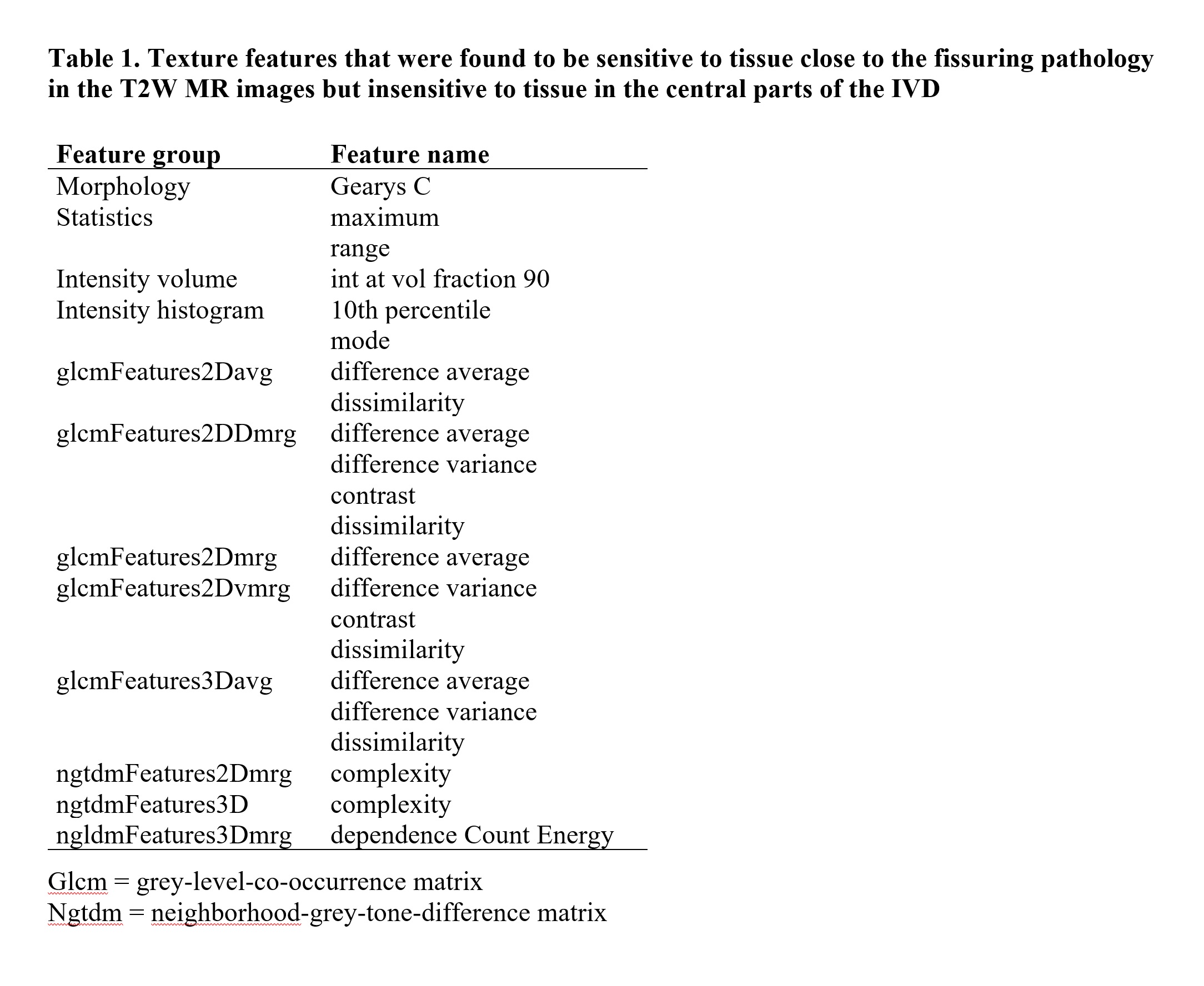
The proposed method could select texture features sensitive to pathology that was not visible to the naked eye in MR images. The method selected 22 out of 480 texture features that were alone capable to characterize the tissue close to the fissure pathology but were insensitive to tissue in other parts of the IVD that also have been affected by the pathology (Table 1). Combined with ANNs and attention mapping, the application of the described method permitted accurate localization of fissuring pathology in 87% of the analyzed IVDs (Figure 2).Discussion
This study demonstrated an easily implemented method to select texture features that can be used to localize pathology in MR images. As the number of texture features required to accurately characterize pathological tissue may differ between studies, the threshold ratios RATIO#1 and RATIO#2 might have to be adjusted. Since the method was applied to determine the location of annular fissures in T2W MR images, the threshold ratios presented were chosen to select texture features that have a substantially higher value in close proximity to the pathology. However, applied to other pathologies and different image contrasts, feature values that are substantially lower close to the pathology might be of equal importance why, in some studies, adding also another threshold of RATIO#1 ≤ 1 or less might be beneficial.Conclusion
The proposed method efficiently selects texture features sensitive only to pathological tissue. Combined with ANNs and attention mapping, the selected features can be used to determine the position of pathological tissue otherwise not visible in MR images.Acknowledgements
No acknowledgement found.References
1. Zeiler, M.D. and R. Fergus. Visualizing and understanding convolutional networks. in European conference on computer vision. 2014. Springer.
2. Waldenberg, C., et al., ISMRM 2021, Abstract: 4204 - Annular tears in the intervertebral disc can be detected and highlighted using conventional MRI, texture analysis and neural networks. International Society for Magnetic Resonance in Medicine, 2021.
3. Waldenberg, C., et al., Differences in IVD characteristics between low back pain patients and controls associated with HIZ as revealed with quantitative MRI. PLoS One, 2019. 14(8): p. e0220952.
4. Pfaehler, E., et al., RaCaT: An open source and easy to use radiomics calculator tool. PLOS ONE, 2019. 14(2): p. e0212223. 5. Zwanenburg, A., et al., Image biomarker standardisation initiative. arXiv preprint arXiv:1612.07003, 2016.
Figures